. 2024 Nov 30;15(12):1464.
doi: 10.3390/mi15121464.

Intestinal Cells-on-Chip for Permeability Studies

Affiliations

Intestinal Cells-on-Chip for Permeability Studies

Marit Keuper-Navis et al. Micromachines (Basel). .

Abstract

Background: To accurately measure permeability of compounds in the intestine, there is a need for preclinical in vitro models that accurately represent the specificity, integrity and complexity of the human small intestinal barrier. Intestine-on-chip systems hold considerable promise as testing platforms, but several characteristics still require optimization and further development.

Methods: An established intestine-on-chip model for tissue explants was adopted for intestinal cell monolayer culture. A 3D-printed culture disc was designed to allow cell culture in static conditions and subsequent permeability studies in a dynamic environment. Membrane characteristics and standardized read-outs were investigated and compared to traditional permeability studies under static conditions.

Results: By starting cultures outside the chip in conventional wells plates, the new cell disc design could support accurate cell monolayer formation for both Caco-2 and human enteroids. When transferred to the chip with laminar flow, there was accurate detection of barrier integrity (FD4 and Cascade Blue) and permeability (atenolol/antipyrine). Both flow and membrane characteristics had a significant impact on permeability outcomes.

Conclusions: This novel intestinal cell-on-chip system offers large flexibility for intestinal permeability studies, although it still requires validation with more compounds to reveal its full potential.

Keywords: cell monolayer; in vitro model; intestinal absorption; intestinal barrier; intestine-on-chip; permeability.


Conflict of interest statement

The authors declare no conflicts of interest. Hossein Eslami Amirabadi is employee of AZAR Innovations, and ARTIC Technologies B.V.; Markus Walles and Birk Poller are employees of Novartis Pharma AG. The paper reflects the views of the scientists, and not the company.

Figures

Figure 1
Figure 1
Schematic overview of the cell-on-chip system. (A) In-house developed 3D-printed disc with porous membrane and rubber ring for cell monolayer culture. Scale bar equals 2 mm. (B) Disc in a carrier for cell seeding and monolayer formation (static culture). (C) The disc with a cell monolayer can be transferred to a chip with dual flow. (D) Complete cell-on-chip system set-up, with eight chips connected to separate apical and basolateral medium reservoirs, with a peristaltic pump to create laminar flow, placed inside an incubator.
Figure 2
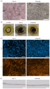
Figure 2
Intestinal cell monolayer culture on disc. (A) Brightfield images of epithelial cell monolayer cultures on a disc, derived from Caco-2 and enteroid cells. (B) Growth area coverage and monolayer viability indicated by MTT staining for Caco-2 and enteroid monolayers, compared to a disc with no cells. (C) Cell monolayer morphology assessed by whole-mount immunofluorescent detection of dapi (blue) and actin (orange). (D) HE staining of a cross-section of Caco-2 and enteroid monolayers, visualizing a single epithelial cell layer on the membrane. Scale bar equals 200 µm for (A) and 50 µm for (C,D).
Figure 3
Figure 3
Epithelial barrier integrity. (A) Apparent permeability values (Papp) for dextran-FITC 4 kDa (FD4) in cells-on-chip containing transparent Transwell membranes, determined between 1 and 2 h of perfusion and between 2 and 4 h of perfusion. Each dot represents a single chip. (B) Papp values for FD4 in cells-on-chip with translucent (dashed) and transparent (open) membranes, between 2 and 4 h of perfusion. (C) Cascade Blue 0.5 kDa (CB) permeability in cells-on-chip with translucent and transparent membranes, between 2 and 4 h of perfusion. n = 3–4 per group, with mean and SEM plotted.
Figure 4
Figure 4
Epithelial barrier permeability. (A) Schematic overview of the transport routes for antipyrine (transcellular) and atenolol (paracellular) over the intestinal epithelial barrier. Papp values for (B) antipyrine and (C) atenolol in intestinal cell monolayers on chip, measured between 2 and 4 h of perfusion, for both Caco-2 and enteroid monolayers compared to no-cell control chip, with (D) corresponding antipyrine/atenolol ratios. Cut-off value for a functional intestinal epithelial barrier, i.e., an A/A ratio > 2, is indicated by a dashed line. For all graphs, the dashed bars represent monolayers on translucent membranes, and the open bars represent transparent membranes. n = 3–4 per group, with mean +/− SEM displayed.
Figure 5
Figure 5
Active drug transport and metabolism in enteroid monolayers. (A) Schematic overview of the main routes for carrier-mediated active transport of rosuvastatin (BCRP) and metformin (OCT1) in the intestinal epithelial barrier, and phase I metabolism of midazolam (CYP3A4) and phase II metabolism of 7OH-coumarin (UGTs) by the epithelial cells. (BD) Papp values for rosuvastatin and metformin in enteroid monolayer cultures, determined between 1 and 2 h of incubation, including inhibitors to block active transport, in (B) static transparent Transwell inserts, (C) chip system with translucent membranes and (D) chip system with transparent membranes. (E) OH-midazolam formation over time in enteroid monolayers in static Transwell cultures. (F,G) 7OH-coumarin-glu formation over time in enteroid monolayers in (F) static Transwell inserters and (G) chip system. The drug cocktail included metformin (50 µM), rosuvastatin (10 µM), midazolam (10 µM) and 7-hydroxycoumarin (12 µM). The inhibitors added were elacridar (5 µM) and ketoconazole (5 µM). n = 4–6 per group, with mean +/− SEM displayed.

References

    1. France M.M., Turner J.R. The mucosal barrier at a glance. J. Cell Sci. 2017;130:307–314. doi: 10.1242/jcs.193482. - DOI - PMC - PubMed
    1. Schoultz I., Keita Å.V. The Intestinal Barrier and Current Techniques for the Assessment of Gut Permeability. Cells. 2020;9:1909. doi: 10.3390/cells9081909. - DOI - PMC - PubMed
    1. Turner J.R. Intestinal mucosal barrier function in health and disease. Nat. Rev. Immunol. 2009;9:799–809. doi: 10.1038/nri2653. - DOI - PubMed
    1. Rahman S., Ghiboub M., Donkers J.M., van de Steeg E., van Tol E.A.F., Hakvoort T.B.M., de Jonge W.J. The Progress of Intestinal Epithelial Models from Cell Lines to Gut-On-Chip. Int. J. Mol. Sci. 2021;22:13472. doi: 10.3390/ijms222413472. - DOI - PMC - PubMed
    1. Arian C.M., Imaoka T., Yang J., Kelly E.J., Thummel K.E. Gutsy science: In vitro systems of the human intestine to model oral drug disposition. Pharmacol. Ther. 2022;230:107962. doi: 10.1016/j.pharmthera.2021.107962. - DOI - PMC - PubMed

LinkOut - more resources