Review
. 2024 Nov 26;13(12):1039.
doi: 10.3390/pathogens13121039.

Advancements and Challenges in the Management of Prosthetic Valve Endocarditis: A Review

Affiliations
Review

Advancements and Challenges in the Management of Prosthetic Valve Endocarditis: A Review

Francesco Nappi. Pathogens. .

Abstract

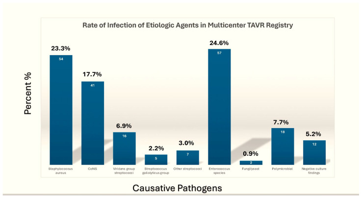
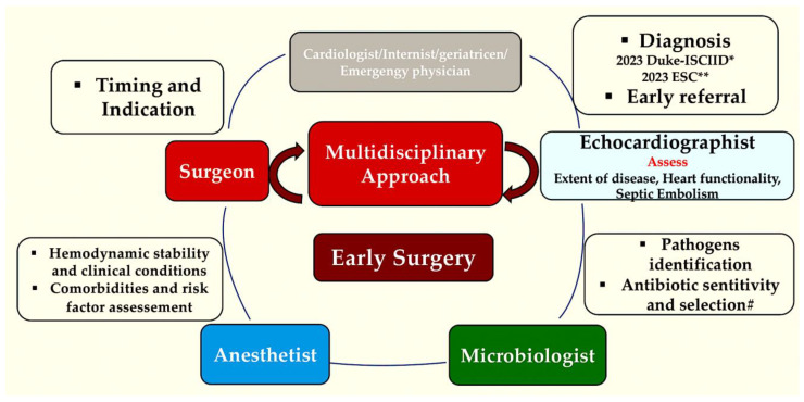
Prosthetic valve endocarditis (PVE) is the medical term used to describe a focus of infection involving a valvular substitute within the heart. It is a significant concern in the field of cardiology, and the epidemiology of PVE has seen notable developments over the last five decades. The disease currently affects an older demographic and is becoming increasingly prevalent in patients with transcatheter-implanted valves. It is imperative that we urgently address the significant challenges posed by PVE. It is a disease that has a wide range of potential aetiologies, clinical presentations, and courses. In developed countries, Staphylococcus aureus is now the predominant causative organism, resulting in an aggressive form of disease that frequently afflicts vulnerable or elderly populations. However, it is clear that Enterococcus species present a significant challenge in the context of PVE following TAVR procedures, given their elevated prevalence. The 2023 Duke/International Society for Cardiovascular Infectious Diseases infective endocarditis diagnostic criteria now include significant developments in microbiological and image-based techniques for diagnostic purposes, specifically the incorporation of fluorine-18 fluorodeoxyglucose positron emission tomography and computed tomography. These developments unequivocally enhance the diagnostic sensitivity for PVE, while maintaining the specificity. They do so in accordance with the results of studies conducted specifically for the purpose of validation. The lack of rigorous scientific studies and a shortage of funding and resources for research have led to a significant gap in our understanding. Randomized controlled trials could provide invaluable insight and guidance for clinical practice, but they are missing, which represents a major gap. It is clear that there is an urgent need for more research. PVE is a life-threatening condition that must be handled by a multidisciplinary endocarditis team at a cardiac centre in order to improve outcomes. The emergence of innovative surgical techniques has empowered clinicians to steer more patients away from surgical procedures, despite the presence of clear indications for them. A select group of patients can now complete parenteral or oral antimicrobial treatment at home. Additionally, antibiotic prophylaxis is the best option for individuals with prosthetic valves who are going to have invasive dental procedures. These individuals should be given antibiotics beforehand.

Keywords: TAVR; infective endocarditis; prosthetic valve endocarditis; vegetation.


Conflict of interest statement

The author declares no conflict of interest.

Figures

Figure 1
Figure 1
Illustration demonstrates the various pathophysiological processes (thrombosis, endocarditis, pannus growth, wear and tear, calcification, and deterioration) that can occur in mechanical and bioprosthetic valves, leading to both structural and nonstructural valve deterioration.
Figure 2
Figure 2
The illustration depicts the prevalence of etiological causative microorganisms documented in a multicentre cohort of the ICE *, as indicated by both the percentage of cases (A) and the number of cases (B). The columns representing the overall population, the NVE group, and the PVE group are coloured blue, orange, and green, respectively. Abbreviations: CoNS, coagulase-negative staphylococci; HACEK, Haemophilus species, Aggregatibacter actino mycetemcomitans, Aggregatibacter aphrophilus (previously, Haemophilus aphrophilus and Haemophilus paraphrophilus), Cardiobacterium hominis, Eikenella corrodens, and Kingella species; * ICE, International Collaboration on Endocarditis; NVE, native valve endocarditis; PVE, prosthetic valve endocarditis [29,30,31].
Figure 2
Figure 2
The illustration depicts the prevalence of etiological causative microorganisms documented in a multicentre cohort of the ICE *, as indicated by both the percentage of cases (A) and the number of cases (B). The columns representing the overall population, the NVE group, and the PVE group are coloured blue, orange, and green, respectively. Abbreviations: CoNS, coagulase-negative staphylococci; HACEK, Haemophilus species, Aggregatibacter actino mycetemcomitans, Aggregatibacter aphrophilus (previously, Haemophilus aphrophilus and Haemophilus paraphrophilus), Cardiobacterium hominis, Eikenella corrodens, and Kingella species; * ICE, International Collaboration on Endocarditis; NVE, native valve endocarditis; PVE, prosthetic valve endocarditis [29,30,31].
Figure 3
Figure 3
Illustration of the documented prevalence of etiological causative pathogens in a multicentre cohort study of the TVAR. Abbreviations: CoNS, coagulase-negative staphylococci; HACEK, Haemophilus species, Aggregatibacter actino mycetemcomitans, Aggregatibacter aphrophilus (formerly, Haemophilus aphrophilus and Haemophilus paraphrophilus), Cardiobacterium hominis, Eikenella corrodens, and Kingella species; values are n (patients). International TAVR Registry [30].
Figure 4
Figure 4
This figure shows the applied strategy of integrated imaging in patients with suspected infective endocarditis IE. In patients included in the subgroup with possible IE after initial evaluation by TTE and TEE, cardiac CT imaging, metabolic imaging, or transverse imaging of the head and viscera by CT scan or MRI is indicated to achieve a precise early diagnosis. For suspected IE 18 FDG-PET/CT or cross-sectional imaging by CT or MRI (or metabolic imaging), scans may assist with the detection of complications, such as abscess, mycotic aneurysm, infarct, or haemorrhage in patients with definite IE. Abbreviations: IE, infective endocarditis; FDG-PET/CT, fluorodeoxyglucose positron emission tomography/computed tomography; MRI, magnetic resonance imaging; TEE, transoesophageal echocardiography; TTE, transthoracic echocardiography. From Nappi et al. [7,36]. * Performed after TEE and discussed in Heart Team.
Figure 5
Figure 5
This illustration presents a comprehensive overview of multimodality imaging in the diagnosis of PVE (AD), elucidating its characteristics, strengths, and limitations, as well as its diagnostic performance in TTE (A,B), TEE (A,B), CT (A,B), 18F-FDG-PET-CT (C,D), and radiolabelled leukocytes SPECT-CT (C,D). Abbreviations: ECG, electrocardiogram; IE, infective endocarditis; PVE, prosthetic valve endocarditis; SPECT-CT, single-photon emission computed tomography/computed tomography; 18F-FDG-PET-CT, fluorine-18fluorodeoxyglucose positron emission tomography/computed tomography [7,11,116,117,118,119,120,121,122,123,124].
Figure 5
Figure 5
This illustration presents a comprehensive overview of multimodality imaging in the diagnosis of PVE (AD), elucidating its characteristics, strengths, and limitations, as well as its diagnostic performance in TTE (A,B), TEE (A,B), CT (A,B), 18F-FDG-PET-CT (C,D), and radiolabelled leukocytes SPECT-CT (C,D). Abbreviations: ECG, electrocardiogram; IE, infective endocarditis; PVE, prosthetic valve endocarditis; SPECT-CT, single-photon emission computed tomography/computed tomography; 18F-FDG-PET-CT, fluorine-18fluorodeoxyglucose positron emission tomography/computed tomography [7,11,116,117,118,119,120,121,122,123,124].
Figure 6
Figure 6
This illustration demonstrates the performance of the 2023 ESC and Duke/ISCVID diagnostic criteria among patients with prosthetic valve endocarditis and compares it with that of previous criteria. Abbreviations: ESC, European Society of Cardiology [7,8,11,125,126,127].
Figure 7
Figure 7
The accompanying illustration depicts the various phases of antibiotic treatment for prosthetic valve infective endocarditis. At present, two phases are under consideration for the treatment of prosthetic valve endocarditis: the first induction phase, which lasts for two weeks and is mandatory parenteral, and a second consolidation phase, in which some patients may be eligible for completion therapy through an outpatient parenteral antibiotic treatment (OPAT) regimen or orally. In this context, the term “free-living bacteria” refers to bacteria in the active replication phase, while “resting bacteria” are bacteria in the quiescent or dormant phase.
Figure 8
Figure 8
(AC) This illustration presents the overall clinical outcomes reported in multicentre cohorts ((A), ICE study; (B) GAMES study) with a comparison of native, prosthetic, and TAVR international registry endocarditis (C). The values are expressed as a percentage (n). The N value refers to patients with infective endocarditis involving the aortic prosthesis [29,30,31].
Figure 8
Figure 8
(AC) This illustration presents the overall clinical outcomes reported in multicentre cohorts ((A), ICE study; (B) GAMES study) with a comparison of native, prosthetic, and TAVR international registry endocarditis (C). The values are expressed as a percentage (n). The N value refers to patients with infective endocarditis involving the aortic prosthesis [29,30,31].
Figure 9
Figure 9
The following illustration presents the key take-home messages and clinical algorithm for the management of left-side endocarditis, based on the 2023 Duke/International Society for Cardiovascular Infectious Disease (ISCVID) criteria as outlined by the ESC in 2023. Abbreviations: BP; bioprosthesis; GMT, guide medical therapy; MP; mechanical prosthesis; MVE, mitral valve endocarditis; NVE, native valve endocarditis; PVE, prosthetic valve endocarditis.
Figure 10
Figure 10
A methodology for the treatment of heart valve endocarditis. TEE ultrasound enables the identification of three categories of patients for whom surgical intervention is recommended and involving elective (grey box), urgent (rose box), and urgent (blue box). The extent of the infectious process is the criterion that guides the choice between surgical options, with the aim of tailoring the conservative or extensive approach towards repair or replacement. The surgical option to be pursued, whether repair or replacement, is determined by the clinical and anatomic findings of the preoperative imaging. In the event of an infection process being confined to a localised area of the valve leaflet, it would be prudent to consider conservative surgery with aortic valve repair (illustrated by the light rose box). In cases of extensive infectious damage with severe pathoanatomical compromise of the valve, surgical replacement of the aortic valve is the recommended course of action (dark grey box). It is imperative that a multidisciplinary team makes a joint decision regarding the timing. In the case of an emergency surgical option (illustrated in the pink box), the surgical procedure to remove the infected valve must be requested within 24 h of the completion of the diagnostic procedure. In the case of the urgent surgical option (blue box), the procedure should be carried out within a few days of the indication being given. In patients who require the elective surgical option (green box), the procedure should be performed after a minimum of one to two weeks of antibiotic administration [22,97,153,154].
Figure 11
Figure 11
The illustrations present a clinical perspective and key messages derived from this review, emphasizing the most significant novel developments pertaining to the management of PVE. A coordinated, multidisciplinary approach to the diagnostic work-up and the necessity for prompt surgical referral, particularly in conditions with a high risk of embolisation and clinical deterioration with indications of heart failure, despite optimal antibiotic therapy, is advocated. PVE is a complex disease, and its management should, therefore, be undertaken on a multidisciplinary basis, with different professionals contributing their expertise to the decision-making process. It is proposed that a diagram should be constructed that highlights the role of each professional. It is recommended that the coordinated efforts of the team members converge towards the early referral of the patient to specialised centres with the aim of performing surgery as soon as it is feasible, according to the patient’s condition. It is advised that the surgical decision is not delayed, especially in cases of complex and extensive endocarditis, where radical and demolition surgical approaches with the use of homograft are advised [14,19,22,153,154,159,160,163,165]. The appropriate expert will be involved according to the individual characteristics, clinical needs, or comorbidities of each patient. * Ref [7]; ** Ref [8]; # Ref [9,10,37,125,126].

Similar articles

References

    1. Nappi F., Martuscelli G., Bellomo F., Avtaar Singh S.S., Moon M.R. Infective Endocarditis in High-Income Countries. Metabolites. 2022;12:682. doi: 10.3390/metabo12080682. - DOI - PMC - PubMed
    1. Cuervo G., Quintana E., Regueiro A., Perissinotti A., Vidal B., Miro J.M., Baddour L.M. The Clinical Challenge of Prosthetic Valve Endocarditis: JACC Focus Seminar 3/4. J. Am. Coll. Cardiol. 2024;83:1418–1430. doi: 10.1016/j.jacc.2024.01.037. - DOI - PubMed
    1. Nappi F., Spadaccio C., Mihos C. Infective endocarditis in the 21st century. Ann. Transl. Med. 2020;8:1620. doi: 10.21037/atm-20-4867. - DOI - PMC - PubMed
    1. Del Val D., Panagides V., Mestres C.A., Miró J.M., Rodés-Cabau J. Infective endocarditis after transcatheter aortic valve replacement: JACC state-of-the-art review. J. Am. Coll. Cardiol. 2023;81:394–412. doi: 10.1016/j.jacc.2022.11.028. - DOI - PubMed
    1. Otto C.M., Nishimura R.A., Bonow R.O., Carabello B.A., Erwin J.P., III, Gentile F., Jneid H., Krieger E.V., Mack M., McLeod C., et al. 2020 ACC/AHA guideline for the management of patients with valvular heart disease: A report of the American College of Cardiology/American Heart Association joint committee on clinical practice guidelines. J. Am. Coll. Cardiol. 2021;77:e25–e197. doi: 10.1016/j.jacc.2020.11.018. - DOI - PubMed

MeSH terms

Substances

LinkOut - more resources