. 2024 Nov 27;13(12):1041.
doi: 10.3390/pathogens13121041.

Functional Analysis of Promoters, mRNA Cleavage, and mRNA Secondary Structure on esxB-esxA in Mycolicibacterium smegmatis

Affiliations

Functional Analysis of Promoters, mRNA Cleavage, and mRNA Secondary Structure on esxB-esxA in Mycolicibacterium smegmatis

Ryan G Peters et al. Pathogens. .

Abstract

The ESX-1 secretion system is critical for the virulence of Mycobacterium tuberculosis as well as for conjugation in the saprophytic model Mycolicibacterium smegmatis. EsxB (CFP-10) and EsxA (ESAT-6) are secreted effectors required for the function of ESX-1 systems. While some transcription factors regulating the expression of esxB and esxA have been identified, little work has addressed their promoter structures or other determinants of their expression. Here, we defined two promoters, one located two genes upstream of esxB and one located immediately upstream, that contribute substantially to the expression of esxB and esxA. We also defined an mRNA cleavage site within the esxB 5' untranslated region (UTR) and found that a single-nucleotide substitution reprogramed the position of this cleavage event without impacting esxB-esxA transcript abundance. We furthermore investigated the impact of a double stem-loop structure in the esxB 5' UTR and found that it does not confer stability on a reporter gene transcript. Consistent with this, there was no detectable correlation between mRNA half-life and secondary structure near the 5' ends of 5' UTRs on a transcriptome-wide basis. Collectively, these data shed light on the determinants of esxB-esxA expression in M. smegmatis as well as provide broader insight into the determinants of mRNA cleavage in mycobacteria and the relationship between 5' UTR secondary structure and mRNA stability.

Keywords: 5′ UTR; ESX-1; EsxA; EsxB; Mycobacterium smegmatis; mRNA cleavage; mRNA processing; mRNA stability; mycobacteria; promoter; transcription start site; transcriptional regulation; type VII secretion system.


Conflict of interest statement

The authors declare no conflicts of interest. The sponsors had no role in the design, execution, interpretation, or writing of this study.

Figures
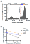
Figure 1
Figure 1
The esxB and esxA transcripts have higher abundance and longer half-lives than the transcripts encoded upstream. (A) RNAseq coverage for the M. smegmatis region encompassing PE35MS, PPE68MS, esxB, and esxA (msmeg_0063-0066). The data are from [15]. The genes of interest are shown to scale above the transcript abundance plot. Two previously mapped transcription start sites (TSSs) and an RNA cleavage site are shown with dashed black and red lines, respectively [20]. The second TSS appears at nearly the same position as the cleavage site in this graphic because they are only 4 nt apart. The coordinates are from NC.008596.1. (B) The degradation rates of the indicated transcripts were assessed by measuring transcript abundance following the addition of rifampicin (RIF) to block transcription. The slopes of the decay curves were compared by linear regression. The slopes of PE35MS and PPE68MS were steeper than the slopes of esxB and esxA (**** p < 0.0001 for each pairwise comparison between the two groups), while the slopes of PE35MS and PPE68MS were not significantly different from each other nor were the slopes of esxB and esxA (p > 0.05 for each pairwise comparison).
Figure 2
Figure 2
A second promoter is present between PPE68ms and esxB. (A) A schematic of a cassette cloned into an episomal plasmid lacking a promoter to test the expression of ESX-1 genes. The cassette contains the tsynA transcriptional terminator [21] to block transcription from spurious promoter sequences in the plasmid backbone. TSS1 and TSS2 were previously mapped in [20]. A previously mapped cleavage site (Martini et al. 2019) is indicated with scissors. The elements in the construct are not drawn to scale. The mutations to the putative promoter for TSS2 are shown. (B) The plasmids harboring variations of the cassette shown in A were transformed into an M. smegmatis strain with a deletion of msmeg_0062 (eccC1b) through msmeg_0066 (esxA). RNA was harvested from biological triplicate cultures in log phase, and qPCR was used to measure the expression of three genes and a region spanning the cleavage site. The data are representative of three independent experiments.
Figure 3
Figure 3
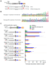
The promoter between PPE68ms and esxB initiates transcription 3–4 nucleotides upstream of a major cleavage site. (A) A schematic of the RNA 5′ ends between PPE68ms and esxB that arise from a previously mapped cleavage site [20] and a promoter defined in Figure 2 and here. (B) An example of Sanger sequencing traces from 5′ RACE used to map 5′ ends in the region between PPE68ms and esxB. The 5′ RACE adapter sequence is shown and indicated with yellow boxes. The RNA sequences being mapped are boxed in purple. The top trace is an example of a clean trace that primarily reflects the previously mapped RNA cleavage site. The bottom trace is an example of a mixed-peak trace where three sequences can be seen: that arising from the cleaved RNA and those arising from two transcripts made from TSS2, which has two possible start positions. The sequencing traces are representative of at least three separately sequenced gel bands.
Figure 4
Figure 4
mRNA cleavage can be reprogrammed by a point mutation but does not appear to affect the expression of PPE68ms, esxB, and esxA. (A) The constructs shown in Figure 2A were subject to a point mutation immediately downstream of the cleavage site (C→G, red font). (B) 5′ RACE was used to map RNA 5′ ends in the vicinity of the cleavage site. Shown are two traces from a strain harboring a construct with the cleavage site C→G mutation. While both traces contain mixed peaks, in the upper trace, a sequence beginning 1 nt upstream of the normal cleaved 5′ end can be seen, while in the lower trace, a sequence beginning 28 nt upstream of the normal cleaved 5′ end can be seen. The sequencing traces are representative of at least three separately sequenced gel bands. (C) The data from Figure 2 are shown with the addition of constructs with a point mutation downstream of the cleavage site. The data are representative of two independent experiments, each performed with biological triplicate cultures.
Figure 5
Figure 5
The secondary structure in the esxB 5′ UTR does not impact transcript half-life. Constructs were made, in which variants of the esxB 5′ UTR were linked to the coding sequence of yfp in integrating plasmids and transformed into wildtype M. smegmatis. (A) The predicted secondary structure of the esxB 5′ UTR and start codon (red) following cleavage. The hairpin structures are indicated in yellow and orange. This is designated “Full UTR” in subsequent experiments. The variants lacked one or both hairpins. The graphic was made with the ViennaRNA Web Services tool forna. (B) The abundance of the yfp transcript fused to the indicated esxB 5′ UTR variants was measured by quantitative PCR and expressed relative to the housekeeping gene sigA. The mean and SD of triplicate samples are shown. The means were compared by ANOVA followed by Dunnett’s multiple comparisons test to compare “Full UTR” to each of the variants. **, p < 0.01; ns, p > 0.05. (C) Half-lives of the yfp transcript when fused to the indicated esxB 5′ UTR variants were measured by quantitative PCR. The mean and 95% CI of triplicate datasets are shown. The upper error bar for the ∆hairpin 1 sample was clipped for visualization purposes. The differences between strains were not statistically significant (linear regression). (D) YFP protein abundance from transcripts with the indicated 5′ UTRs was measured by flow cytometry. A promoterless yfp gene was used as a control for autofluorescence. The median fluorescence from each of three biological replicates was determined and the mean and SD of those medians are shown. The means were compared by ANOVA, followed by Dunnett’s multiple comparisons test to compare “Full UTR” to each of the variants. ***, p < 0.001; **, p < 0.01; ns, p > 0.05.
Figure 6
Figure 6
The extent of secondary structure near the 5′ ends of 5′ UTRs does not generally correlate with transcript stability. The half-lives of ~1800 leadered M. smegmatis genes were previously reported and plotted here as a function of two metrics of 5′ UTR secondary structure [34]. Relationships were assessed by Spearman’s correlation. (A) The 20 nt at the 5′ end of each 5′ UTR was computationally folded by RNAfold (Vienna RNA Package) as described [34]. The minimum free energy (MFE) of the MFE structure for each 5′ UTR is plotted. (B) The 5′ third of each 5′ UTR was computationally folded in 20 nt windows with 10 nt overlaps using RNAfold (Vienna RNA Package) as described [34]. For each 5′ UTR, the mean MFE of the resulting structures is plotted.

References

    1. World Health Organization . Global Tuberculosis Report. WHO; Geneva, Switzerland: 2023.
    1. Bosserman R.E., Champion P.A. Esx Systems and the Mycobacterial Cell Envelope: What’s the Connection? J. Bacteriol. 2017;199:e00131-17. doi: 10.1128/JB.00131-17. - DOI - PMC - PubMed
    1. Passos B.B.S., Araujo-Pereira M., Vinhaes C.L., Amaral E.P., Andrade B.B. The role of ESAT-6 in tuberculosis immunopathology. Front. Immunol. 2024;15:1383098. doi: 10.3389/fimmu.2024.1383098. - DOI - PMC - PubMed
    1. Parbhoo T., Mouton J.M., Sampson S.L. Phenotypic adaptation of Mycobacterium tuberculosis to host-associated stressors that induce persister formation. Front. Cell. Infect. Microbiol. 2022;12:956607. doi: 10.3389/fcimb.2022.956607. - DOI - PMC - PubMed
    1. Rivera-Calzada A., Famelis N., Llorca O., Geibel S. Type VII secretion systems: Structure, functions and transport models. Nat. Rev. Microbiol. 2021;19:567–584. doi: 10.1038/s41579-021-00560-5. - DOI - PubMed

MeSH terms

LinkOut - more resources