. 2024 Dec 14;13(12):1108.
doi: 10.3390/pathogens13121108.

Susceptibility of Mouse Brain to MCMV Infection and Neuroinflammation During Ontogeny

Affiliations

Susceptibility of Mouse Brain to MCMV Infection and Neuroinflammation During Ontogeny

Fran Krstanović et al. Pathogens. .

Abstract

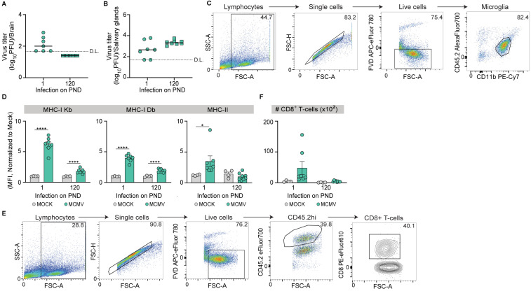
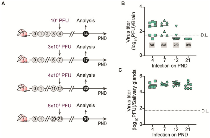
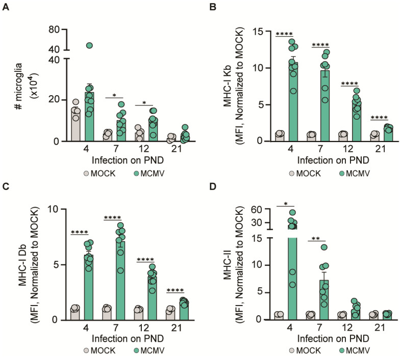
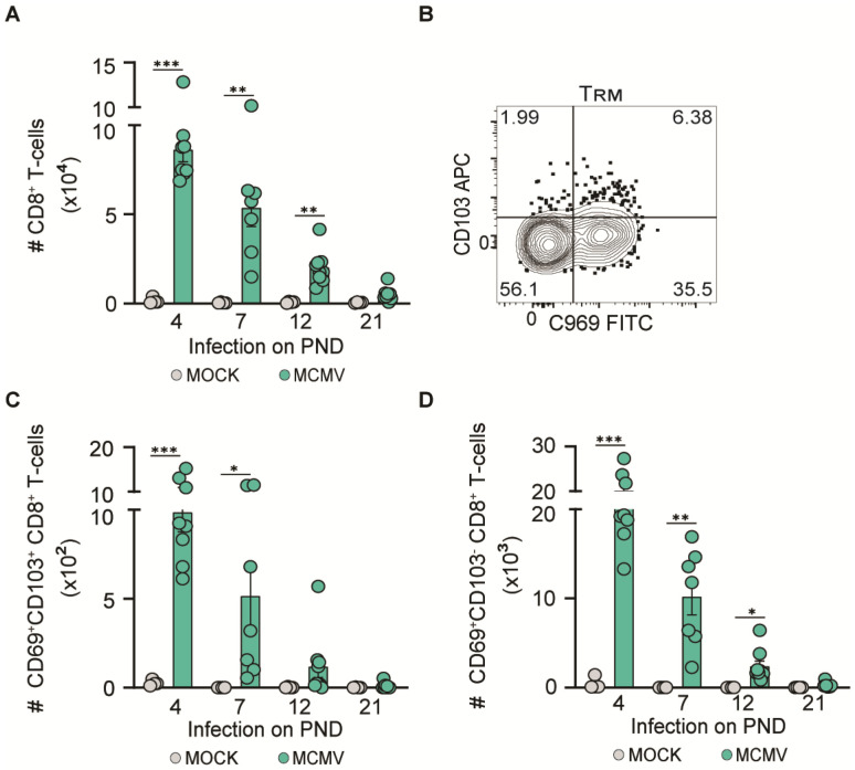
Human cytomegalovirus (HCMV) rarely infects the brain following infection of adult individuals. However, the virus readily infects the brain during congenital HCMV (cHCMV) infection, frequently causing severe neurodevelopmental and neurological sequelae. Interestingly, although the incidence of cHCMV infection is 0.5-1%, the proportion of congenitally infected individuals in which the virus manages to gain access to the brain is unknown. In this study, we used infection of mice with mouse cytomegalovirus (MCMV), the most commonly used experimental system for modeling HCMV disease in humans, to determine the impact of age on the susceptibility of the brain to cytomegalovirus infection and infection-mediated neuroinflammation. We demonstrate that infection of mice during various stages of neonatal development can lead to CMV neuroinvasion and inflammation. In contrast, MCMV infection does not result in MCMV neuroinvasion and neuroinflammation in weanling and adult mice. The obtained results establish a basis for elucidating the mechanisms of CMV neuroinvasion and the deleterious inflammatory response during ontogeny.

Keywords: congenital CMV; cytomegalovirus; neuroinflammation.


Conflict of interest statement

The authors declare no conflicts of interest.

Figures

Figure 1
Figure 1
MCMV brain infection and inflammatory responses in newborn and adult mice. Newborn (postnatal day 1 (PND 1)) and adult (PND 120) C57BL/6 mice were infected with MCMV i.p. and sacrificed 10 days post-infection (dpi). Viral titers in (A) the brain and (B) salivary glands were determined by the plaque assay. Results for individual mice are shown (circles and squares); horizontal bars indicate the median values; D.L., detection limit. (C) Gating strategy for microglia (live CD45.2intCD11b+). (D) Normalized expression levels of MHC-I Kb, MHC-I Db, and MHC-II on microglia were determined 10 dpi. (E) The gating strategy for CD8+ T-cells. (F) Absolute numbers of CD8+ T-cells were determined at 10 dpi. Mean values ± SEM are shown (n = 7–8 mice/time point). Asterisks indicate groups with significant differences in mean MHC-I and MHC-II expression values. * p ≤ 0.05, **** p ≤ 0.0001.
Figure 2
Figure 2
MCMV infection of mouse brain during ontogeny. (A) Schematic representation of the experiment. Weight-adjusted doses of MCMV (plaque-forming units, PFUs) used for the infection of mice of different ages are indicated. Viral titers in (B) the brain and (C) salivary glands 10 days post-infection were determined by the plaque assay (n = 7–9 mice/time point). Numbers in grey boxes indicate the proportion of mice which contained detectable amounts of infection virus in the brain. Results for individual mice are shown; horizontal bars indicate the median values; D.L., detection limit.
Figure 3
Figure 3
Microglia activation in MCMV-infected brains. C57BL/6 mice were infected with a weight-adjusted dose of MCMV i.p. on a different PND. The number of microglia (A) and the expression of (B) MHC-I Kb, (C) MHC-I Db, and (D) MHC-II on microglia were determined at 10 days post-infection. Mean values ± SEM are shown (n = 7–9 mice/time point). Asterisks indicate groups with significant differences in mean MHC-I and MHC-II expression values. * p < 0.05; ** p < 0.01; **** p ≤ 0.0001.
Figure 4
Figure 4
CD8+ T-cells migrate to the infected brain. C57BL/6 mice were infected with a weight-adjusted dose of MCMV i.p. at an indicated PND. The number of (A) CD8+ T-cells, (C) CD69+CD103+ CD8+ T-cells, and (D) CD69+CD103 CD8+ T-cells in the brain was determined 10 days post-infection. Mean values ± SEM are shown (n = 7–9 mice/time point). (B) Representative dot plot of CD103 and CD69 expression using brain CD8+ T-cells. Asterisks indicate groups with significant differences in mean MHC-I and MHC-II expression values. * p < 0.05; ** p < 0.01; *** p < 0.001.

Similar articles

References

    1. Al-Haddad B.J.S., Oler E., Armistead B., Elsayed N.A., Weinberger D.R., Bernier R., Burd I., Kapur R., Jacobsson B., Wang C., et al. The Fetal Origins of Mental Illness. Am. J. Obstet. Gynecol. 2019;221:549–562. doi: 10.1016/j.ajog.2019.06.013. - DOI - PMC - PubMed
    1. Fowler K., Mucha J., Neumann M., Lewandowski W., Kaczanowska M., Grys M., Schmidt E., Natenshon A., Talarico C., Buck P.O., et al. A Systematic Literature Review of the Global Seroprevalence of Cytomegalovirus: Possible Implications for Treatment, Screening, and Vaccine Development. BMC Public Health. 2022;22:1659. doi: 10.1186/s12889-022-13971-7. - DOI - PMC - PubMed
    1. Gandhi M.K., Khanna R. Human Cytomegalovirus: Clinical Aspects, Immune Regulation, and Emerging Treatments. Lancet Infect. Dis. 2004;4:725–738. doi: 10.1016/S1473-3099(04)01202-2. - DOI - PubMed
    1. Goodrum F. Human Cytomegalovirus Latency: Approaching the Gordian Knot. Annu. Rev. Virol. 2016;3:333–357. doi: 10.1146/annurev-virology-110615-042422. - DOI - PMC - PubMed
    1. Griffiths P., Baraniak I., Reeves M. The Pathogenesis of Human Cytomegalovirus. J. Pathol. 2015;235:288–297. doi: 10.1002/path.4437. - DOI - PubMed