. 2024 Nov 30;17(12):1615.
doi: 10.3390/ph17121615.

Suppression of NNK Metabolism by Anthocyanin-Rich Haskap Berry Supplementation Through Modulation of P450 Enzymes

Affiliations

Suppression of NNK Metabolism by Anthocyanin-Rich Haskap Berry Supplementation Through Modulation of P450 Enzymes

Madumani Amararathna et al. Pharmaceuticals (Basel). .

Abstract

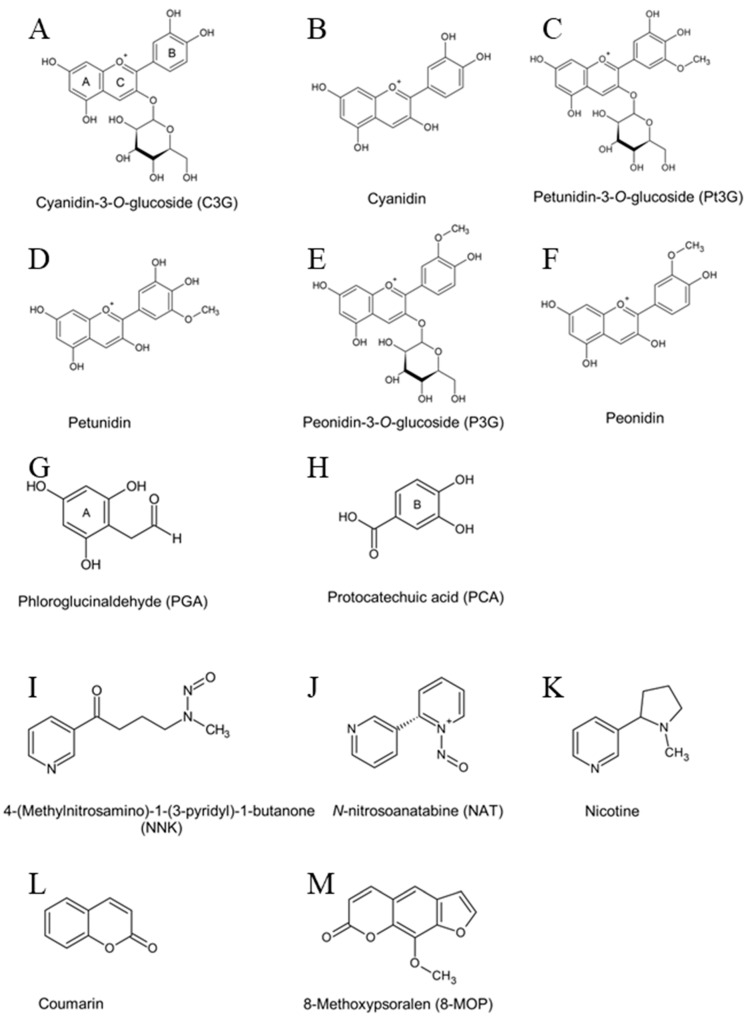
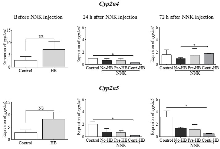
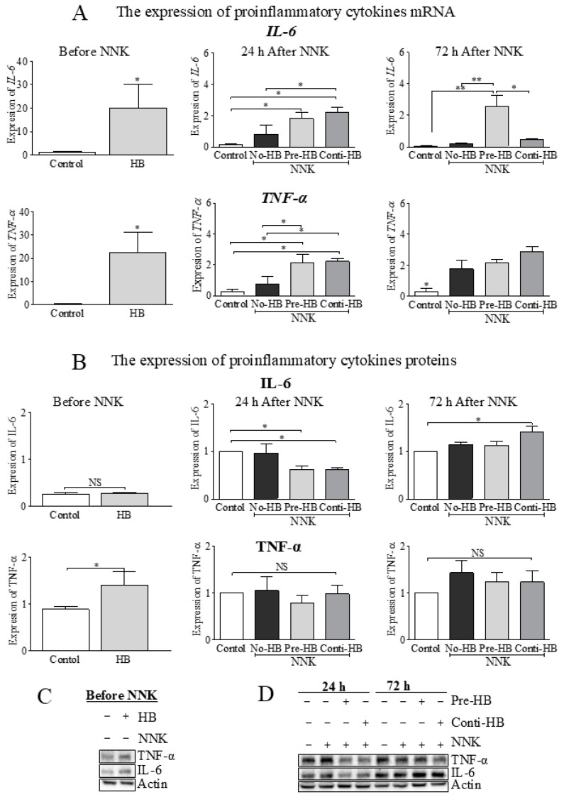
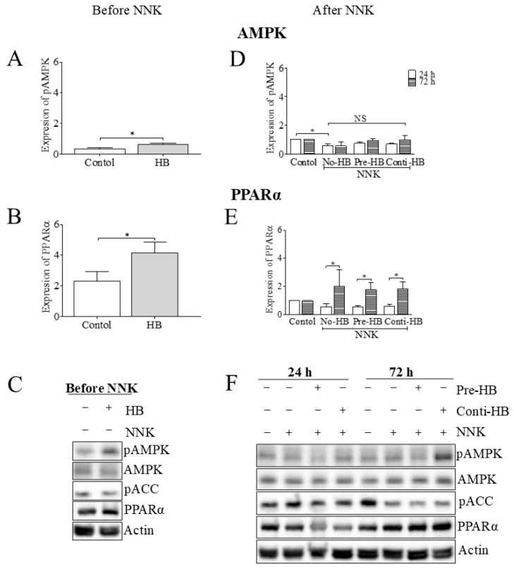
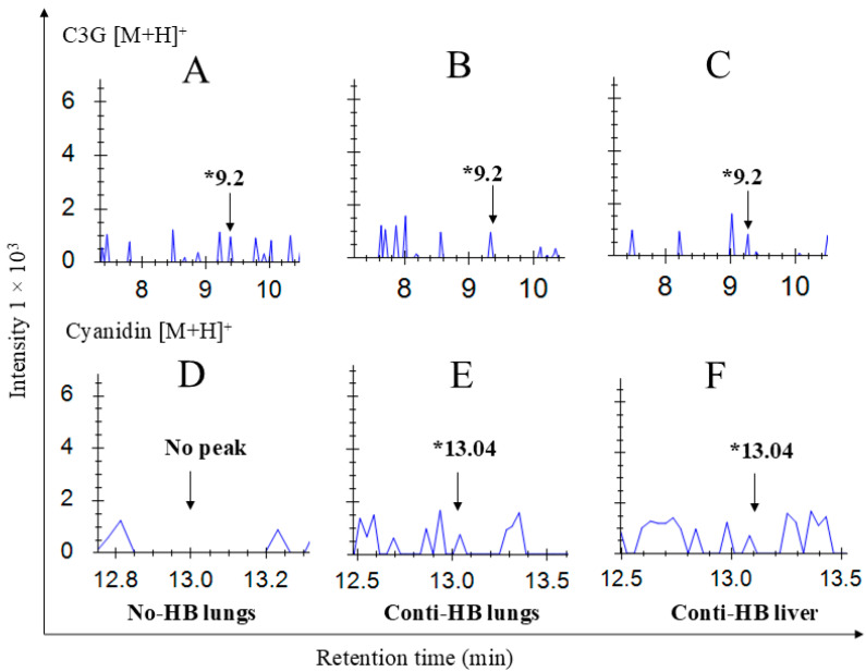
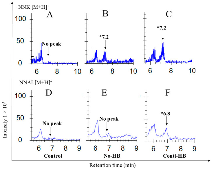
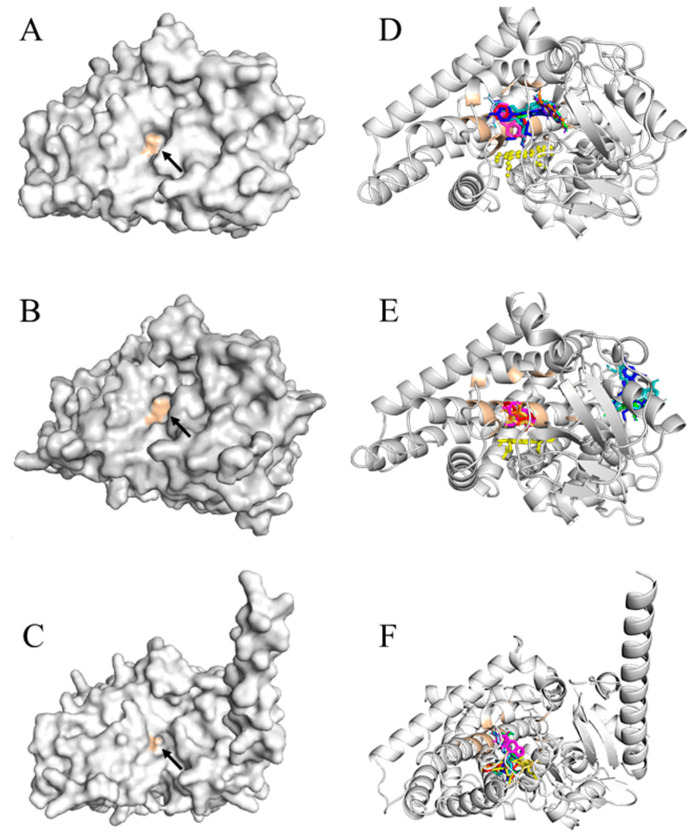
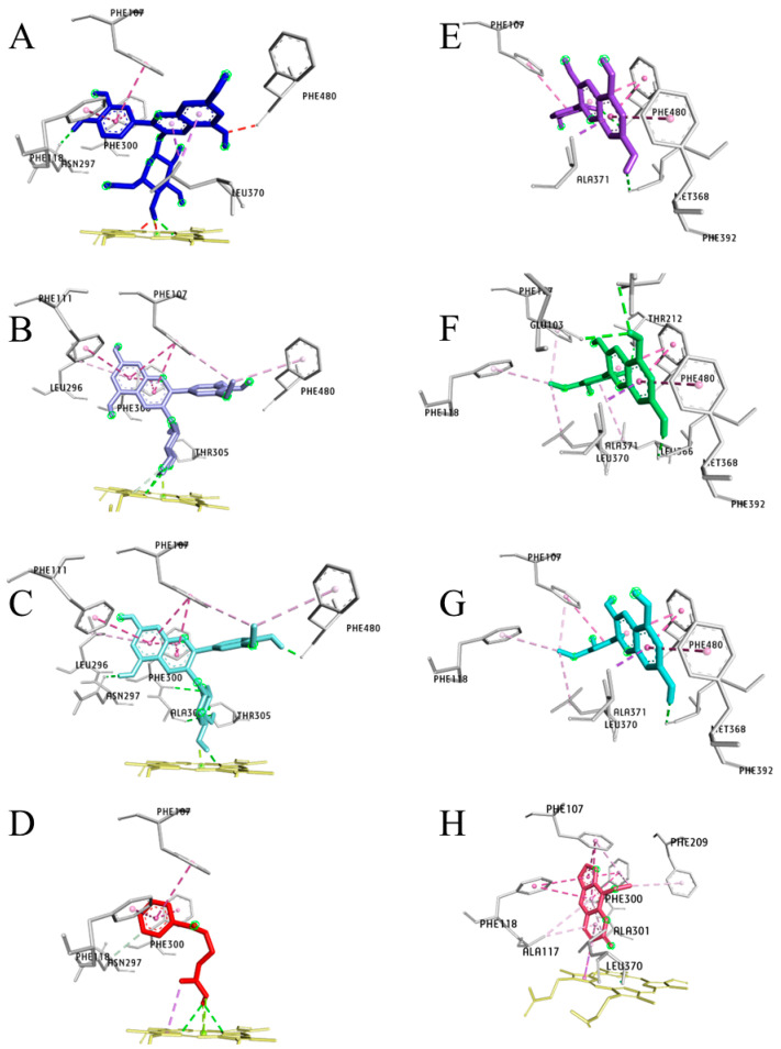
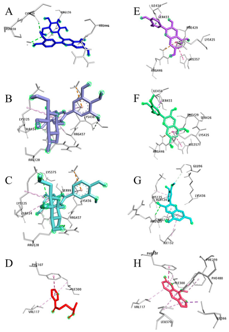
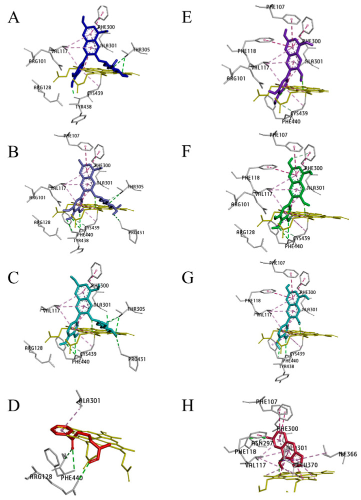
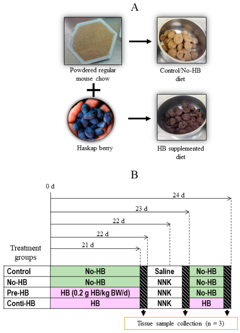
Oral supplementation of anthocyanins-rich haskap (Lonicera caerulea) berry (HB) reduces 4-(methylnitrosamino)-1-(3-pyridyl)-1-butanone (NNK)-induced lung tumorigenesis, cytotoxicity, DNA damage, and modulated inflammation in vitro and in vivo. The procarcinogen NNK is metabolically activated by cytochrome P450 (P450) enzymes, producing reactive metabolites that induce lung carcinogenesis. Hypothesis: Therefore, we hypothesized that the HB-modulated protective effect against NNK could be due to its ability to suppress P450 enzymes. Methods: HB (6 mg of cyanidin-3-O-glucoside [C3G] in 0.2 g of HB/mouse/day) was given to A/J mice as a dietary supplement following subsequent administration of NNK (100 mg/kg body weight). The liver tissues of mice were analyzed to determine the expression of P450s and metabolites. Results: HB upregulated the expression of cyp2a4 and cyp2a5 mRNA and nuclear receptor/transcription factor (PPARα) in NNK-deprived hepatic tissues. With NNK, HB downregulated the expression of cyp2a4 and cyp2a5 and facilitated the formation of non-carcinogenic NNK metabolites. Molecular docking indicated a high binding affinity and strong hydrophobic interactions between C3G and its major metabolites, peonidin-3-O-glucoside, petunidin-3-O-glucoside, peonidin and cyanidin with Cyp2a5 and with human P450 homologue CYP2A13. Conclusions: HB could be a potential dietary supplement to inhibit the P450 activated NNK carcinogenic metabolites formation. Hence, inhibiting the activation of NNK by lung CYP2A13 through dietary HB supplementation could be a strategy to reduce lung carcinogenesis among smokers. Understanding the effect of HB on the activity of CYP2A13 in human studies is necessary before recommending these natural compounds as therapeutics.

Keywords: blue honeysuckle; carcinogen; cytochrome P450; hepatic metabolism; lung cancer; smoking.


Conflict of interest statement

The authors declare no conflicts of interest. The funders had no role in designing the study, in the collection, analyses, or interpretation of data, in the writing of the manuscript, or in the decision to publish the results.

Figures

Figure 1
Figure 1
The chemical structures of haskap berry anthocyanins and their metabolites (AH), tobacco-specific carcinogens (IK), a common ligand (L), and an inhibitor (M) of cytochrome 450 enzymes. A, A-ring, B, B-ring, and C, C-ring of the C3G structure; a, the methyl carbon atom of NNK; b, the methylene carbon atom of NNK.
Figure 2
Figure 2
The effect of HB and NNK on the expression of hepatic cyp mRNA. Hepatic cyp2a4 and cyp2a5 mRNA in HB-fed A/J mice (6 mg of C3G in 0.2 g of HB/mouse/day for 21 days) compared to regular chow-fed A/J mice before (left column), 24 h (middle column) and 72 h (right column) after NNK (100 mg/kg body weight, i.p.) treatment. Pre-HB mice received HB before NNK, Conti-HB mice received HB before and after NNK, No-HB mice did not receive HB, and the controls were administered an equal volume of saline i.p. Each data set is expressed as mean ± SD. * The indicated groups were significantly different (p < 0.05). One-way ANOVA followed by Tukey’s pairwise comparison. NS = not significantly different. HB, haskap berry; NNK, 4-(methylnitrosamino)-1-(3-pyridyl)-1-butanone.
Figure 3
Figure 3
The effect of HB and NNK on the expression of pro-inflammatory cytokines in hepatic tissues of the A/J mice. (A) RT-PCR was performed to detect the expression of hepatic IL-6 and TNF-α mRNA before (left column), 24 h (middle column) and 72 h (right column) after NNK injection. (B) Western blotting was conducted to measure the levels of IL-6 and TNF-α proteins before (left column) and after (middle and right columns) the NNK exposure. (C) The expression of proteins before NNK and (D) 24 h and 72 h after NNK injection. One-way ANOVA with the Bonferroni test (at α = 0.05) for mean comparison and t-test were performed. * and ** indicate statistical differences at p ≤ 0.05 and 0.01, respectively, with mean ± SD. NS = not significantly different. HB, haskap berry; NNK, 4-(methylnitrosamino)-1-(3-pyridyl)-1-butanone; Pre-HB mice received HB before NNK; Conti-HB mice received HB before and after NNK; No-HB mice did not receive HB, and the controls were administered an equal volume of saline i.p.
Figure 4
Figure 4
The effect of HB and NNK on lipogenic enzyme expression in hepatic tissues of the A/J mice. (AC) Western blotting was conducted to measure phosphorylated AMPK and PPARα before NNK exposure, and (DF) 24 h and 72 h after NNK exposure, respectively. Two-way ANOVA with the Bonferroni test (at α = 0.05) for mean comparison and t-test were performed. * Indicates statistical differences at p ≤ 0.05 with mean ± SD. NS = not significantly different. HB, haskap berry; NNK, 4-(methylnitrosamino)-1-(3-pyridyl)-1-butanone; Pre-HB mice received HB before NNK; Conti-HB mice received HB before and after NNK; No-HB mice did not receive HB, and the controls were administered an equal volume of saline i.p.
Figure 5
Figure 5
Detection of cyanidin-3-O-glucoside (C3G) and its primary metabolites present in lung and liver tissues of A/J mice. Lung and liver tissues were collected following 24 h of NNK injection and analyzed by UPLC/ESI/Q-TOF/MS for anthocyanin metabolites. C3G and cyanidin were detected in HB-fed mice tissues after 9.2 min and 13.04 min, respectively. Chromatograms shown are (A) lungs of mice from No-HB, (B) lungs of mice from Conti-HB, (C) liver of Conti-HB, and cyanidin of (D) No-HB, (E) lung of Conti-HB, and (F) liver of Conti-HB. HB, haskap berry; NNK, 4-(methylnitrosamino)-1-(3-pyridyl)-1-butanone; Conti-HB mice received HB before and after NNK; No-HB mice did not receive HB; * the retention time of each compound.
Figure 6
Figure 6
Detection of NNK and its major metabolites present in the serum of A/J mice. Blood samples were collected from the control, No-HB and Conti-HB groups following 24 h NNK challenge and analyzed by UPLC/ESI/Q-TOF/MS. Chromatograms are (AC) NNK and (DF) NNAL. HB, haskap berry; NNK, 4-(methylnitrosamino)-1-(3-pyridyl)-1-butanone; NNAL, 4-(methylnitrosamino)-1-(3-pyridyl)-1-butanol; * the retention time of each compound; Conti-HB mice received HB before and after NNK; No-HB mice did not receive HB, and the controls were administered an equal volume of saline i.p.
Figure 7
Figure 7
Ligand binding pocket of cytochrome P450 target proteins with bound ligand complexes. Panels to the left: (A) CYP2A13, (B) CYP2A6, and (C) Cyp2a5 enzymes are illustrated as surface colored by chain (in grey) with the active binding site (in tint color) indicated in black arrows. Panels to the right: (D) CYP2A13, (E) CYP2A6, and (F) Cyp2a5 proteins are represented in the ribbon model, and ligands are represented in the stick model. Cyanidin-3-O-glucoside, blue; cyanidin, purple, petunidin-3-O-glucoside, teal; petunidin, green; peonidin-3-O-glucoside, pale cyan; peonidin, cyan; phloroglucinaldehyde, yellow orange; protocatechuic acid, orange; 4-(methylnitrosamino)-1-(3-pyridyl)-1-butanone (NNK), red; 8-methoxypsoralen, magenta; HEME group, yellow. Figures (AF) were generated using the PyMOL Molecular Graphics System, Version 2.0 Schrödinger, LLC, Mannheim, Germany.
Figure 8
Figure 8
The highest-affinity C3G and its key metabolites are depicted along with NNK and 8-MOP, a known cytochrome 450 enzyme inhibitor in the three-dimensional plots and show the interaction with the active site of CYP2A13 enzyme. All the ligands (A) cyanidin-3-O-glucoside (C3G), (B) petunidin-3-O-glucoside (Pt3G), (C) peonidin-3-O-glucoside (P3G), (D) 4-(methylnitrosamino)-1-(3-pyridyl)-1-butanone (NNK), (E) cyanidin, (F) petunidin, (G) peonidin, and (H) 8-methoxypsoralen (8-MOP) are bound to the active site of CYP2A13 at their highest binding energy state. Color coding of ligand-amino acid interactions, green, conventional hydrogen bonds; pink, Pi bonds; purple, Sigma bonds; red, donor-donor bonds. Figure (AH) are generated using BIOVIA Discovery Studio Visualizer V21.1.0.20198.
Figure 9
Figure 9
The high-affinity C3G and its metabolites are illustrated along with NNK and 8-MOP, a known cytochrome 450 enzyme inhibitor in the three-dimensional plots and show the interaction with the active site of the CYP2A6 enzyme. The ligands (A) cyanidin-3-O-glucoside (C3G), (B) petunidin-3-O-glucoside (Pt3G), (C) peonidin-3-O-glucoside (P3G), (E) cyanidin, (F) petunidin, and (G) peonidin are bound outside the active site of the CYP2A6 at their highest binding energy state. (D) 4-(methylnitrosamino)-1-(3-pyridyl)-1-butanone (NNK), and (H) 8-methoxypsoralen (8-MOP) are bound to the active site of CYP2A6. Color coding of ligand-amino acid interactions, green, conventional hydrogen bonds; pink, Pi bonds; purple, Sigma bonds; orange, Pi-Cation bond. Figures (AH) are generated using BIOVIA Discovery Studio Visualizer V21.1.0.20198.
Figure 10
Figure 10
The high-affinity of C3G and its primary metabolites are shown along with NNK and 8-MOP, a known cytochrome 450 enzyme inhibitor in the three-dimensional plots and show the interaction with the active site of Cyp2a5 enzyme. All the ligands (A) cyanidin-3-O-glucoside (C3G), (B) petunidin-3-O-glucoside (Pt3G), (C) peonidin-3-O-glucoside (P3G), (D) 4-(methylnitrosamino)-1-(3-pyridyl)-1-butanone (NNK), (E) cyanidin, (F) petunidin, (G) peonidin and (H) 8-methoxypsoralen (8-MOP) are bound to the active site of CYP2A5 at their highest binding energy state. Colour coding of ligand-amino acid interactions, green, conventional hydrogen bonds; pink, Pi bonds; purple, Sigma bonds; red, donor-donor bonds. Figures (AH) are generated using BIOVIA Discovery Studio Visualizer V21.1.0.20198.
Figure 11
Figure 11
Experimental procedure of A/J mouse model. (A) Haskap berry (HB)-rich diet (6 mg of C3G in 0.2 g of HB/mouse/day) was prepared by mixing powdered regular mouse chow with freeze-dried HB powder and formed into pellets. A control/no-HB diet was made using regular mouse chow without mixing HB. (B) Experimental groups and timeline. Control/No-HB diet was given to the control and No-HB groups throughout the experimental period. The Pre-HB group received an HB-rich diet (6 mg of C3G in 0.2 g of HB/mouse/day) only before the NNK injection, while the Conti-HB group received HB diet even after the NNK injection. A single intraperitoneal injection of NNK (100 mg/kg BW) was used to induce lung carcinogenesis in the no-HB, pre-HB and Conti-HB groups, and saline was used as a shame for the control group. Three mice from each group were euthanized after 21 days before the NNK injection, 24 h and 72 h after the NNK injection. Blood and liver tissues were collected for analysis.

Similar articles

Cited by

References

    1. Li Y., Hecht S.S. Metabolism and DNA adduct formation of tobacco-specific N-nitrosamines. Int. J. Mol. Sci. 2022;23:5109. doi: 10.3390/ijms23095109. - DOI - PMC - PubMed
    1. Paço L., Hackett J.C., Atkins W.M. Nanodisc-embedded cytochrome P450 P3A4 binds diverse ligands by distributing confor-mational dynamics to its flexible elements. J. Inorg. Biochem. 2023;244:112211. doi: 10.1016/j.jinorgbio.2023.112211. - DOI - PMC - PubMed
    1. Abdelmonem H., Abdelaal N.M., Anwer E.K.E., Rashwan A.A., Hussein M.A., Ahmed T.F., Khashana R., Hanna M.M. Decoding the role of CYP450 enzymes in metabolism and disease: A comprehensive review. Biomedicines. 2024;12:1467. doi: 10.3390/biomedicines12071467. - DOI - PMC - PubMed
    1. Deng X., Pu Q., Wang E., Yu C. Celery extract inhibits mouse CYP2A5 and human CYP2A6 activities via different mechanisms. Oncol. Lett. 2016;12:5309–5314. doi: 10.3892/ol.2016.5317. - DOI - PMC - PubMed
    1. Sellers E.M., Ramamoorthy Y., Zeman M.V., Djordjevic M.V., Tyndale R.F. The effect of methoxsalen on nicotine and 4-(methylnitrosamino)-1-(3-pyridyl)-1-butanone (NNK) metabolism in vivo. Nicotine Tob. Res. 2003;5:891–899. doi: 10.1080/14622200310001615231. - DOI - PubMed

LinkOut - more resources