. 2024 Dec 5;17(12):1633.
doi: 10.3390/ph17121633.

Study of Interactions Between Gadolinium-Based Contrast Agents and Collagen by Taylor Dispersion Analysis and Frontal Analysis Continuous Capillary Electrophoresis

Affiliations

Study of Interactions Between Gadolinium-Based Contrast Agents and Collagen by Taylor Dispersion Analysis and Frontal Analysis Continuous Capillary Electrophoresis

Chutintorn Somnin et al. Pharmaceuticals (Basel). .

Abstract

Background: Gadolinium-based contrast agents (GBCA) are widely used in magnetic resonance imaging (MRI) to enhance image contrast by interacting with water molecules, thus improving diagnostic capabilities. However, understanding the residual accumulation of GBCA in tissues after administration remains an area of active research. This highlights the need for advanced analytical techniques capable of investigating interactions between GBCAs and biopolymers, such as type I collagen, which are abundant in the body.

Objective: This study explores the interactions of neutral and charged GBCAs with type I collagen under physiological pH conditions (pH 7.4) using Taylor dispersion analysis (TDA) and frontal analysis continuous capillary electrophoresis (FACCE).

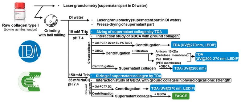
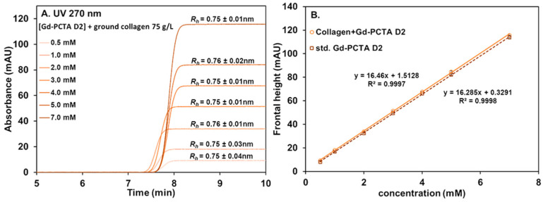
Methods: Collagen from bovine achilles tendon was ground using a vibratory ball mill to achieve a more uniform particle size and increased surface area. Laser granulometry was employed to characterize the size distributions of both raw and ground collagen suspensions in water. TDA was used to assess the hydrodynamic radius (Rh) of the soluble collagen fraction present in the supernatant.

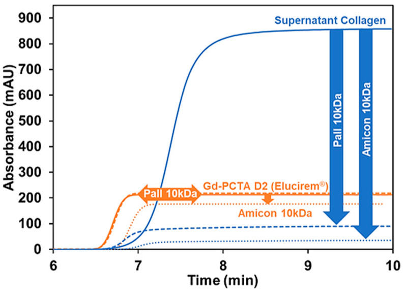
Results: From the TDA and FACCE results, it was shown that there were no significant interactions between the tested GBCAs and either the ground collagen or its soluble fraction at pH 7.4. Interestingly, we also observed that collagen interacts with filtration membranes, indicating that careful selection of membrane material, or the absence of filtration in the experimental protocol, is essential in interaction studies involving collagen.

Conclusion: These findings bring valuable insights into the behavior of GBCAs in biological systems with potential implications for clinical applications.

Keywords: Taylor dispersion analysis; collagen; frontal analysis continuous capillary electrophoresis; gadolinium-based contrast agents; interaction.


Conflict of interest statement

The authors declare no conflicts of interest.

Figures

Figure 1
Figure 1
Presentation of the workflow analysis for the study of GBCA/collagen interactions.
Figure 2
Figure 2
Laser diffraction granulometry of raw collagen and ground collagen 5 g/L suspended in water. The span number is a parameter that indicates the width of particle size distribution. For raw collagen, a significant fraction of the material flocculated before the laser analysis.
Figure 3
Figure 3
Frontal Taylorgrams (black) and its 1st derivative (blue) of 3.65 g/L supernatant collagen in 10 mM tris pH 7.4 with UV detection at 200 nm (A). Deconvolution of 1st derivative Taylorgram by Gaussian fitting with two Gaussian curves (B). Fit is plotted as a red dotted line. Experimental conditions: fused silica capillary of 65 cm total length (56.5 cm to UV detector) × 50 µm i.d. eluent: 10 mM tris buffer (pH 7.4). Mobilization pressure: 100 mbar. Experiments were performed at 37 °C. Supernatant collagen was prepared following the procedure described in Section 2.3.
Figure 4
Figure 4
Frontal Taylorgrams of Gd-PCTA D2 (0.5–7.0 mM) in the presence of ground collagen (75 g/L) and after centrifugation (A). Linear calibration curves of Gd-PCTA D2 obtained by frontal TDA in the absence (dotted line) and in the presence of ground collagen after centrifugation (solid line) at 270 nm (B). Experimental conditions: fused silica capillary of 65 cm total length (56.5 cm to UV detector) × 50 µm i.d. eluent: 10 mM tris buffer (pH 7.4). Mobilization pressure: 100 mbar. UV detection at 270 nm. Incubation of mixture: 37 °C 1000 rpm for 4 h. TDA experiments were performed at 37 °C.
Figure 5
Figure 5
Frontal Taylorgrams showing the potential retention of supernatant collagen (3.65 g/L, in blue) and Gd-PCTA D2 (2.5 mM, in orange) before and after filtration using Amicon® and Pall® devices with MWCO of 10 kDa. Experimental conditions: fused silica capillary of 65 cm total length (56.5 cm to UV detector) × 50 µm i.d. eluent: 10 mM tris buffer (pH 7.4). Mobilization pressure: 100 mbar. UV detection at 200 nm. Incubation of mixture: 37 °C 1000 rpm for 4 h. Sample volume: 60 µL. TDA experiments were performed at 37 °C.
Figure 6
Figure 6
The first derivative of the experimental mixture (black trace) and the sum of the individual (yellow) of 1.22 g/L supernatant collagen (blue) and 2.5 mM Gd-PCTA D2 (orange). Experimental conditions: fused silica capillary of 65 cm total length (56.5 cm to UV detector) × 50 µm i.d. eluent: 10 mM tris buffer, pH 7.4. Mobilization pressure: 100 mbar. UV detection at 200 nm. Incubation of mixture: 37 °C 1000 rpm for 4 h. Sample volume: 60 µL. TDA experiments were performed at 37 °C.
Figure 7
Figure 7
Frontal electropherograms showing the electrophoretic mobility of individual Gd-PCTA D2, Gd-BOPTA, Gd-DOTA, and supernatant collagen in the effective mobility scale (A). Timescale frontal electropherograms of 2.5 mM Gd-PCTA D2, Gd-BOPTA, Gd-DOTA standards (plain lines) and their mixtures in the presence of 1.825 g/L supernatant collagen (dotted lines) (B). Experimental conditions: PDADMAC coated capillary of 65 cm total length (56.5 cm to UV detector) × 50 µm i.d. eluent: 150 mM tris with 36 mM NaCl buffer (pH 7.4). UV detection at 200 and 270 nm. Incubation of mixture: 37 °C 1000 rpm for 4 h. Applied voltage: −15 kV (from inlet) for standard GBCA and applied co-pressure +50 mbar (from inlet) for supernatant collagen and the mixture. Sample volume: 120 µL. FACCE experiments were performed at 37 °C.

Similar articles

Cited by

References

    1. Dumas S., Jacques V., Sun W.-C., Troughton J.S., Welch J.T., Chasse J.M., Schmitt-Willich H., Caravan P. High relaxivity Magnetic Resonance Imaging Contrast Agents part 1. Investig. Radiol. 2010;45:600–612. doi: 10.1097/RLI.0b013e3181ee5a9e. - DOI - PMC - PubMed
    1. Lacampagne A., Gannier F., Argibay J., Garnier D., Guennec J.-Y.L. The stretch-activated ion channel blocker gadolinium also blocks L-type calcium channels in isolated ventricular myocytes of the guinea-pig. Biochim. Biophys. Acta Biomembr. 1994;1191:205–208. doi: 10.1016/0005-2736(94)90250-X. - DOI - PubMed
    1. Lauffer R.B. Paramagnetic metal complexes as water proton relaxation agents for NMR imaging: Theory and design. Chem. Rev. 1987;87:901–927. doi: 10.1021/cr00081a003. - DOI
    1. Port M., Idée J.-M., Medina C., Robic C., Sabatou M., Corot C. Efficiency, thermodynamic and kinetic stability of marketed gadolinium chelates and their possible clinical consequences: A critical review. BioMetals. 2008;21:469–490. doi: 10.1007/s10534-008-9135-x. - DOI - PubMed
    1. Caroline R., Sarah C., De Marie-Christine G., Jean-Marc I., Marc P. The role of phosphate on Omniscan® dechelation: An in vitro relaxivity study at pH 7. BioMetals. 2011;24:759–768. doi: 10.1007/s10534-011-9422-9. - DOI - PubMed

LinkOut - more resources