. 2024 Dec 6;17(12):1645.
doi: 10.3390/ph17121645.

An Azomethine Derivative, BCS3, Targets XIAP and cIAP1/2 to Arrest Breast Cancer Progression Through MDM2-p53 and Bcl-2-Caspase Signaling Modulation

Affiliations

An Azomethine Derivative, BCS3, Targets XIAP and cIAP1/2 to Arrest Breast Cancer Progression Through MDM2-p53 and Bcl-2-Caspase Signaling Modulation

Reetuparna Acharya et al. Pharmaceuticals (Basel). .

Abstract

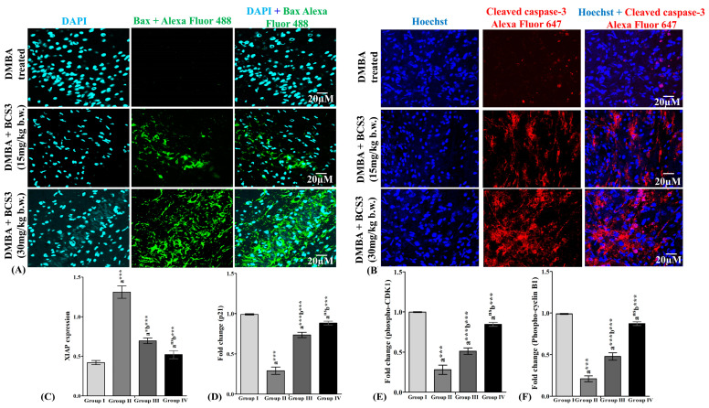
Background: Breast cancer influences more than 2 million women worldwide annually. Since apoptotic dysregulation is a cancer hallmark, targeting apoptotic regulators encompasses strategic drug development for cancer therapy. One such class of apoptotic regulators is inhibitors of apoptosis proteins (IAP) which are a class of E3 ubiquitin ligases that actively function to support cancer growth and survival. Methods: The current study reports design, synthesis, docking analysis (based on binding to IAP-BIR3 domains), anti-proliferative and anti-tumor potential of the azomethine derivative, 1-(4-chlorophenyl)-N-(4-ethoxyphenyl)methanimine (BCS3) on breast cancer (in vitro and in vivo) and its possible mechanisms of action. Results: Strong selective cytotoxic activity was observed in MDA-MB-231, MCF-7, and MDA-MB-468 breast cancer cell lines that exhibited IC50 values, 1.554 µM, 5.979 µM, and 6.462 µM, respectively, without affecting normal breast cells, MCF-10A. For the evaluation of the cytotoxic potential of BCS3, immunofluorescence, immunoblotting, and FACS (apoptosis and cell cycle) analyses were conducted. BCS3 antagonized IAPs, thereby causing MDM2-p53 and Bcl-2-Caspase-mediated intrinsic and extrinsic apoptosis. It also modulated p53 expression causing p21-CDK1/cyclin B1-mediated cell cycle arrest at S and G2/M phases. The in vitro findings were consistent with in vivo findings as observed by reduced tumor volume and apoptosis initiation (TUNEL assay) by IAP downregulation. BCS3 also produced potent synergistic effects with doxorubicin on tumor inhibition. Conclusions: Having witnessed the profound anti-proliferative potential of BCS3, the possible adverse effects related to anti-cancer therapy were examined following OECD 407 guidelines which confirmed its systemic safety profile and well tolerability. The results indicate the promising effect of BCS3 as an IAP antagonist for breast cancer therapy with fewer adverse effects.

Keywords: 7,12-dimethylbenz(a)anthracene; apoptosis; breast cancer; caspases; inhibitors of apoptosis protein; molecular docking.


Conflict of interest statement

The authors declare no conflicts of interest.

Figures

Figure 1
Figure 1
The in silico molecular docking studies of BCS3 with IAPs that represent (A) 2D and 3D docked poses of BCS3 with inhibitors of apoptosis proteins (XIAP (PDB ID: 3CLX), cIAP1 (PDB ID: 3MUP) and cIAP2 (PDB ID: 3M0A). In silico ADME prediction representing (B) BOILED-Egg model of BCS3 (C) SwissADME bioavailability radar report of BCS3 and (D) Prediction of possible metabolites of BCS3 by Phase I biotransformation.
Figure 2
Figure 2
Cytotoxic effects of various concentrations of BCS3 on the proliferation of breast cancer cells, (A) MDA-MB-231, (B) MCF-7, and (C) MDA-MB-468 through MTT assay for 24 h. (D) Time-course study of proliferation of MDA-MB-231 cells after incubation with varying doses of BCS3 (0.8, 1.6, and 3.2 µM) up to 96 h. Cell viability of the DMSO vehicle control group was set at 100%. Effect of varying doses of BCS3 (0.8, 1.6, and 3.2 µM) on the relative protein expressions of inhibitory apoptotic proteins (IAPs) as quantified by western blotting analysis in MDA-MB-231 cells (E) XIAP, (F) cIAP1, and cIAP2, respectively. Respective graphs represent each band of cIAP1 and cIAP2 measured by densitometry and normalized to corresponding p-97. (G) Protein analysis of MDM2 by ELISA in MDA-MB-231 cells. (H) Relative protein expression of p53 quantified by western blotting in MDA-MB-231 cells. The respective graph represents each band of XIAP, cIAP1, cIAP2, and p53 measured by densitometry and normalized to corresponding p-97. Mean ± SEM was calculated by replicating the experiment thrice (n = 3). Significant differences are shown as * p < 0.05, ** p < 0.01, *** p < 0.001, with the control group.
Figure 3
Figure 3
Relative protein expression of (A) MCL-1 quantified by western blotting in MDA-MB-231 cells. (B) Protein analysis of cytochrome c by ELISA in MDA-MB-231 cells. Relative protein expression of (C) SMAC, (D) Survivin, and (E) Apaf-1 quantified by western blotting in MDA-MB-231 cells. The respective graph represents each band of MCL-1, SMAC, Survivin, and Apaf-1 measured by densitometry and normalized to corresponding p-97 and β-actin. Cytotoxicity assessment through measurement of (F) ROS, (G) ATP content, and (H) lactate dehydrogenase (LDH) activity in MDA-MB-231 cells for 24 h post-treatment with varying doses of BCS3 (0.8, 1.6, and 3.2 µM). Mean ± SEM was calculated by replicating the experiment thrice (n = 3). Significant differences are shown as * p < 0.05; ** p < 0.01 and *** p < 0.001with the control group.
Figure 4
Figure 4
Analysis of mitochondrial membrane potential of BCS3 on MDA-MB-231 cells verified using (A) JC-1 and (B) JC-10 levels. Mean ± SEM was calculated by replicating the experiment thrice (n = 3). Significant differences are shown as * p < 0.05; ** p < 0.01 and *** p < 0.001 with the control group. (C) Determination of apoptotic cell death in MDA-MB-231 cells documented by annexin V-Alexa Fluor 647 (a647)/PI double staining and FACS analysis after treatment with BCS3 (0.8, 1.6, and 3.2 µM). Annexin V[+] and PI[−]: apoptotic cells (N4); annexin V[+] and PI[+]: Late apoptotic cells (N2); annexin V[−] and PI[+]: necrotic cells (N1) and annexin V[−] and PI[−]: Living cells (N3). (D) Bar diagram depicting the percentage of apoptosis caused by a number of early and late apoptotic cellular populations after BCS3 treatment. Representation of western blot analysis showing the protein expression of (E) cleaved caspase-8, (F) cleaved caspase-3, and cleaved caspase-9 after 72 h of treatment with BCS3 (0.8, 1.6, and 3.2 µM). Respective graphs represent each band of caspases (3 and 9) measured by densitometry and normalized to corresponding α-tubulin and p-97. Mean ± SEM was calculated by replicating the experiment thrice (n = 3). Significant differences are shown as * p < 0.05; ** p < 0.01 and *** p < 0.001 with a control group.
Figure 5
Figure 5
Immunofluorescence analysis depicting the elevation of (A) cleaved caspase-8 and (B) cleaved caspase-3 in vitro in MDA-MB-231 cells after 72 h of treatment with varying doses of BCS3 (1.6 and 3.2 µM) where Alexa 633 (red fluorescence) denotes caspase-3 and -8 expressions, respectively, and Hoechst (blue fluorescence) denotes cell nuclei locations. The scale bars of immunofluorescence analysis are represented as 10 µm.
Figure 6
Figure 6
(A) Immunofluorescence analysis depicting the elevation of Bax in vitro in MDA-MB-231 cells after 72 h of treatment with varying doses of BCS3 (1.6 and 3.2 µM) where FITC (green fluorescence) denotes Bax expression and DAPI (light blue fluorescence) denotes cell nuclei locations. (B) Immunofluorescence analysis depicting the depletion of the Bcl-2 protein in vitro in MDA-MB-231 cells after 72 h of treatment with varying doses of BCS3 (1.6 and 3.2 µM) where Alexa 647 (red fluorescence) denotes Bcl-2 expression and DAPI (light blue fluorescence) denotes cell nuclei locations. The scale bars are represented as 20 µm.
Figure 7
Figure 7
Flow cytometric analysis indicating (A) cell cycle progression of untreated MDA-MB-231 cells, the effect of different concentrations of BCS3 (0.8 µM, 1.6 µM and 3.2 µM) on MDA-MB-231 cells, (B) percentage of cell cycle distribution protein analysis of (C) p21, (D) phospho-CDK1, and (E) phospho-cyclin B1 by ELISA. Mean ± SEM was calculated through replicating the experiment thrice (n = 3). Significant differences are shown as * p < 0.05; ** p < 0.01 and *** p < 0.001 with the control group.
Figure 8
Figure 8
(A) Effect of BCS3 on body weight (in grams) of DMBA-induced breast cancer in experimental rats. (B) Effect of BCS3 on tumor volume of DMBA-induced breast cancer in experimental rats. (C) Histopathological observation (400×) of hematoxylin and eosin (H&E)-stained mammary tissues where scale bare represents the 50 µm section. Yellow arrows represent normal acini and ductules in normal groups. Black arrows show ductal hyperplasia in DMBA-treated group with abnormal tissue architecture. BCS3-treated groups show restoration of ductal architecture by yellow arrows. (D) Representation of TUNEL fluorescent images of a section of tumor excised from experimental animals from each group. Cell nuclei locations are indicated with Hoechst (blue fluorescence) and apoptotic cell location is indicated by Alexa Fluor 594 (purple fluorescence). The scale bars of immunofluorescence analysis are represented as 100 µm. (E) Evaluation of the rate of tumor cell apoptosis in mammary tissues of rats from each experimental group in accordance with images of TUNEL assay. Comparisons: a-Groups II, III, and IV with Group I; b-Groups III and IV compared to Group II; *** p < 0.001, ** p < 0.01 and * p < 0.05. Group I: Control; Group II: induced control (DMBA, 20 mg in 0.5 mL of olive oil); Group III: DMBA (20 mg) + BCS3 (15 mg/kg, b.w); and Group IV: DMBA (20 mg) + BCS3 (30 mg/kg, b.w). DMBA: 7,12-dimethylbenz(a)anthracene.
Figure 9
Figure 9
(A) Immunofluorescence analysis of Bax in mammary tissues belonging to cancer-bearing animals. Expression of Bax was observed to enhance upon BCS3 treatment which was observed by Alexa Fluor 488 (green fluorescence) representing Bax expression and DAPI (light-blue fluorescence) denoting cell nuclei locations. The scale bars of immunofluorescence analysis are represented as 20 µm. (B) Immunofluorescence analysis of cleaved caspase-3 in mammary tissues belonging to cancer-bearing animals. Expression of cleaved caspase-3 was observed to enhance upon BCS3 treatment which was observed by Alexa Fluor 647 (red fluorescence) representing cleaved caspase-3 expression and Hoechst (blue fluorescence) denoting cell nuclei locations. The scale bars of immunofluorescence analysis are represented as 20 µm. (C) Effect of BCS3 on protein expression of XIAP. Effect of BCS3 on expression of cell cycle proteins (D) p21, (E) phospho-CDK1, and (F) phospho-cyclin B1 on mammary tissues of experimental animals. Comparisons: a-Groups II, III, and IV with Group I; b-Groups III and IV compared to Group II; *** p < 0.001, * p < 0.05, and ns p > 0.05. Group I: Control; Group II: induced control (DMBA, 20 mg in 0.5 mL of olive oil); Group III: DMBA (20 mg) + BCS3 (15 mg/kg, b.w); and Group IV: DMBA (20 mg) + BCS3 (30 mg/kg, b.w). DMBA: 7,12-dimethylbenz(a)anthracene.
Figure 10
Figure 10
(A) Dose-inhibition response curve for BCS3 and doxorubicin output; (B) dose-response matrix (inhibition)/heat map for BCS3 and doxorubicin where the degree of red is positively related to inhibition ratio; (C) drug interaction landscape; and (D) synergy plot of combined treatment of BCS3 and doxorubicin calculated with zero interaction potency (ZIP) reference model of Synergyfinder.
Figure 11
Figure 11
Representative photomicrograph of vital organs (liver, kidney, brain, and lungs) of normal experimental rats from repeated dose sub-acute oral toxicity study. Black arrows represent the presence of lipid droplets caused by hepatic steatosis. The kidney represents cloudy swelling (yellow arrows) and leukocytic infiltration as an effect of repeated administration of 300 mg/kg/day BCS3 dose. Group I: vehicle control group; Group II: 30 mg/kg/day of BCS3; Group III: 300 mg/kg/day of BCS3 (28 days); Group IV: satellite control group; and Group V: 300 mg/kg of BCS3 (satellite groups: 42 days); n = 5.

Similar articles

Cited by

References

    1. Bray F., Laversanne M., Sung H., Ferlay J., Siegel R.L., Soerjomataram I., Jemal A. Global cancer statistics 2022: GLOBOCAN estimates of incidence and mortality worldwide for 36 cancers in 185 countries. CA Cancer J Clin. 2024;74:229–263. doi: 10.3322/caac.21834. - DOI - PubMed
    1. Yersal O., Barutca S. Biological subtypes of breast cancer: Prognostic and therapeutic implications. World J. Clin. Oncol. 2014;5:412–424. doi: 10.5306/wjco.v5.i3.412. - DOI - PMC - PubMed
    1. Arnold M., Morgan E., Rumgay H., Mafra A., Singh D., Laversanne M., Vignat J., Gralow J.R., Cardoso F., Siesling S., et al. Current and future burden of breast cancer: Global statistics for 2020 and 2040. Breast. 2022;66:15–23. doi: 10.1016/j.breast.2022.08.010. - DOI - PMC - PubMed
    1. Eckelman B.P., Salvesen G.S., Scott F.L. Human inhibitor of apoptosis proteins: Why XIAP is the black sheep of the family. EMBO Rep. 2006;7:988–994. doi: 10.1038/sj.embor.7400795. - DOI - PMC - PubMed
    1. Obexer P., Ausserlechner M.J. X-linked inhibitor of apoptosis protein—A critical death resistance regulator and therapeutic target for personalized cancer therapy. Front. Oncol. 2014;4:197. doi: 10.3389/fonc.2014.00197. - DOI - PMC - PubMed

LinkOut - more resources