. 2024 Dec 9;17(12):1656.
doi: 10.3390/ph17121656.

The Comparative Effect of Morphine on Proliferation of Cancer Cell Lines Originating from Different Organs: An In Vitro Study

Affiliations

The Comparative Effect of Morphine on Proliferation of Cancer Cell Lines Originating from Different Organs: An In Vitro Study

Lydia Whitham et al. Pharmaceuticals (Basel). .

Abstract

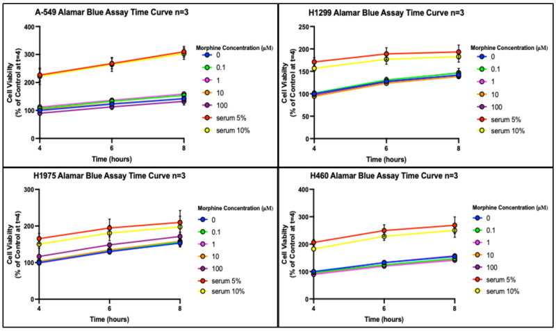
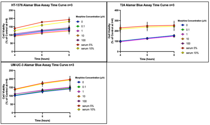
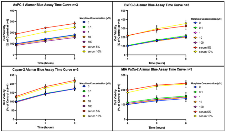
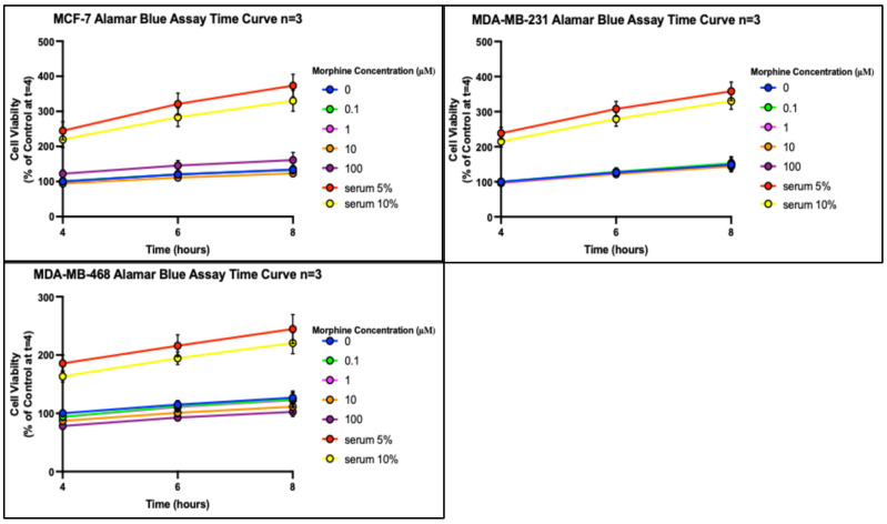
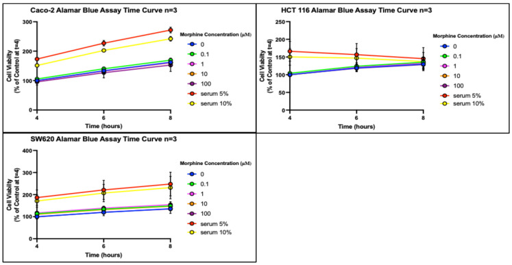
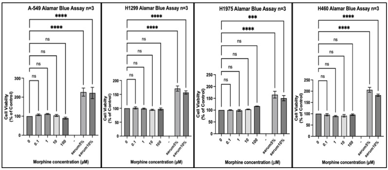
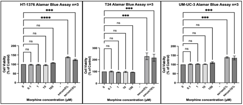
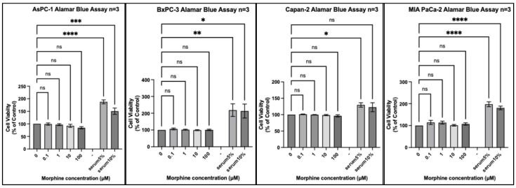
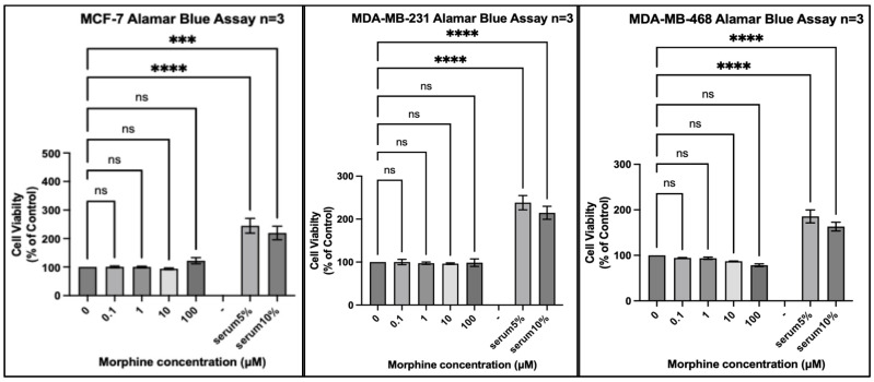
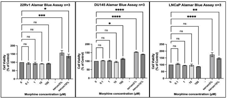
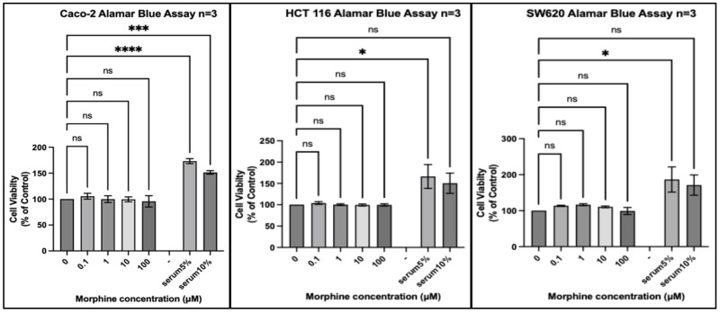
Background/Objectives: Opium consumption was recently classified by the International Agency for Research on Cancer (IARC) monograph as carcinogenic to humans based on strong evidence for cancers of the larynx, lung, and urinary bladder, and limited evidence for cancers of the oesophagus, stomach, pancreas, and pharynx. This poses the question of a potential pro-cancer effect of pharmaceutical opioid analgesics. In vitro studies employing a variety of experimental conditions suggest that opioid alkaloids have proliferative or antiproliferative effects. We set out to reconcile this discrepancy and explore the hypothesis that opioids promote cancer cell proliferation in an organ-dependent fashion. Methods: Using strictly controlled conditions, we tested the effect of morphine on the proliferation of a series of human cancer cell lines isolated from organs where cancer risk was linked causally to opium consumption in human studies (i.e., lung, bladder, and larynx), or control organs where no link between cancer risk and opium consumption has been reported in human studies (i.e., breast, colon, prostate). Results: Our results showed a minimal effect on proliferation on any cell line and no trend supporting an organ-specific effect of morphine. Conclusions: This argues against a direct effect of opioids on tumour cell proliferation to support their organ-specific effect.

Keywords: cancer cell lines; morphine; proliferation.


Conflict of interest statement

The authors declare no conflicts of interest.

Figures

Figure A1
Figure A1
Proliferation of lung cancer cell lines (A-549, H1299, H1975, and H460) in response to morphine. Cells were exposed to indicated concentrations of morphine or serum for 48 h. Resazurin reduction was quantified after 4, 6, and 8 h by fluorescence at an excitation of 560 nm and an emission of 590 nm. Results are expressed as a percentage of the viability of control cells unexposed to morphine or serum at time = 4 h. Data are shown as mean ± SEM, n = 3 independent experiments.
Figure A2
Figure A2
Proliferation of bladder cancer cell lines (HT-3376, T24, and UM-UC-3) in response to morphine. Cells were exposed to indicated concentrations of morphine or serum for 48 h. Resazurin reduction was quantified after 4, 6, and 8 h by fluorescence at an excitation of 560 nm and an emission of 590 nm. Results are expressed as a percentage of the viability of control cells unexposed to morphine or serum at time = 4 h. Data are shown as mean ± SEM, n = 3 independent experiments.
Figure A3
Figure A3
Proliferation of pancreas cancer cell lines (AsPC-1, BxPC-3, Capan-2, and MIA PaCa-2) in response to morphine. Cells were exposed to indicated concentrations of morphine or serum for 48 h. Resazurin reduction was quantified after 4, 6, and 8 h by fluorescence at an excitation of 560 nm and an emission of 590 nm. Results are expressed as a percentage of the viability of control cells unexposed to morphine or serum at time = 4 h. Data are shown as mean ± SEM, n = 3 independent experiments.
Figure A4
Figure A4
Proliferation of breast cancer cell lines (MCF-7, MDA-MB-231, and MDA-MB-468) in response to morphine. Cells were exposed to indicated concentrations of morphine or serum for 48 h. Resazurin reduction was quantified after 4, 6, and 8 h by fluorescence at an excitation of 560 nm and an emission of 590 nm. Results are expressed as a percentage of the viability of control cells unexposed to morphine or serum at time = 4 h. Data are shown as mean ± SEM, n = 3 independent experiments.
Figure A5
Figure A5
Proliferation of prostate cancer cell lines (22Rv1, DU145, and LNCaP) in response to morphine. Cells were exposed to indicated concentrations of morphine or serum for 48 h. Resazurin reduction was quantified after 4, 6, and 8 h by fluorescence at an excitation of 560 nm and an emission of 590 nm. Results are expressed as a percentage of the viability of control cells unexposed to morphine or serum at time = 4 h. Data are shown as mean ± SEM, n = 3 independent experiments.
Figure A6
Figure A6
Proliferation of colon cancer cell lines (Caco-2, HCT 116, and SW620) in response to morphine. Cells were exposed to indicated concentrations of morphine or serum for 48 h. Resazurin reduction was quantified after 4, 6, and 8 h by fluorescence at an excitation of 560 nm and an emission of 590 nm. Results are expressed as a percentage of the viability of control cells unexposed to morphine or serum at time = 4 h. Data are shown as mean ± SEM, n = 3 independent experiments.
Figure A7
Figure A7
Gene expression of mu (left), delta (centre), and kappa (right) opioid receptors expressed in transcripts per million (TPM), taken from the PCTA.
Figure 1
Figure 1
Proliferation of lung cancer cell lines (A-549, H1299, H1975, and H460) in response to morphine. Cells were exposed to indicated concentrations of morphine or serum for 48 h. Resazurin reduction was quantified after 4 h by fluorescence at an excitation of 560 nm and an emission of 590 nm. Results are expressed as a percentage of the viability of control cells unexposed to morphine or serum. Data are shown as mean ± SEM, n = 3 independent experiments. ns, not significant; *** p < 0.001; **** p < 0.0001.
Figure 2
Figure 2
Proliferation of bladder cancer cell lines (HT-1376, T24, and UM-UC-3) in response to morphine. Cells were exposed to indicated concentrations of morphine or serum for 48 h. Resazurin reduction was quantified after 4 h by fluorescence at an excitation of 560 nm and an emission of 590 nm. Results are expressed as a percentage of the viability of control cells unexposed to morphine or serum. Data are shown as mean ± SEM, n = 3 independent experiments. ns, not significant; *** p < 0.001; **** p < 0.0001.
Figure 3
Figure 3
Proliferation of pancreas cancer cell lines (AsPC-1, BxPC-3, Capan-2, and MIA PaCa-2) in response to morphine. Cells were exposed to indicated concentrations of morphine or serum for 48 h. Resazurin reduction was quantified after 4 h by fluorescence at an excitation of 560 nm and an emission of 590 nm. Results are expressed as a percentage of the viability of control cells unexposed to morphine or serum. Data are shown as mean ± SEM, n = 3 independent experiments. ns, not significant; * p < 0.05; ** p < 0.01; *** p < 0.001; **** p < 0.0001.
Figure 4
Figure 4
Proliferation of breast cancer cell lines (MCF-7, MDA-MB-231, and MDA-MB-468) in response to morphine. Cells were exposed to indicated concentrations of morphine or serum for 48 h. Resazurin reduction was quantified after 4 h by fluorescence at an excitation of 560 nm and an emission of 590 nm. Results are expressed as a percentage of the viability of control cells unexposed to morphine or serum. Data are shown as mean ± SEM, n = 3 independent experiments. ns, not significant; *** p < 0.001; **** p < 0.0001.
Figure 5
Figure 5
Proliferation of prostate cancer cell lines (22Rv1, DU145, and LNCaP) in response to morphine. Cells were exposed to indicated concentrations of morphine or serum for 48 h. Resazurin reduction was quantified after 4 h by fluorescence at an excitation of 560 nm and an emission of 590 nm. Results are expressed as a percentage of the viability of control cells unexposed to morphine or serum. Data are shown as mean ± SEM, n = 3 independent experiments. ns, not significant; * p < 0.05; ** p < 0.01; *** p < 0.001; **** p < 0.0001.
Figure 6
Figure 6
Proliferation of colon cancer cell lines (Caco-2, HCT 116, and SW620) in response to morphine. Cells were exposed to indicated concentrations of morphine or serum for 48 h. Resazurin reduction was quantified after 4 h by fluorescence at an excitation of 560 nm and an emission of 590 nm. Results are expressed as a percentage of the viability of control cells unexposed to morphine or serum. Data are shown as mean ± SEM, n = 3 independent experiments. ns, not significant; * p < 0.05; *** p < 0.001; **** p < 0.0001.

Similar articles

  • Opium, Street Opium, and Cancer Risk.
    López-Lázaro M. López-Lázaro M. Curr Pharm Des. 2022;28(25):2039-2042. doi: 10.2174/1381612828666220607104805. Curr Pharm Des. 2022. PMID: 35674306
  • Potential impact of controlling opium use prevalence on future cancer incidence in Iran.
    Nemati S, Dardashti AR, Mohebbi E, Kamangar F, Malekzadeh R, Zendehdel K, Sheikh M. Nemati S, et al. EClinicalMedicine. 2024 Jun 3;73:102650. doi: 10.1016/j.eclinm.2024.102650. eCollection 2024 Jul. EClinicalMedicine. 2024. PMID: 38881571 Free PMC article.
  • Palliative Treatment of Cancer-Related Pain [Internet].
    Kongsgaard U, Kaasa S, Dale O, Ottesen S, Nordøy T, Hessling SE, von Hofacker S, Bruland ØS, Lyngstadaas A. Kongsgaard U, et al. Oslo, Norway: Knowledge Centre for the Health Services at The Norwegian Institute of Public Health (NIPH); 2005 Dec. Report from Norwegian Knowledge Centre for the Health Services (NOKC) No. 09-2005. Oslo, Norway: Knowledge Centre for the Health Services at The Norwegian Institute of Public Health (NIPH); 2005 Dec. Report from Norwegian Knowledge Centre for the Health Services (NOKC) No. 09-2005. PMID: 29320015 Free Books & Documents. Review.
  • The carcinogenicity of opium consumption: a systematic review and meta-analysis.
    M Filho A, Turner MC, Warnakulasuriya S, Richardson DB, Hosseini B, Kamangar F, Pourshams A, Sewram V, Cronin-Fenton D, Etemadi A, Glass DC, Rahimi-Movaghar A, Sheikh M, Malekzadeh R, Schubauer-Berigan MK. M Filho A, et al. Eur J Epidemiol. 2023 Apr;38(4):373-389. doi: 10.1007/s10654-023-00969-7. Epub 2023 Feb 11. Eur J Epidemiol. 2023. PMID: 36773182 Free PMC article.
  • Opioids, Opioid Antagonists.
    [No authors listed] [No authors listed] 2020 Nov 24. LiverTox: Clinical and Research Information on Drug-Induced Liver Injury [Internet]. Bethesda (MD): National Institute of Diabetes and Digestive and Kidney Diseases; 2012–. 2020 Nov 24. LiverTox: Clinical and Research Information on Drug-Induced Liver Injury [Internet]. Bethesda (MD): National Institute of Diabetes and Digestive and Kidney Diseases; 2012–. PMID: 31643200 Free Books & Documents. Review.

Cited by

References

    1. Snijders R.A.H., Brom L., Theunissen M., van den Beuken-van Everdingen M.H.J. Update on Prevalence of Pain in Patients with Cancer 2022: A Systematic Literature Review and Meta-Analysis. Cancers. 2023;15:591. doi: 10.3390/cancers15030591. - DOI - PMC - PubMed
    1. McGuire D.B. Occurrence of cancer pain. J. Natl. Cancer Inst. Monogr. 2004;2004:51–56. doi: 10.1093/jncimonographs/lgh015. - DOI - PubMed
    1. Abdel Shaheed C., Hayes C., Maher C.G., Ballantyne J.C., Underwood M., McLachlan A.J., Martin J.H., Narayan S.W., Sidhom M.A. Opioid analgesics for nociceptive cancer pain: A comprehensive review. CA Cancer J. Clin. 2024;74:286–313. doi: 10.3322/caac.21823. - DOI - PubMed
    1. Cohen S.P., Vase L., Hooten W.M. Chronic pain: An update on burden, best practices, and new advances. Lancet. 2021;397:2082–2097. doi: 10.1016/S0140-6736(21)00393-7. - DOI - PubMed
    1. Mathieson S., Wertheimer G., Maher C.G., Christine Lin C.W., McLachlan A.J., Buchbinder R., Pearson S.A., Underwood M. What proportion of patients with chronic noncancer pain are prescribed an opioid medicine? Systematic review and meta-regression of observational studies. J. Intern. Med. 2020;287:458–474. doi: 10.1111/joim.13026. - DOI - PubMed

LinkOut - more resources