. 2024 Dec 20;17(12):1729.
doi: 10.3390/ph17121729.

Pentagalloyl Glucose from Bouea macrophylla Suppresses the Epithelial-Mesenchymal Transition and Synergizes the Doxorubicin-Induced Anticancer and Anti-Migration Effects in Triple-Negative Breast Cancer

Affiliations

Pentagalloyl Glucose from Bouea macrophylla Suppresses the Epithelial-Mesenchymal Transition and Synergizes the Doxorubicin-Induced Anticancer and Anti-Migration Effects in Triple-Negative Breast Cancer

Jiraporn Kantapan et al. Pharmaceuticals (Basel). .

Abstract

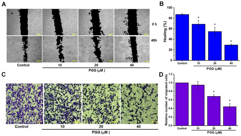
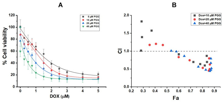
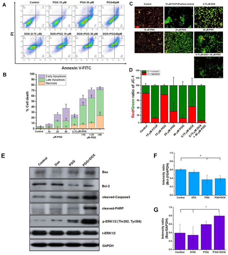
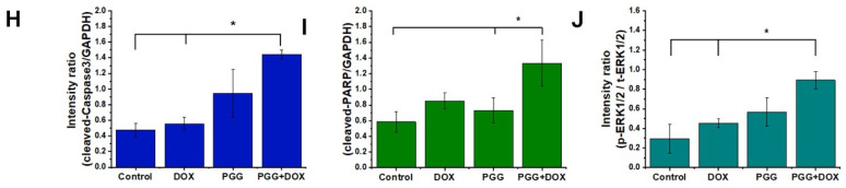
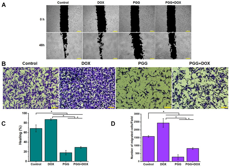
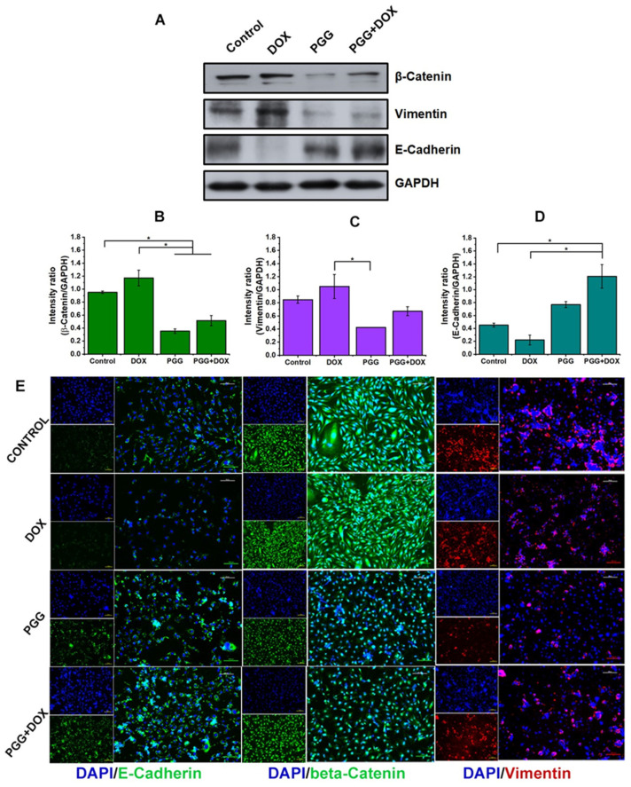
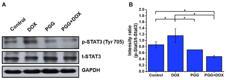
Background: Triple-negative breast cancer (TNBC) represents an aggressive form of breast cancer with few available therapeutic options. Chemotherapy, particularly with drugs like doxorubicin (DOX), remains the cornerstone of treatment for this challenging subtype. However, the clinical utility of DOX is hampered by adverse effects that escalate with higher doses and drug resistance, underscoring the need for alternative therapies. This study explored the efficacy of pentagalloyl glucose (PGG), a natural polyphenol derived from Bouea macrophylla, in enhancing DOX's anticancer effects and suppressing the epithelial-mesenchymal transition (EMT) in TNBC cells. Methods: This study employed diverse methodologies to assess the effects of PGG and DOX on TNBC cells. MDA-MB231 triple-negative breast cancer cells were used to evaluate cell viability, migration, invasion, apoptosis, mitochondrial membrane potential, and protein expression through techniques including MTT assays, wound healing assays, flow cytometry, Western blotting, and immunofluorescence. Results: Our findings demonstrate that PGG combined with DOX significantly inhibits TNBC cell proliferation, migration, and invasion. PGG enhances DOX-induced apoptosis by disrupting the mitochondrial membrane potential and activating caspase pathways; consequently, the activation of caspase-3 and the cleavage of PARP are increased. Additionally, the study shows that the combination treatment upregulates ERK signaling, further promoting apoptosis. Moreover, PGG reverses DOX-induced EMT by downregulating mesenchymal markers (vimentin and β-catenin) and upregulating epithelial markers (E-cadherin). Furthermore, it effectively inhibits STAT3 phosphorylation, associated with cell survival and migration. Conclusions: These results highlight the potential of PGG as an adjuvant therapy in TNBC treatment. PGG synergizes with DOX, which potentiates its anticancer effects while mitigating adverse reactions.

Keywords: doxorubicin; epithelial–mesenchymal transition (EMT); pentagalloyl glucose (PGG); synergistic; triple-negative breast cancer (TNBC).


Conflict of interest statement

The authors declare no conflicts of interest. The funders had no role in the design of the study; the collection, analysis, or interpretation of data; the writing of the manuscript; or the decision to publish the results.

Figures

Figure 1
Figure 1
The inhibitory effect of PGG and DOX on cell proliferation in TNBC cells, as assessed using the MTT assay. (AC) Dose–response curves illustrating the cytotoxic effects of PGG on MCF-10A cells (A), DOX on MDA-MB231 cells (B), and PGG on MDA-MB231 cells (C) after 48 h of treatment. (D,E) Clonogenic survival analysis indicating the number of colonies formed by MDA-MB231 cells following treatment with varying concentrations of PGG (0, 2.5, 5, 10, 20, and 40 µM). Representative colony images are shown in (D), with quantification graphs in (E). The results are presented as the mean ± SD from three independent experiments. * p < 0.05 indicates a statistically significant difference compared to the control, while *** p < 0.001 denotes a highly significant difference from the control. PGG, pentagalloyl glucose; DOX, doxorubicin.
Figure 2
Figure 2
PGG inhibits the migration and invasion capabilities of TNBC cells. (A) Representative microscopic images from wound healing assays performed on MDA-MB231 cells treated with varying concentrations of PGG, captured at 0 and 48 h. (B) Quantification of wound closure percentages, demonstrating the impact of PGG treatment. (C) Transwell chamber images showing cell migration following treatment with various concentrations of PGG. (D) Quantification of migrated cells: migrating cells were counted in five high-power fields and averaged. The results are presented as the mean ± SD from three independent experiments. * p < 0.05 indicates a statistically significant difference compared to the control. Scale bar = 100 µm (A,C). PGG, pentagalloyl glucose.
Figure 3
Figure 3
Impact of combined treatment with 10, 20, and 40 µM PGG and varying concentrations of DOX on the viability of MDA-MB231 cells after 48 h, as assessed using the MTT assay (A). Fa-CI plot analysis depicting the interaction between DOX and PGG in MDA-MB231 cells. The dashed line at CI = 1 signifies an additive effect, while CI values less than, equal to, or greater than 1 indicate synergy, additivity, or antagonism, respectively (B). The effect (Fa) represents the degree of fractional inhibition associated with each combination index. PGG, pentagalloyl glucose; DOX, doxorubicin.
Figure 4
Figure 4
The impact on apoptosis of PGG and DOX as monotherapies or in combination in TNBC cells. (A) Representative dot plots show the apoptotic response of MDA-MB231 cells to the indicated treatments. (B) Quantitative data represent the percentage of total cell death, as determined using flow cytometry. (C,D) The effects of PGG on mitochondrial membrane potential in MDA-MB231 cells were assessed using JC-1 staining: (C) representative images display JC-1 fluorescence across different treatment groups after 24 h, with FCCP as the positive control. Monomeric JC-1 exhibits green fluorescence, while aggregated JC-1 emits red fluorescence; (D) quantification of the red/green fluorescence ratio shown in a histogram. Data are presented as the mean ± SD of three independent experiments. (E) Western blot analysis of apoptotic markers, including Bax, Bcl-2, caspase-3, PARP, p-ERK, and t-ERK. The uncropped Western blot images are provided in Figure S1. (FJ) The relative protein density values were quantified, with expression levels normalized to GAPDH as the loading control. The results are presented as the mean ± SD from three independent experiments. * p < 0.05 indicates a statistically significant difference compared to the control. Scale bar = 100 µm (C). PGG, pentagalloyl glucose; DOX, doxorubicin; PARP, poly ADP ribose polymerase; ERK, extracellular signal-regulated kinase.
Figure 4
Figure 4
The impact on apoptosis of PGG and DOX as monotherapies or in combination in TNBC cells. (A) Representative dot plots show the apoptotic response of MDA-MB231 cells to the indicated treatments. (B) Quantitative data represent the percentage of total cell death, as determined using flow cytometry. (C,D) The effects of PGG on mitochondrial membrane potential in MDA-MB231 cells were assessed using JC-1 staining: (C) representative images display JC-1 fluorescence across different treatment groups after 24 h, with FCCP as the positive control. Monomeric JC-1 exhibits green fluorescence, while aggregated JC-1 emits red fluorescence; (D) quantification of the red/green fluorescence ratio shown in a histogram. Data are presented as the mean ± SD of three independent experiments. (E) Western blot analysis of apoptotic markers, including Bax, Bcl-2, caspase-3, PARP, p-ERK, and t-ERK. The uncropped Western blot images are provided in Figure S1. (FJ) The relative protein density values were quantified, with expression levels normalized to GAPDH as the loading control. The results are presented as the mean ± SD from three independent experiments. * p < 0.05 indicates a statistically significant difference compared to the control. Scale bar = 100 µm (C). PGG, pentagalloyl glucose; DOX, doxorubicin; PARP, poly ADP ribose polymerase; ERK, extracellular signal-regulated kinase.
Figure 5
Figure 5
The impact of PGG combined with DOX on the migratory behavior of TNBC cells. (A) Representative microscopic images from wound healing assays and (C) quantification of wound closure percentages illustrating the effects of treatments with PGG (40 µM), DOX (0.75 µM), and their combination. (B) Transwell chamber images showing cell migration following treatment with PGG (40 µM), DOX (0.75 µM), or a combination of both. (D) Quantitative analysis of the number of migrating cells. The results are presented as the mean ± SD from three independent experiments. * p < 0.05 indicates a statistically significant difference compared to the control and DOX treatment alone. Scale bar = 100 µm (A,B). PGG, pentagalloyl glucose; DOX, doxorubicin.
Figure 6
Figure 6
Reversal of EMT and suppression of EMT marker expression by PGG in TNBC cell lines. MDA-MB231 cells were treated for 48 h, as indicated, and the expression of EMT markers was analyzed using Western blot. (A) Representative Western blot images showing the levels of β-catenin, vimentin, E-cadherin, and GAPDH. The uncropped Western blot images are provided in Figure S2. (BD) Quantification of band intensities from the Western blot analysis. Relative protein levels were quantified and normalized to GAPDH as the loading control. The results are presented as the mean ± SD from three independent experiments. * p < 0.05 indicates a statistically significant difference compared to the control and DOX-only treatment. (E) Immunofluorescence staining of β-catenin (green), E-cadherin (green), and vimentin (red) in MDA-MB231 cells, with nuclei counterstained using DAPI (blue). Scale bar = 100 µm.
Figure 7
Figure 7
The effects of PGG and DOX, either alone or in combination, on STAT3 signaling proteins in TNBC cells. (A) Representative Western blot images showing the expression levels of phosphorylated STAT3 (p-STAT3), total STAT3 (t-STAT3), and GAPDH after treatment with PGG (40 µM), DOX (0.75 µM), or their combination for 48 h in MDA-MB231 cells. The uncropped Western blot images are provided in Figure S3. (B) Bar graph depicting the fold change in protein expression. The relative protein densities were quantified and normalized to the GAPDH loading control. The results are presented as the mean ± SD from three independent experiments. * p < 0.05 indicates a statistically significant difference compared to the control and DOX treatment alone. PGG, pentagalloyl glucose; DOX, doxorubicin; STAT3, signal transducer and activator of transcription 3; p, phosphorylated; t, total; GAPDH, glyceraldehyde 3-phosphate dehydrogenase.

Similar articles

References

    1. Yin L., Duan J.J., Bian X.W., Yu S.C. Triple-negative breast cancer molecular subtyping and treatment progress. Breast Cancer Res. 2020;22:61. doi: 10.1186/s13058-020-01296-5. - DOI - PMC - PubMed
    1. Cao L., Niu Y. Triple negative breast cancer: Special histological types and emerging therapeutic methods. Cancer Biol. Med. 2020;17:293–306. doi: 10.20892/j.issn.2095-3941.2019.0465. - DOI - PMC - PubMed
    1. Zhou Y.D., Li J., Du L., Mahdi F., Le T.P., Chen W.L., Swanson S.M., Watabe K., Nagle D.G. Biochemical and anti-triple negative metastatic breast tumor cell properties of Psammaplins. Mar. Drugs. 2018;16:442. doi: 10.3390/md16110442. - DOI - PMC - PubMed
    1. Nedeljković M., Damjanović A. Mechanisms of chemotherapy resistance in triple-negative breast cancer-How we can rise to the challenge. Cells. 2019;8:957. doi: 10.3390/cells8090957. - DOI - PMC - PubMed
    1. Nath A., Mitra S., Mistry T., Pal R., Nasare V.D. Molecular targets and therapeutics in chemoresistance of triple-negative breast cancer. Med. Oncol. 2021;39:14. doi: 10.1007/s12032-021-01610-x. - DOI - PubMed

LinkOut - more resources