. 2024 Nov 28;12(12):2445.
doi: 10.3390/microorganisms12122445.

Concomitant Potentially Contagious Factors Detected in Poland and Regarding Acanthamoeba Strains, Etiological Agents of Keratitis in Humans

Affiliations

Concomitant Potentially Contagious Factors Detected in Poland and Regarding Acanthamoeba Strains, Etiological Agents of Keratitis in Humans

Lidia Chomicz et al. Microorganisms. .

Abstract

Background: Diseases in humans caused by amphizoic amoebae that can result in visual impairment and even blindness, have recently been identified more frequently worldwide. Etiologically complex incidents of keratitis, including those connected with Acanthamoeba strains detected in Poland, were evaluated in this study.

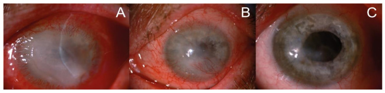
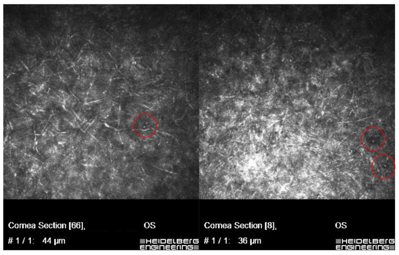
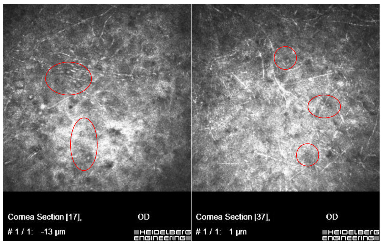
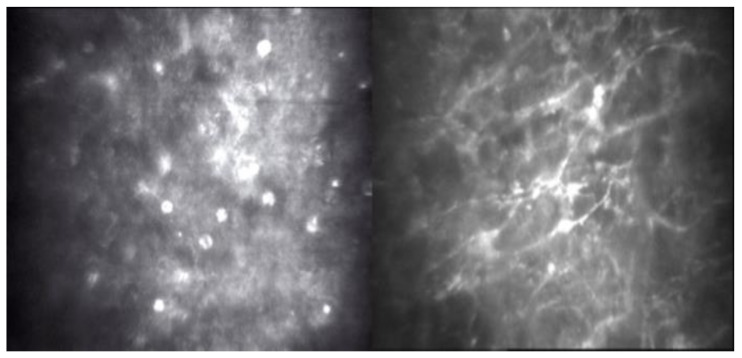
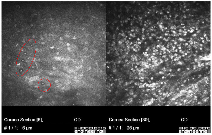
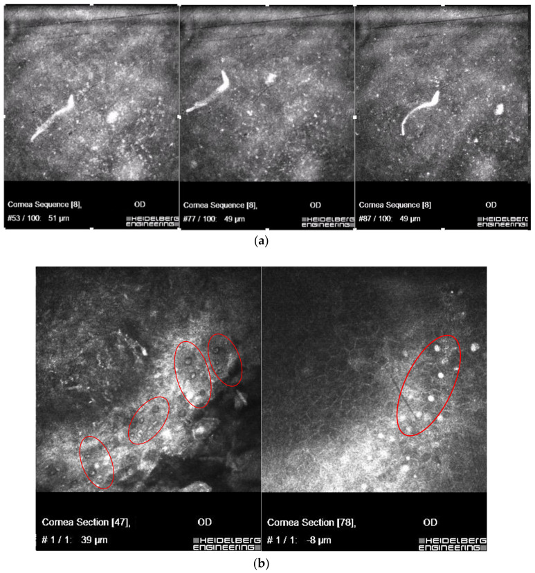
Methods: Corneal samples from cases resistant to antimicrobial therapy assessed for epidemiological, microbiological and parasitological aspects were investigated by phase-contrast microscope, slit lamp and by confocal microscopy. In vitro techniques were applied for detection of bacteria and fungi, and corneal isolates cultured under axenic condition using BSC medium-for detection of Acanthamoeba spp.; molecular techniques were applied for amoeba species identification.

Results: Most etiologically complicated keratitis cases, detected in ~84% of incidents, was due to exposure of contact lenses to tap water or pool water; trophozoites and cysts of Acanthamoeba, concomitant bacteriae, e.g., Pseudomonas aeruginosa, fungi and microfilariae were identified in contact lens users.

Conclusions: In samples from contact lens wearers where microbial keratitis is identified along with some connection with the patient's exposure to contaminated water environments, a risk of Acanthamoeba spp. infections should be considered. Understanding the complicated relationship between Acanthamoeba spp., co-occurring pathogens including associated endosymbionts is needed. In vivo confocal microscopy and in vitro cultivation were necessary to identify potentially contagious concomitant factors affecting the complex course of the keratitis.

Keywords: Acanthamoeba keratitis; cellular and molecular diagnostics; co-occurring infections; concomitant factors; confocal microscopy; corneal isolates; in vitro methods; infectious strains; microbiota; non-invasive in vivo methods.


Conflict of interest statement

The authors declare no conflicts of interest.

Figures

Figure 1
Figure 1
Representative slit lamp images of the patient’s eye affected by Acanthamoeba keratitis. (A) At the beginning of the treatment: corneal oedema, ring-shaped extensive infiltration covering 2/3 of the corneal surface, severe ciliary congestion and corneal neovascularization; (B) after 6 weeks of therapy: significant reduction of corneal edema, decrease in the magnitude of infiltration, more intense corneal neovascularization and improvement in the corneal structure; (C) at the end of the treatment: the translucent, stable cornea with regressed vessels. Corneal scar in anterior corneal stroma is covered with healthy epithelium, the anterior chamber is clean, and there are no pathological post-inflammatory vessels in the iris.
Figure 2
Figure 2
In vivo confocal microscopy scans showing co-infection of Fusarium spp. with Acanthamoeba.
Figure 3
Figure 3
In vivo confocal microscopy scans showing co-infection of Aspergillus spp. with Acanthamoeba (cysts in red circles).
Figure 4
Figure 4
In vivo confocal microscopy scans showing co-infection of filamentous fungi with Acanthamoeba.
Figure 5
Figure 5
In vivo confocal microscopy scans showing co-infection of bacteria Pseudomonas aeruginosa and Acanthamoeba (cysts shown in the left side of the picture). Right side of the picture shows massive inflammatory response.
Figure 6
Figure 6
In vivo confocal microscopy scans showing co-infection of live microfilaria (a), with Acanthamoeba (in red circles) (b).

Similar articles

References

    1. Rodriguez-Zaragoza S. Ecology of free-living amoebae. Crit. Rev. Microbiol. 1994;20:225–241. doi: 10.3109/10408419409114556. - DOI - PubMed
    1. Martinez A.J., Visvesvara G.S. Free-living, amphizoic and opportunistic amebas. Brain Pathol. 1997;7:583–598. doi: 10.1111/j.1750-3639.1997.tb01076.x. - DOI - PMC - PubMed
    1. Marciano-Cabral F., Cabral G. Acanthamoeba spp. as agents of disease in humans. Clin. Microbiol. Rev. 2003;16:273–307. doi: 10.1128/CMR.16.2.273-307.2003. - DOI - PMC - PubMed
    1. Shuster F.L., Visvesvara G.S. Free-living amoebae as opportunistic and non-opportunistic pathogens of humans and animals. Int. J. Parasitol. 2004;34:1001–1027. doi: 10.1016/j.ijpara.2004.06.004. - DOI - PubMed
    1. Naginton J., Watson P.G., Playfair T.J., McGill J., Jones B.R., Steele A.D. Amoebic infection of the eye. Lancet. 1974;2:1537–1540. doi: 10.1016/S0140-6736(74)90285-2. - DOI - PubMed

LinkOut - more resources