. 2024 Dec 18;12(12):2625.
doi: 10.3390/microorganisms12122625.

Unexpected Genetic Diversity of Nostocales (Cyanobacteria) Isolated from the Phyllosphere of the Laurel Forests in the Canary Islands (Spain)

Affiliations

Unexpected Genetic Diversity of Nostocales (Cyanobacteria) Isolated from the Phyllosphere of the Laurel Forests in the Canary Islands (Spain)

Nereida M Rancel-Rodríguez et al. Microorganisms. .

Abstract

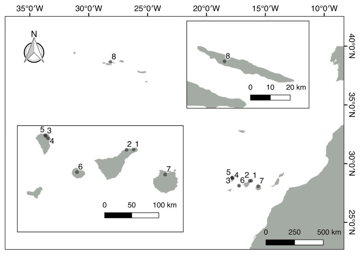
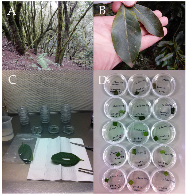
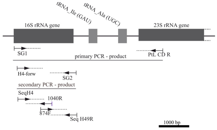
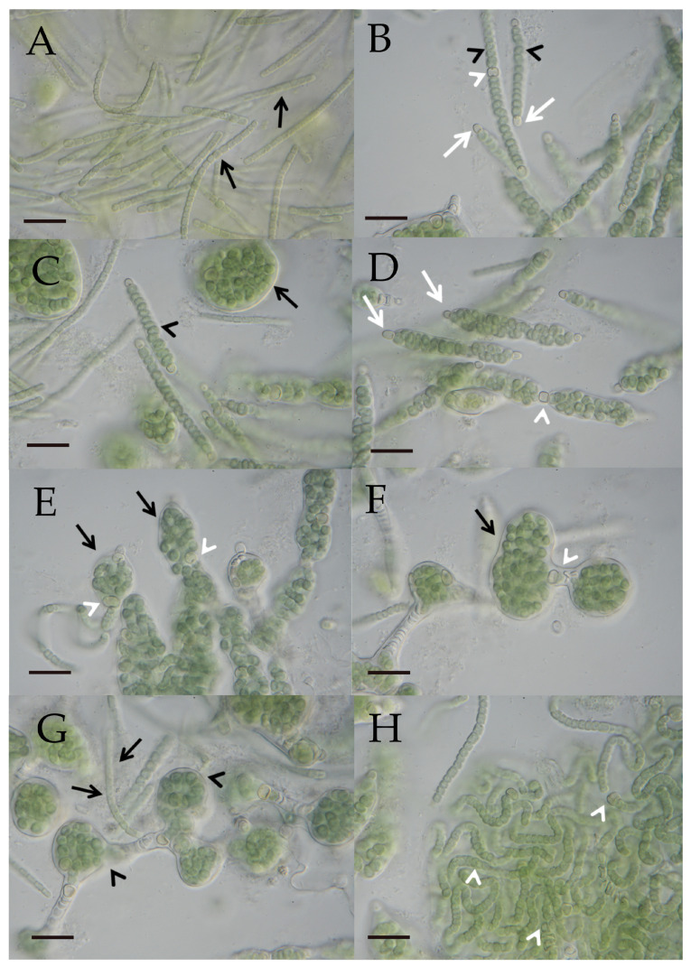
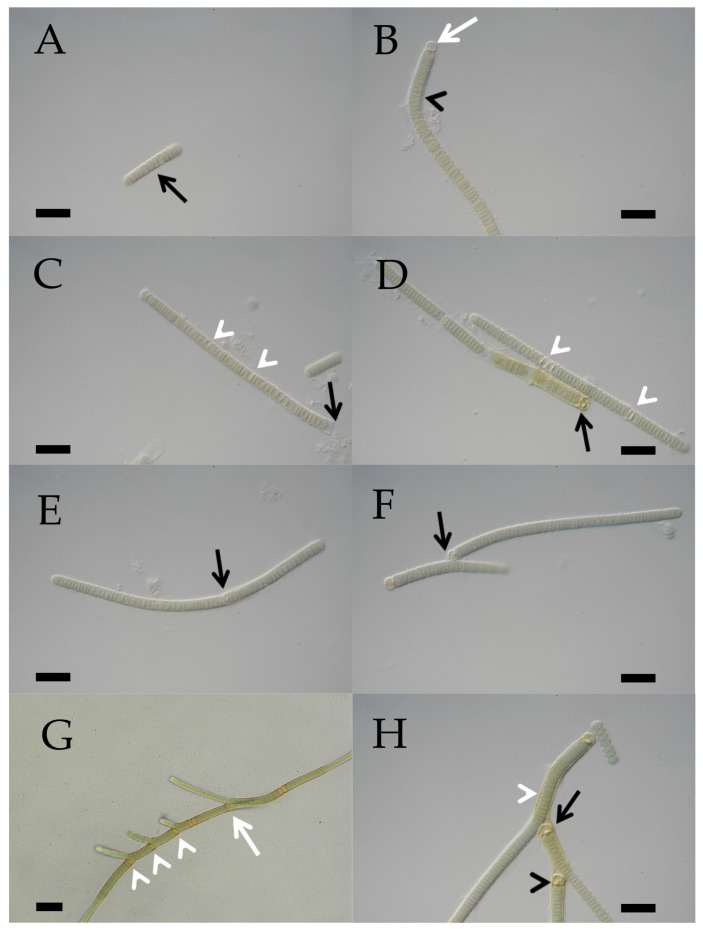
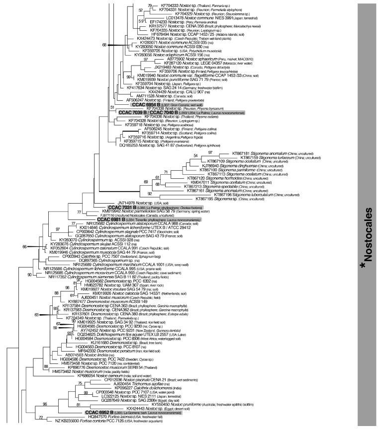
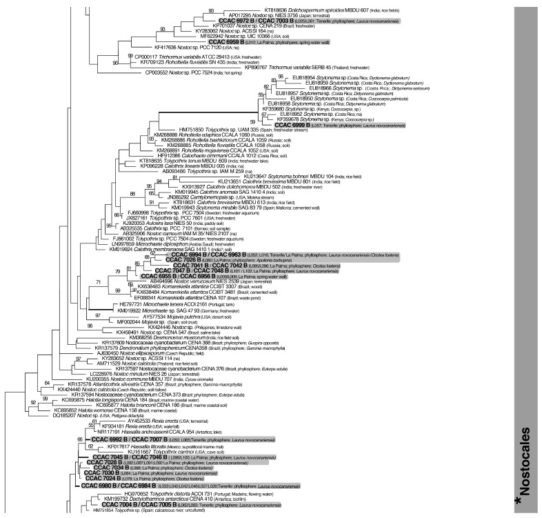
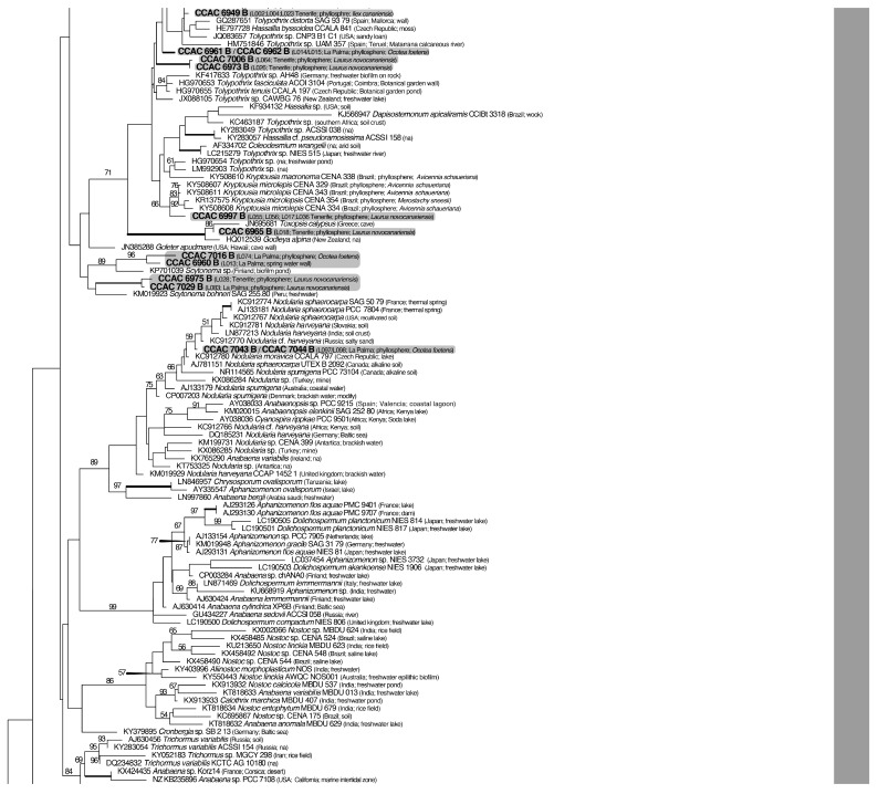
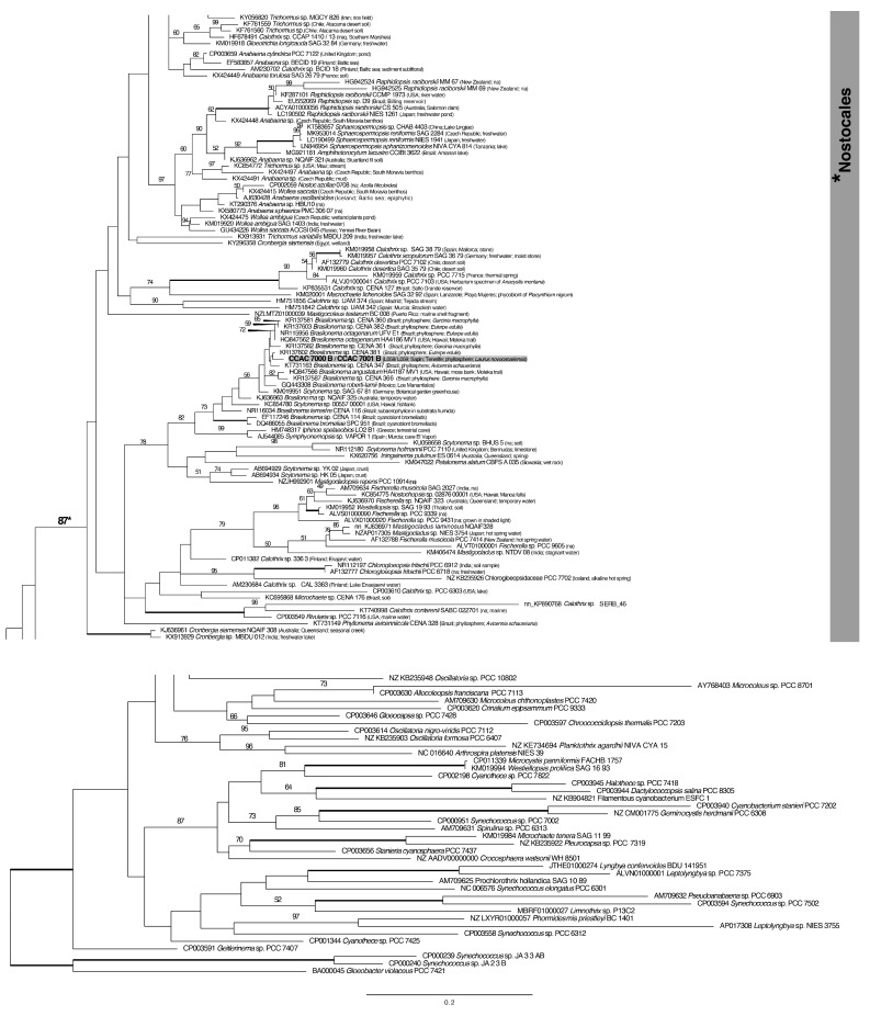
A total of 96 strains of Nostocales (Cyanobacteria) were established from the phyllosphere of the laurel forests in the Canary Islands (Spain) and the Azores (Portugal) using enrichment media lacking combined nitrogen. The strains were characterized by light microscopy and SSU rRNA gene comparisons. Morphologically, most strains belonged to two different morphotypes, termed "Nostoc-type" and "Tolypothrix-type". Molecular phylogenetic analysis of 527 SSU rRNA gene sequences of cyanobacteria (95 sequences established during this study plus 392 sequences from Nostocales and 40 sequences from non-heterocyte-forming cyanobacteria retrieved from the databases) revealed that none of the SSU rRNA gene sequences from the phyllosphere of the laurel forests was identical to a database sequence. In addition, the genetic diversity of the isolated strains was high, with 42 different genotypes (44% of the sequences) recognized. Among the new genotypes were also terrestrial members of the genus Nodularia as well as members of the genus Brasilonema. It is concluded that heterocyte-forming cyanobacteria represent a component of the phyllosphere that is still largely undersampled in subtropical/tropical forests.

Keywords: biodiversity; clonal culture; epiphyllous; heterocyte-forming cyanobacteria; rRNA sequence comparison.


Conflict of interest statement

The authors declare no conflicts of interest.

Figures

Figure 1
Figure 1
Sampling sites of laurel forests in the Canary Islands and Sao Jorge (the Azores). Numbers refer to sampling sites (localities) in Table 1.
Figure 2
Figure 2
(AD) Photographic documentation of sampling of leaves and set-up of enrichment cultures. (A) A typical view of the laurel forest of La Gomera (Canary Islands). The tree trunks are heavily colonized by epiphytes. (B) Two sampled leaves of different sizes from Laurus novocanariensis, with their upper surfaces partially covered by epiphylls. (C) Leaf material being prepared for enrichment cultures in a laminar flow hood. (D) Inoculated leaf disks in Petri dishes containing culture media for enrichment of heterocyte-forming cyanobacteria.
Figure 3
Figure 3
Overview of PCR and sequencing strategies for cyanobacterial 16S rRNA genes. The position of the primers used for amplification and sequencing of the 16S rRNA gene and the primary and secondary PCR products are shown. The general structure of the ribosomal rDNA operon with the 16S rRNA gene, two tRNA genes, and 23S rDNA gene is depicted schematically at the top of the figure. The primer sequences are shown in Table 2. Abbreviations used for the primers in the figure refer to the following primer designations and sequences in Table 2: 16S_SG1_short_forw (SG1), 16S H4_forw (H4-forw), ptLSU C-D_rev (PtL CD R), SG2_rev (SG2), Seq_16S_H4_forw (SeqH4), Seq_16S_1040_rev (1040R), Seq_16S_pos874_forw (874F), and Seq_16S_49_rev (Seq H49R).
Figure 4
Figure 4
(AH) Photographic documentation of the “Nostoc”-type morphotype (L066/CCAC 7008B/BEA 1768B) in a developmental sequence. (A) Several hormogonia (black arrows) characterized by barrel-shaped cells. Scale bar = 10 µm. (B) Differentiated filaments show lenticular to sublenticular vegetative cells (black arrowheads). Spherical to subspherical terminal heterocytes (white arrows) first appear on both ends of a filament, followed by a lenticular to sublenticular intercalary heterocyte (white arrowhead). Scale bar = 10 µm. (C) A common sheath develops surrounding the filament (black arrowhead). Within the sheath, vegetative cells continue to divide, expanding the sheath, with the filament eventually forming a globular structure (black arrow). Scale bar = 10 µm. (D) Terminal heterocytes (white arrows) and the initial intercalary heterocyte (white arrowhead) lack a sheath. Scale bar = 10 µm. (E,F) Curled filaments within expanded sheaths (black arrows) held together by intercalary heterocytes (white arrowheads). Scale bars = 10 µm. (G) When the flexible sheath (black arrowheads) breaks open, hormogonia (black arrows) emerge from the globular structures and start the developmental cycle again. Scale bar = 10 µm. (H) Filaments with intercalary heterocytes (white arrowheads) derived from a broken globular structure. Scale bar = 10 µm.
Figure 5
Figure 5
(AH) Photographic documentation of the “Tolypothrix”-type morphotype (L088/CCAC 7034B/BEA 1790B) in a developmental sequence. (A) Hormogonia are characterized by lenticular to sublenticular cells (black arrow): note the slight polarity of the filament. Scale bar = 10 µm. (B) An older filament with lenticular to sublenticular vegetative cells (black arrowhead) and a spherical to subspherical terminal heterocyte (white arrow). Scale bar = 10 µm. (C) A firm sheath (black arrow) surrounds the straight filament. Early developmental stages of differentiation of intercalary heterocytes from vegetative cells (white arrowheads). Scale bar = 10 µm. (D) Lenticular to sublenticular differentiated intercalary heterocytes (white arrowheads; the left arrowhead depicts two adjacent intercalary heterocytes). The intercalary heterocytes remain enclosed in the firm sheath (unlike the situation in the “Nostoc”-type morphotype). A yellowish firm sheath surrounds vegetative cells of an older filament near a terminal heterocyte (black arrow). Scale bar = 10 µm. (E) Very early stage of the formation of a false branch. A vegetative cell adjacent to an intercalary heterocyte dissociates from the heterocyte and starts to bulge the sheath (black arrow). The intercalary heterocyte of the filament thus becomes a new terminal heterocyte. Scale bar = 10 µm. (F) A false branch attached to a heterocyte (black arrow). Scale bar = 10 µm. (G) Several false branches arising from vegetative cells adjacent to intercalary heterocytes (white arrowheads) or a necrotic cell (white arrow). Scale bar = 20 µm. (H) A primary (black arrow) and a secondary (black arrowhead) false branch share the same firm sheath (white arrowhead). Scale bar = 10 µm.
Figure 6
Figure 6
Phylogeny of heterocyte-forming cyanobacteria (Nostocales) from the phyllosphere of the laurel forests in the Canary Islands and the Azores using 16S rDNA sequence comparisons. The split tree (shown in 6 sections) was constructed with a database of 527 sequences, including 95 sequences generated during this study. The 16S rDNA dataset, with 1448 positions, was analyzed with maximum likelihood (RAxML). The model GTR + I + Γ and the parameters were estimated by RAxML (for further details, see Section 2). The sequences generated during this study are highlighted by a grey background and strain numbers (CCAC) in bold (strains with identical sequences are provided with their L numbers as well as the location and plant host in parentheses). Bootstrap values > 50% are shown; the bold value with an asterisk denotes support for the monophyly of the Nostocales. All tree branches in bold received maximal support. The complete merged tree is shown in Supplementary Figure S1.
Figure 6
Figure 6
Phylogeny of heterocyte-forming cyanobacteria (Nostocales) from the phyllosphere of the laurel forests in the Canary Islands and the Azores using 16S rDNA sequence comparisons. The split tree (shown in 6 sections) was constructed with a database of 527 sequences, including 95 sequences generated during this study. The 16S rDNA dataset, with 1448 positions, was analyzed with maximum likelihood (RAxML). The model GTR + I + Γ and the parameters were estimated by RAxML (for further details, see Section 2). The sequences generated during this study are highlighted by a grey background and strain numbers (CCAC) in bold (strains with identical sequences are provided with their L numbers as well as the location and plant host in parentheses). Bootstrap values > 50% are shown; the bold value with an asterisk denotes support for the monophyly of the Nostocales. All tree branches in bold received maximal support. The complete merged tree is shown in Supplementary Figure S1.

Similar articles

References

    1. Morales D., Jimenez M.S., Gonzalez-Rodríguez A.M., Cermák J. Laurel forest in Tenerife, Canary Islands. I. The site, stand structure and stand leaf area distribution. Trees. 1996;11:34–40. doi: 10.1007/s004680050055. - DOI
    1. Nogué S., De Nascimento L., Fernández-Palacios J.M., Whittaker R.J., Willis K.J. The ancient forests of La Gomera, Canary Islands, and their sensitivity to environmental change. J. Ecol. 2013;101:368–377. doi: 10.1111/1365-2745.12051. - DOI
    1. Fernández-Palacios J.M. Climatic responses of plant species on Tenerife, The Canary Islands. J. Veg. Sci. 1992;3:595–603. doi: 10.2307/3235826. - DOI
    1. Del Arco Aguilar M.J., Rodriguez Delgado O. Vegetation of the Canary Islands. In: del Arco Aguilar M.J., Rodriguez Delgado O., editors. Vegetation of the Canary Islands. Plant and Vegetation. Volume 16. Springer; Cham, Switzerland: 2018. pp. 83–319. - DOI
    1. Fernández–Palacios J.M., De Nicolás J.P. Altitudinal pattern of vegetation variation on Tenerife. J. Veg. Sci. 1995;6:183–190. doi: 10.2307/3236213. - DOI

LinkOut - more resources