. 2024 Dec 23;12(12):2670.
doi: 10.3390/microorganisms12122670.

Gram Negative Biofilms: Structural and Functional Responses to Destruction by Antibiotic-Loaded Mixed Polymeric Micelles

Affiliations

Gram Negative Biofilms: Structural and Functional Responses to Destruction by Antibiotic-Loaded Mixed Polymeric Micelles

Tsvetozara Damyanova et al. Microorganisms. .

Abstract

Biofilms are a well-known multifactorial virulence factor with a pivotal role in chronic bacterial infections. Their pathogenicity is determined by the combination of strain-specific mechanisms of virulence and the biofilm extracellular matrix (ECM) protecting the bacteria from the host immune defense and the action of antibacterials. The successful antibiofilm agents should combine antibacterial activity and good biocompatibility with the capacity to penetrate through the ECM. The objective of the study is the elaboration of biofilm-ECM-destructive drug delivery systems: mixed polymeric micelles (MPMs) based on a cationic poly(2-(dimethylamino)ethyl methacrylate)-b-poly(ε-caprolactone)-b-poly(2-(dimethylamino)ethyl methacrylate) (PDMAEMA35-b-PCL70-b-PDMAEMA35) and a non-ionic poly(ethylene oxide)-b-poly(propylene oxide)-b-poly(ethylene oxide) (PEO100-b-PPO65-b-PEO100) triblock copolymers, loaded with ciprofloxacin or azithromycin. The MPMs were applied on 24 h pre-formed biofilms of Escherichia coli and Pseudomonas aeruginosa (laboratory strains and clinical isolates). The results showed that the MPMs were able to destruct the biofilms, and the viability experiments supported drug delivery. The biofilm response to the MPMs loaded with the two antibiotics revealed two distinct patterns of action. These were registered on the level of both bacterial cell-structural alterations (demonstrated by scanning electron microscopy) and the interaction with host tissues (ex vivo biofilm infection model on skin samples with tests on nitric oxide and interleukin (IL)-17A production).

Keywords: biocompatibility; biofilm destruction; cationic polymers; drug delivery; ex vivo skin model; extracellular vesicles; mixed polymer micelles; nanotubules.


Conflict of interest statement

The authors declare no conflict of interest.

Figures
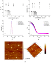
Figure 1
Figure 1
(a) Hydrodynamic diameter, Dh, and (b) ζ-potential variations as a function of the micellar concentration of SCPMs and MPMs based on PDMAEMA35-PCL70-PDMAEMA35 and Pluronic F127 triblock copolymers. (c) Size distribution curves (d) DLS correlation functions and (e) representative AFM micrograph of MPMs prepared from PDMAEMA35-PCL70-PDMAEMA35 and Pluronic F127 triblock copolymers at a molar ratio of 1:1 in the concentration range of 1 to 0.125 mg mL−1. The PDI values ranged in the 0.11–0.19 interval. All DLS measurements were performed at 25 °C.
Figure 2
Figure 2
Variations of encapsulation efficiency (a) and drug loading content (b) as a function of the composition of SCPMs and MPMs based on PDMAEMA35-PCL70-PDMAEMA35 and Pluronic F127 triblock copolymers. The loading was performed at polymer-to-drug mass ratio of 10:1.
Figure 3
Figure 3
Hydrodynamic diameter, Dh, (a,c,e) and ζ potential (b,d,f) of empty or loaded with antibiotics MPMs based on PDMAEMA35-PCL70-PDMAEMA35 and Pluronic F127 triblock copolymers in the concentration range of 1 to 0.125 mg mL−1. Measurements were performed at 25 °C at pH 7. Each data point represents the arithmetic mean ± SD of three separate experiments.
Figure 3
Figure 3
Hydrodynamic diameter, Dh, (a,c,e) and ζ potential (b,d,f) of empty or loaded with antibiotics MPMs based on PDMAEMA35-PCL70-PDMAEMA35 and Pluronic F127 triblock copolymers in the concentration range of 1 to 0.125 mg mL−1. Measurements were performed at 25 °C at pH 7. Each data point represents the arithmetic mean ± SD of three separate experiments.
Figure 4
Figure 4
Drug release profiles of MPMs based on PDMAEMA35-PCL70-PDMAEMA35 and Pluronic F127 triblock copolymers, prepared at a 10:1 polymer-to-drug mass ratio, determined by HPLC. MPMs were formed at molar ratios of 3:1 (a), 1:1 (b), and 1:3 (c). The release was performed at 37 °C in phosphate buffer pH 7.4. Each data point represents the arithmetic mean ± SD of three separate experiments.
Figure 5
Figure 5
Cytotoxicity of the SCPMs and MPMs based on PDMAEMA35-PCL70-PDMAEMA35 and Pluronic F127 triblock copolymers loaded with CF (a) or AZ (b) at a 10:1 polymer-to-drug mass ratio. The micelles were applied for 4 h in concentrations of 0.5, 0.25, and 0.125 mg mL−1 onto confluent cultured HaCaT. The results are presented as percentage of the control—cells cultivated parallelly in DMEM. The data are the means of four repeats and are presented as the mean ± SD. Differences between control (DMEM) and treated with micelles cells are accepted as statistically significant (*) when p < 0.05 and (**) when p < 0.001.
Figure 6
Figure 6
Reduction of the biomass of mature 24 h biofilms as a result of treatment for 4 or 24 h with 0.25 mg mL−1 of empty or antibiotics-loaded MPMs based on PDMAEMA35-PCL70-PDMAEMA35 and Pluronic F127 triblock copolymers. The results were calculated as percentage of the biofilm at the start of each experiment. (a) E. coli 25922; (b) P. aeruginosa PAO1. Results for biofilms treated with dH2O are included since the micelles were dispersed in dH2O. Each data point represents the mean ± SD of six repeats.
Figure 7
Figure 7
Viability of the biofilms after treatment for 24 h with 0.25 mg mL−1 of empty or antibiotics-loaded MPMs based on PDMAEMA35-PCL70-PDMAEMA35 and Pluronic F127 triblock copolymers. Viability was estimated by the reduction of resazurin using the Alamar Blue reagent (Invitrogen). The results were calculated as percentage of the untreated control (biofilm cultivated parallelly in M63 medium in the absence of the tested agents). dH2O bars are included to show the effect of treatment with dH2O alone_ the medium in which the micelles were dispersed. (a) E. coli 25922; (b) P. aeruginosa PAO1. Each data point represents the mean ± SD of six repeats. p < 0.05 (*); p < 0.001 (***), ANOVA test.
Figure 8
Figure 8
Reduction of biofilms of pathogenic strains of E. coli treated with empty or antibiotic-loaded MPMs 3:1 (a) and of P. aeruginosa treated with empty or antibiotic-loaded MPMs 1:1 (b). The results were calculated as percentage of the “0” controls, i.e., the amount of biofilms of the strains before the start of the treatments. Each data point represents the mean ± SD of six repeats.
Figure 9
Figure 9
Scanning electron microscopy of biofilms of E. coli 25922 (AH) and P. aeruginosa PAO1 (IP). Arrows: white—infolds of the cell wall; yellow—outer membrane vesicles; red—tunneling nanotubules. (A) E. coli 48 h control biofilm; (B,E,F,F1) E. coli 24 h biofilm treated for a further 24 h with empty MPMs 3:1; yellow asterisk mark slimy covering of cells in some areas of the treated biofilm. (G,G1) E. coli 24 h biofilm treated for a further 24 h with CF-loaded MPMs 3:1; (H,H1) E. coli 24 h biofilm treated for a further 24 h with AZ-loaded MPMs 3:1. (I,M) P. aeruginosa 48 h control biofilm; (J,N) P. aeruginosa 24 h biofilm treated for a further 24 h with empty MPMs 1:1; (K,O) P. aeruginosa 24 h biofilm treated for a further 24 h with CF-loaded MPMs 1:1; (L,P,P1) P. aeruginosa 24 h biofilm treated for a further 24 h with AZ-loaded MPMs 1:1; white asterisks, cells with extensively blebbed surfaces.
Figure 10
Figure 10
Histological sections of skin explants infected with P. aeruginosa PAO1 biofilm. (A) Untreated 24 h ex vivo biofilm. (B,C) Mature 24 h biofilms on skin explants were treated for 24 h with 0.25 mg mL−1 of MPMs 1:1 loaded with CF (B) or AZ (C). Bar = 10 µm.
Figure 11
Figure 11
Effect of MPMs loaded with CF or AZ on NO (a) and IL-17A (b) production in ex vivo murine skin explant P. aeruginosa PAO1 biofilm model. Murine skin explants were infected with P. aeruginosa for 24 h for the development of biofilm. Afterwards the skin explants were treated with 50 µL of either 0.5 or 0.25 mg mL−1 MPMs loaded with CF or AZ. Control samples, infected or uninfected with P. aeruginosa biofilm, were treated in parallel with either PBS or dH2O (the solvent for the MPM samples). Data represents mean ± SD from 3 samples/group * p < 0.05, ** p < 0.01, *** p < 0.001 when comparing the biofilm groups to the control PBS one, ANOVA test; ## p < 0.05 when comparing the non-biofilm groups to the control PBS one, ANOVA test.

Similar articles

References

    1. Pinto R.M., Soares F.A., Reis S., Nunes C., Van Dijck P. Innovative Strategies Toward the Disassembly of the EPS Matrix in Bacterial Biofilms. Front. Microbiol. 2020;11:952. doi: 10.3389/fmicb.2020.00952. - DOI - PMC - PubMed
    1. Damyanova T., Dimitrova P.D., Borisova D., Topouzova-Hristova T., Haladjova E., Paunova-Krasteva T. An Overview of Biofilm-Associated Infections and the Role of Phytochemicals and Nanomaterials in Their Control and Prevention. Pharmaceutics. 2024;16:162. doi: 10.3390/pharmaceutics16020162. - DOI - PMC - PubMed
    1. Wu J., Zhang B., Lin N., Gao J. Recent Nanotechnology-Based Strategies for Interfering with the Life Cycle of Bacterial Biofilms. Biomater. Sci. 2023;11:1648–1664. doi: 10.1039/D2BM01783K. - DOI - PubMed
    1. Ciofu O., Moser C., Jensen P.Ø., Høiby N. Tolerance and Resistance of Microbial Biofilms. Nat. Rev. Microbiol. 2022;20:621–635. doi: 10.1038/s41579-022-00682-4. - DOI - PubMed
    1. Stoitsova S., Paunova-Krasteva T., Dimitrova P.D., Damyanova T. The Concept for the Antivirulence Therapeutics Approach as Alternative to Antibiotics: Hope or Still a Fiction? Biotechnol. Biotechnol. Equip. 2022;36:697–705. doi: 10.1080/13102818.2022.2106887. - DOI

LinkOut - more resources