. 2024 Dec 11;16(24):4271.
doi: 10.3390/nu16244271.

Lutein Maintains Bone Mass In Vitro and In Vivo Against Disuse-Induced Bone Loss in Hindlimb-Unloaded Mice

Affiliations

Lutein Maintains Bone Mass In Vitro and In Vivo Against Disuse-Induced Bone Loss in Hindlimb-Unloaded Mice

Yuki Tanaka et al. Nutrients. .

Abstract

Background: Lutein, a carotenoid, exhibits various biological activities such as maintaining the health of the eye, skin, heart, and bone. Recently, we found that lutein has dual roles in suppressing bone resorption and promoting bone formation. In this study, we examined the effects of lutein in a disuse-induced osteoporosis model using hindlimb-unloaded (HLU) mice.

Methods: Osteoclast differentiation was assessed by coculturing mouse primary osteoblasts and bone marrow cells or culturing a mouse osteoclast precursor cell line. The bone-resorbing activity was determined by mouse calvarial organ cultures. An in situ docking simulation was conducted to reveal the interaction of lutein and IκB kinase (IKK) β protein. HLU mice were fed a 1% lutein-containing diet for two weeks, and the femoral bone mass was measured by μCT.

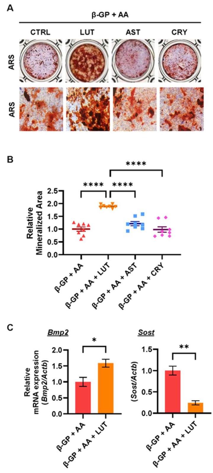
Results: Osteoclast differentiation is significantly inhibited by lutein, astaxanthin, and β-cryptoxanthin. In contrast, only lutein promoted osteoblastic calcified bone nodule formation. To elucidate the molecular role of lutein, we functionally analyzed the NF-κB complex, a molecule involved in bone metabolism, especially in osteoclasts. Docking simulations showed that lutein binds to IKK, thus inhibiting the activation of NF-κB. In a cell culture analysis, the phosphorylation of p65, the active form of NF-κB in osteoblasts, was suppressed by lutein treatment. In vivo, a μCT analysis of the bone microarchitecture showed that lutein improves several bone parameters while maintaining bone mass.

Conclusions: Lutein is effective in maintaining bone mass by controlling both bone resorption and formation, which is applied to prevent disuse-induced osteoporosis.

Keywords: astaxanthin; beta-cryptoxanthin; bone formation; bone resorption; disuse osteoporosis; lutein; osteoclast.


Conflict of interest statement

The authors declare no conflicts of interest in association with the present study.

Figures

Figure 1
Figure 1
Effects of LUT, CRY and AST on osteoclastic bone resorption. (A) Chemical structures of lutein (LUT), β-cryptoxanthin (CRY), and astaxanthin (AST) were described. (B,C) POBs and BMCs were cocultured with IL-1 (2 ng/mL) and LUT, CRY, or AST (20 μM, each) for 7 days. Images show TRAP-stained multinucleated osteoclasts (B). The number of TRAP-stained multinucleated osteoclasts was counted (C). (D) Calvarial bones from neonatal mice were cultured with IL-1 (2 ng/mL) and each carotenoid (20 μM). Bone-resorbing activity was determined by measuring the concentration of calcium leached from bone in conditioned medium. (E) The mRNA expression of Tnfsf11 (encoding RANKL) was quantified by RT-qPCR. (F,G) Raw264.7 cells were cultured with or without sRANKL (100 ng/mL) and LUT (3, 10, and 30 μM) for 5 days. Images show TRAP-stained multinucleated osteoclasts (F). The number of TRAP-stained multinucleated osteoclasts was counted (G). (H) The mRNA expression of Ctsk (encoding cathepsin K) was analyzed by RT-qPCR. The data are expressed as the mean ± SEM of 8 cultures (C), 5 bones (D), 4 cultures (G), or triplicate from a representative experiment (E,F). The Actb gene was used for normalization. Asterisks indicate significant differences between 2 groups: ** p < 0.01, *** p < 0.001, **** p < 0.0001 by a one-way ANOVA followed by post hoc Tukey’s test.
Figure 2
Figure 2
Effects of LUT, CRY and AST on osteoblastic bone formation. (A,B) POBs were cultured with β-glycerophosphate (β-GP) and ascorbic acid (AA) and LUT, CRY, or AST (20 μM, each) for 14 days. Images of alizarin red S (ARS) and alkaline phosphatase (ALP) double-staining are shown (A). The ARS-positive area was measured as the bone mineralized area (B). (C) The mRNA expression of Bmp2 and Sost (encoding sclerostin) was analyzed by RT-qPCR. The data are expressed as the mean ± SEM of 8 cultures (B) or triplicate from a representative experiment (C). The Actb gene was used for normalization. Asterisks indicate a significant difference between 2 groups: * p < 0.05, ** p < 0.01, **** p < 0.0001 by a one-way ANOVA followed by post hoc Tukey’s test (B) and by a two-tailed Welch’s t test (C).
Figure 3
Figure 3
Mechanistic analysis of the effects of lutein on osteoclast differentiation and bone mineralization. (A) The overall image of the molecular docking results. (B,C) Three-dimensional (B) and 2-dimensional images (C) of the docking site. The dashed lines indicate interactions between hydrophobic regions and charged areas of the molecules. Numbers indicate the distance (Å) between IKKβ residue and lutein. (D) The effect of lutein on the kinase activity of IKKβ in an in vitro experiment. The data are expressed as the mean ± SEM of 3 wells. Asterisks indicate a significant difference between 2 groups: *** p < 0.001 and **** p < 0.0001 by a one-way ANOVA followed by post hoc Tukey’s test. (E,F) Mouse POBs were treated with IL-1 or β-GP + AA and lutein for 24 h. Whole-cell lysates were collected, and phospho-p65 and β-actin were detected by Western blotting. The blot images are shown in upper images, and the relative blot intensity of phospho-p65 was shown in lower graphs.
Figure 4
Figure 4
Lutein intake ameliorates disuse-induced bone loss in HLU mice. (A) Reconstructed images using μCT of a horizontal section and the distal femur from a longitudinal section. The scale bar represents 1 mm. (BG) The bone microarchitecture parameters, BV/TV (%) (B), Tb.N (1/mm) (C), Tb.Sp (μm) (D), BS/BV (1/mm) (E), Conn.D (1/mm3), and TBPf (1/mm) were calculated by μCT. The data are expressed as the mean ± SEM of 5 mice. Asterisks indicate a significant difference between 2 groups: * p < 0.05, ** p < 0.01, *** p < 0.001, and **** p < 0.0001 by a one-way ANOVA followed by post hoc Tukey’s test.
Figure 5
Figure 5
Model illustrating the effects of lutein on bone resorption and bone formation. Three carotenoids, including astaxanthin, β-cryptoxanthin, and lutein, inhibited osteoclast differentiation and bone resorption, whereas only lutein promoted osteoblastic calcified bone nodule formation. Mechanistically, lutein can directly bind to IKK protein and suppress its kinase activity, attenuating NF-κB transcriptional activation. The inhibition of NF-κB by lutein results in the downregulation of osteoclast markers in osteoclasts and RANKL in osteoblasts, which, in turn, inhibits osteoclastic bone resorption. In contrast, the inhibition of NF-κB by lutein can elevate the expression of Bmp2 and suppress the expression of Sost, leading to osteoblastic calcified bone nodule formation.

References

    1. Bolamperti S., Villa I., Rubinacci A. Bone Remodeling: An Operational Process Ensuring Survival and Bone Mechanical Competence. Bone Res. 2022;10:48. doi: 10.1038/s41413-022-00219-8. - DOI - PMC - PubMed
    1. Natesan V., Kim S.-J. Metabolic Bone Diseases and New Drug Developments. Biomol. Ther. 2022;30:309–319. doi: 10.4062/biomolther.2022.007. - DOI - PMC - PubMed
    1. Tanaka S., Tanaka Y., Ishiguro N., Yamanaka H., Takeuchi T. RANKL: A Therapeutic Target for Bone Destruction in Rheumatoid Arthritis. Mod. Rheumatol. 2017;28:9–16. doi: 10.1080/14397595.2017.1369491. - DOI - PubMed
    1. Inada M., Matsumoto C., Uematsu S., Akira S., Miyaura C. Membrane-Bound Prostaglandin E Synthase-1-Mediated Prostaglandin E2 Production by Osteoblast Plays a Critical Role in Lipopolysaccharide-Induced Bone Loss Associated with Inflammation. J. Immunol. 2006;177:1879–1885. doi: 10.4049/jimmunol.177.3.1879. - DOI - PubMed
    1. Steeve K.T., Marc P., Sandrine T., Dominique H., Yannick F. IL-6, RANKL, TNF-Alpha/IL-1: Interrelations in Bone Resorption Pathophysiology. Cytokine Growth Factor Rev. 2004;15:49–60. doi: 10.1016/j.cytogfr.2003.10.005. - DOI - PubMed

LinkOut - more resources