. 2024 Dec 16;16(24):4338.
doi: 10.3390/nu16244338.

Curcumin and Its Potential to Target the Glycolytic Behavior of Lactate-Acclimated Prostate Carcinoma Cells with Docetaxel

Affiliations

Curcumin and Its Potential to Target the Glycolytic Behavior of Lactate-Acclimated Prostate Carcinoma Cells with Docetaxel

Dongsic Choi et al. Nutrients. .

Abstract

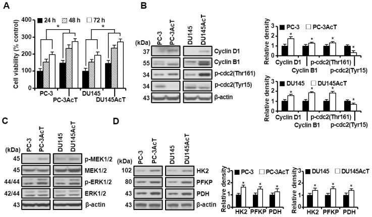
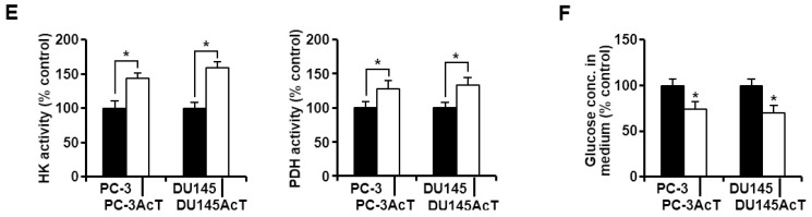
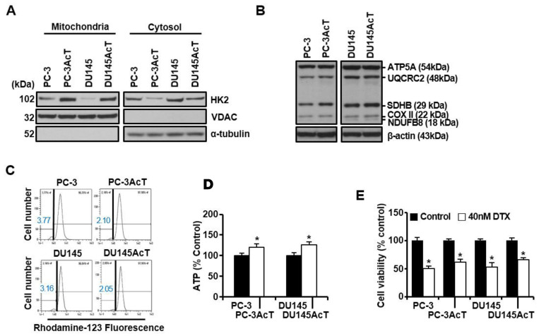
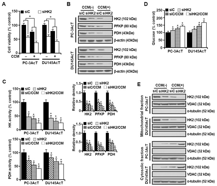
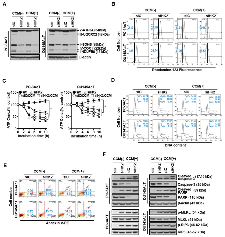
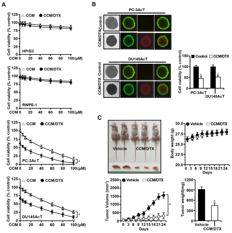
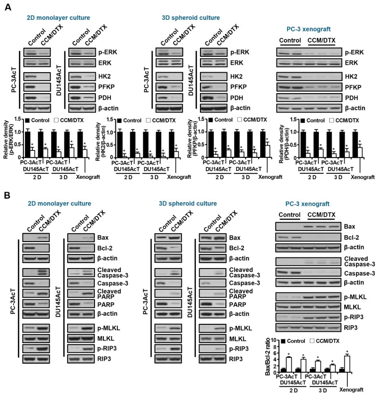
Background: Dysregulated cellular metabolism is known to be associated with drug resistance in cancer treatment. Methods: In this study, we investigated the impact of cellular adaptation to lactic acidosis on intracellular energy metabolism and sensitivity to docetaxel in prostate carcinoma (PC) cells. The effects of curcumin and the role of hexokinase 2 (HK2) in this process were also examined. Results: PC-3AcT and DU145AcT cells that preadapted to lactic acid displayed increased growth behavior, increased dependence on glycolysis, and reduced sensitivity to docetaxel compared to parental PC-3 and DU145 cells. Molecular analyses revealed activation of the c-Raf/MEK/ERK pathway, upregulation of cyclin D1, cyclin B1, and p-cdc2Thr161, and increased levels and activities of key regulatory enzymes in glycolysis, including HK2, in lactate-acclimated cells. HK2 knockdown resulted in decreased cell growth and glycolytic activity, decreased levels of complexes I-V in the mitochondrial electron transport chain, loss of mitochondrial membrane potential, and depletion of intracellular ATP, ultimately leading to cell death. In a xenograft animal model, curcumin combined with docetaxel reduced tumor size and weight, induced downregulation of glycolytic enzymes, and stimulated the upregulation of apoptotic and necroptotic proteins. This was consistent with the in vitro results from 2D monolayer and 3D spheroid cultures, suggesting that the efficacy of curcumin is not affected by docetaxel. Conclusions: Overall, our findings suggest that metabolic plasticity through enhanced glycolysis observed in lactate-acclimated PC cells may be one of the underlying causes of docetaxel resistance, and targeting glycolysis by curcumin may provide potential for drug development that could improve treatment outcomes in PC patients.

Keywords: apoptosis; chemoresistance; curcumin; glycolysis; lactic acid; necroptosis; prostate cancer cells.


Conflict of interest statement

The authors declare no conflicts of interest.

Figures

Figure 1
Figure 1
Increased glycolytic flux in PC-3AcT and DU145AcT cells pre-adapted to lactic acid. Cellular responses were examined after culturing cells in DMEM containing 3.8 μM lactic acid for the indicated time (or 48 h, otherwise). (A) Percent cell viability. (BD) Western blot analysis of cell cycle-regulatory (B), MEK/ERK signaling (C), and key regulatory enzymes in glycolysis (D). (E) Activities of hexokinase and pyruvate dehydrogenase. (F) Changes in glucose concentration in culture medium. Data are expressed as the mean ± standard deviation of three independent experiments. Statistical significance comparing respective PC-3 or DU145 cells was considered at * p < 0.05 using one-way ANOVA and Tukey’s post hoc correction. HK, hexokinase; PFKP, phosphofructokinase platelet; PDH, pyruvate dehydrogenase.
Figure 1
Figure 1
Increased glycolytic flux in PC-3AcT and DU145AcT cells pre-adapted to lactic acid. Cellular responses were examined after culturing cells in DMEM containing 3.8 μM lactic acid for the indicated time (or 48 h, otherwise). (A) Percent cell viability. (BD) Western blot analysis of cell cycle-regulatory (B), MEK/ERK signaling (C), and key regulatory enzymes in glycolysis (D). (E) Activities of hexokinase and pyruvate dehydrogenase. (F) Changes in glucose concentration in culture medium. Data are expressed as the mean ± standard deviation of three independent experiments. Statistical significance comparing respective PC-3 or DU145 cells was considered at * p < 0.05 using one-way ANOVA and Tukey’s post hoc correction. HK, hexokinase; PFKP, phosphofructokinase platelet; PDH, pyruvate dehydrogenase.
Figure 2
Figure 2
Mitochondrial localization of HK2 and effect of docetaxel treatment on PC-3AcT and DU145AcT cells. Cells were cultured in DMEM containing 3.8 μM lactic acid with or without docetaxel (40 nM) for 48 h. (A) Western blot analysis of HK2 in mitochondrial and cytosolic fractions. (B) Western blot analysis of complexes I–V in the mitochondrial electron transport chain. (C) Measurement of mitochondrial membrane potential after staining cells with rhodamine123. (D) Changes in intracellular ATP concentration. (E) Percent cell viability for cells treated with or without 40 nM docetaxel. (F) Annexin V-PE binding assay for cells treated with or without 40 nM docetaxel. (G) Measurements of mitochondrial membrane potential for cells treated with or without 40 nM docetaxel. Data are expressed as the mean ± standard deviation of three independent experiments. Statistical significance comparing respective PC-3 or DU145 cells was considered at * p < 0.05 using one-way ANOVA and Tukey’s post hoc correction. HK2, hexokinase 2; VDAC, voltage-dependent anion channel; NDUFB8, NADH-ubiquinone oxidoreductase subunit B8 (complex I); SDHB, succinate dehydrogenase complex iron sulfur subunit B (complex II); UQCRC2, ubiquinone-cytochrome C reductase core protein 2 (complex III); COX II, mitochondrial cytochrome C oxidase subunit II (complex IV); ATP5A, ATP synthase F1 subunit alpha (complex V); DTX, docetaxel.
Figure 2
Figure 2
Mitochondrial localization of HK2 and effect of docetaxel treatment on PC-3AcT and DU145AcT cells. Cells were cultured in DMEM containing 3.8 μM lactic acid with or without docetaxel (40 nM) for 48 h. (A) Western blot analysis of HK2 in mitochondrial and cytosolic fractions. (B) Western blot analysis of complexes I–V in the mitochondrial electron transport chain. (C) Measurement of mitochondrial membrane potential after staining cells with rhodamine123. (D) Changes in intracellular ATP concentration. (E) Percent cell viability for cells treated with or without 40 nM docetaxel. (F) Annexin V-PE binding assay for cells treated with or without 40 nM docetaxel. (G) Measurements of mitochondrial membrane potential for cells treated with or without 40 nM docetaxel. Data are expressed as the mean ± standard deviation of three independent experiments. Statistical significance comparing respective PC-3 or DU145 cells was considered at * p < 0.05 using one-way ANOVA and Tukey’s post hoc correction. HK2, hexokinase 2; VDAC, voltage-dependent anion channel; NDUFB8, NADH-ubiquinone oxidoreductase subunit B8 (complex I); SDHB, succinate dehydrogenase complex iron sulfur subunit B (complex II); UQCRC2, ubiquinone-cytochrome C reductase core protein 2 (complex III); COX II, mitochondrial cytochrome C oxidase subunit II (complex IV); ATP5A, ATP synthase F1 subunit alpha (complex V); DTX, docetaxel.
Figure 3
Figure 3
Effect of HK2 knockdown alone or in combination with curcumin on glucose metabolism in PC-3AcT and Du145AcT cells. Cells were transfected with 10 nM HK2-targeting siRNA (siHK2) or stealth RNAi control (siC) for 24 h. They were then treated with or without curcumin (40 μM) in DMEM containing 3.8 μM lactic acid for 48 h. (A) Percent cell viability. (B) Western blot analysis of key regulatory enzymes in glycolysis. (C) Activities of hexokinase and pyruvate dehydrogenase. (D) Changes in glucose concentration in culture medium. (E) Western blot analysis of HK2 in mitochondrial and cytosolic fractions. The bar graph represents densitometric analysis of Western blot images normalized to β-actin. Data are expressed as the mean ± standard deviation of three independent experiments. Statistical significance comparing the respective siC group was considered at * p < 0.05 using one-way ANOVA and Tukey’s post hoc correction. CCM, curcumin; HK, hexokinase; PFKP, phosphofructokinase platelet; PDH, pyruvate dehydrogenase.
Figure 4
Figure 4
Effects of HK2 knockdown alone or in combination with curcumin on mitochondrial function and programmed cell death in PC-3AcT and Du145AcT cells. Cells were transfected with 10 nM HK2-targeting siRNA (siHK2) or stealth RNAi control (siC) for 24 h. They were then treated with or without curcumin (40 μM) in DMEM containing 3.8 μM lactic acid for 48 h. (A) Western blot analysis of complexes I–V in mitochondrial electron transport chain. (B) Measurements of mitochondrial membrane potential after staining cells with rhodamine123. (C) Changes in intracellular ATP concentration. (D) Cell cycle analysis. (E) Annexin V-PE binding assay. (F) Western blot analysis of apoptosis- and necroptosis-related proteins. Data are expressed as the mean ± standard deviation of three independent experiments. Statistical significance comparing the respective siC group was considered at * p < 0.05 using one-way ANOVA and Tukey’s post hoc correction. CCM, curcumin; NDUFB8, NADH-ubiquinone oxidoreductase subunit B8 (complex I); SDHB, succinate dehydrogenase complex iron sulfur subunit B (complex II); UQCRC2, ubiquinone-cytochrome C reductase core protein 2 (complex III); COX II, mitochondrial cytochrome C oxidase subunit II (complex IV); ATP5A, ATP synthase F1 subunit alpha (complex V).
Figure 5
Figure 5
Growth-inhibiting effect of co-treatment with curcumin and docetaxel. (A) Cell viability in 2D monolayer culture. Cells were cultured in DMEM containing 3.8 μM lactic acid with or without curcumin (40 μM) and docetaxel (40 nM) for 48 h. (B) Vitality staining of spheroids: from left to right: (i) phase-contrast image, (ii) fluorescent images of fluorescein diacetate(+) living cells in green, (iii) propidium iodide(+) dead cells in red, and (iv) merged; and spheroid cell viability. Spheroids were then treated with or without curcumin (40 µM) and docetaxel (40 nM) for 48 h in DMEM containing 3.8 μM lactic acid. (C) Representative mice, body weight, tumor volume, and tumor weight in PC-3-xenografted nude mice model. Mice (0.3–0.4 cm wide and 0.3–0.4 cm long) were injected intratumorally with vehicle or curcumin (15 mg/kg) and docetaxel (0.5 mg/kg) three times per week for 24 days. Data are expressed as the mean ± standard deviation of three independent experiments. Statistical significance comparing the respective control group was considered at * p < 0.05 using one-way ANOVA and Tukey’s post hoc correction. CCM/DTX, co-treatment with curcumin and docetaxel.
Figure 6
Figure 6
Effects of co-treatment with curcumin and docetaxel on expression of glycolysis-, apoptosis-, and necroptosis-related key proteins in 2D monolayer, 3D spheroid cultures, and nude mice xenograft models. Proteins were extracted from cells, spheroids, and tumors described in Figure 5, separated on 4–12% NuPAGE gels, and subjected to Western blot analysis. (A) Expression levels of key regulatory enzymes of glycolysis. (B) Expression levels of proteins related to apoptosis and necroptosis. The bar graph represents densitometric analysis of Western blot images normalized to β-actin. Data are expressed as the mean ± standard deviation of three independent experiments. Statistical significance comparing the respective control group was considered at * p < 0.05 using one-way ANOVA and Tukey’s post hoc correction. CCM/DTX, co-treatment with curcumin and docetaxel.

Similar articles

Cited by

References

    1. Vaupel P., Multhoff G. Revisiting the Warburg effect: Historical dogma versus current understanding. J. Physiol. 2021;599:1745–1757. doi: 10.1113/JP278810. - DOI - PubMed
    1. Pfeiffer T., Schuster S., Bonhoeffer S. Cooperation and competition in the evolution of ATP-producing pathways. Science. 2001;292:504–507. doi: 10.1126/science.1058079. - DOI - PubMed
    1. Locasale J.W., Cantley L.C. Altered metabolism in cancer. BMC Biol. 2010;25:88. doi: 10.1186/1741-7007-8-88. - DOI - PMC - PubMed
    1. de la Cruz-López K.G., Castro-Muñoz L.J., Reyes-Hernández D.O., García-Carrancá A., Manzo-Merino J. Lactate in the regulation of tumor microenvironment and therapeutic approaches. Front. Oncol. 2019;1:1143. doi: 10.3389/fonc.2019.01143. - DOI - PMC - PubMed
    1. Chetta P., Sriram R., Zadra G. Lactate as key metabolite in prostate cancer progression: What are the clinical implications? Cancers. 2023;15:3473. doi: 10.3390/cancers15133473. - DOI - PMC - PubMed