

Active Vitamin D Ameliorates Arsenite-Induced Thyroid Dysfunction in Sprague-Dawley Rats by Inhibiting the Toll-like Receptor 4/NF-KappaB-Mediated Inflammatory Response
- PMID: 39771102
- PMCID: PMC11728788
- DOI: 10.3390/toxics12120887
Active Vitamin D Ameliorates Arsenite-Induced Thyroid Dysfunction in Sprague-Dawley Rats by Inhibiting the Toll-like Receptor 4/NF-KappaB-Mediated Inflammatory Response
Abstract
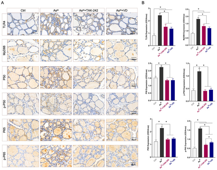
Arsenic, a well-known environmental endocrine disruptor, exerts interference on the body's endocrine system. Our previous investigations have demonstrated that chronic exposure to sodium arsenite (NaAsO2) can induce thyroid damage and dysfunction in Sprague-Dawley (SD) rats. Vitamin D (VD) is an indispensable fat-soluble vitamin that plays a crucial role in maintaining thyroid health. In recent years, numerous studies have demonstrated the association between VD deficiency and the development of various thyroid disorders. However, the precise intervention roles and mechanisms of VD in arsenic-induced thyroid injury remain elusive. This study aimed to investigate the intervention effect of VD on NaAsO2-induced thyroid dysfunction in SD rats. The results demonstrated that exposure to NaAsO2 activates the TLR4/NF-κB signaling pathway in thyroid tissue of rats, leading to apoptosis of thyroid cells and subsequent inflammatory damage and disruption of serum thyroid hormone secretion. Supplementation with TAK-242 (a TLR4 inhibitor) and VD effectively inhibits the activation of the TLR4/NF-κB signaling pathway in rat thyroid tissue exposed to NaAsO2, thereby reducing the inflammatory damage and dysfunction caused by arsenic exposure. In conclusion, the findings of this study offer innovative insights into the application of VD in the prevention and treatment of thyroid dysfunction caused by arsenic exposure.
Keywords: TLR4/NF-κB signaling pathway; sodium arsenite; thyroid hormone; thyrotoxicity; vitamin D.
Conflict of interest statement
The authors declare no conflicts of interest.
Figures


Similar articles
-
The activation of TLR4-MyD88 signaling promotes hepatic dysfunction and fibrotic changes in SD rats resulting from prolonged exposure to sodium arsenite.Int Immunopharmacol. 2024 Oct 25;140:112823. doi: 10.1016/j.intimp.2024.112823. Epub 2024 Jul 30. Int Immunopharmacol. 2024. PMID: 39083929
-
Prolonged exposure to NaAsO2 induces thyroid dysfunction and inflammatory injury in Sprague‒Dawley rats, involvement of NLRP3 inflammasome‒mediated pyroptosis.Arch Toxicol. 2024 Nov;98(11):3673-3687. doi: 10.1007/s00204-024-03837-9. Epub 2024 Aug 9. Arch Toxicol. 2024. PMID: 39120795
-
Alterations of Bax/Bcl-2 ratio contribute to NaAsO2 induced thyrotoxicity in human thyroid follicular epithelial cells and SD rats.Ecotoxicol Environ Saf. 2023 Oct 1;264:115449. doi: 10.1016/j.ecoenv.2023.115449. Epub 2023 Sep 6. Ecotoxicol Environ Saf. 2023. PMID: 37683429
-
Omega-3 polyunsaturated fatty acid supplementation attenuates microglial-induced inflammation by inhibiting the HMGB1/TLR4/NF-κB pathway following experimental traumatic brain injury.J Neuroinflammation. 2017 Jul 24;14(1):143. doi: 10.1186/s12974-017-0917-3. J Neuroinflammation. 2017. PMID: 28738820 Free PMC article.
-
Molecular disturbances and thyroid gland dysfunction in rats chronically exposed to a high dose of NaAsO₂: Insights from proteomic and phosphoproteomic analyses.J Hazard Mater. 2025 Feb 15;484:136746. doi: 10.1016/j.jhazmat.2024.136746. Epub 2024 Dec 3. J Hazard Mater. 2025. PMID: 39637814
Cited by
-
The Addition of Pumpkin Flour Impacts the Functional and Bioactive Properties of Soft Wheat Composite Flour Blends.Foods. 2025 Jan 14;14(2):243. doi: 10.3390/foods14020243. Foods. 2025. PMID: 39856909 Free PMC article.
References
Grants and funding
- 82060604/National Natural Science Foundation of China
- [2023] No. 103/the Excellent Young Talents Plan of Guizhou Medical University
- [2021] No. 5611/the Outstanding Young Scientific and Technological Talent Projects of Guizhou Province
- [2020] No. 4Y153; [2019] No. 1264/Guizhou Provincial Science and Technology Projects
LinkOut - more resources
Full Text Sources
Miscellaneous
