. 2024 Dec 13;12(12):906.
doi: 10.3390/toxics12120906.

Multigenerational Consequences of Prenatal Exposure to Benzophenone-3 Demonstrate Sex- and Region-Dependent Neurotoxic and Pro-Apoptotic Effects in Mouse Brain

Affiliations

Multigenerational Consequences of Prenatal Exposure to Benzophenone-3 Demonstrate Sex- and Region-Dependent Neurotoxic and Pro-Apoptotic Effects in Mouse Brain

Karolina Przepiórska-Drońska et al. Toxics. .

Abstract

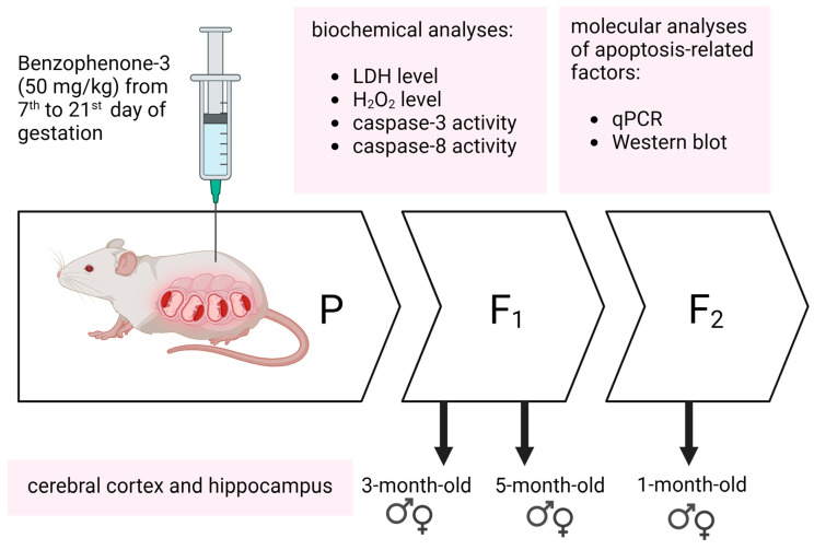
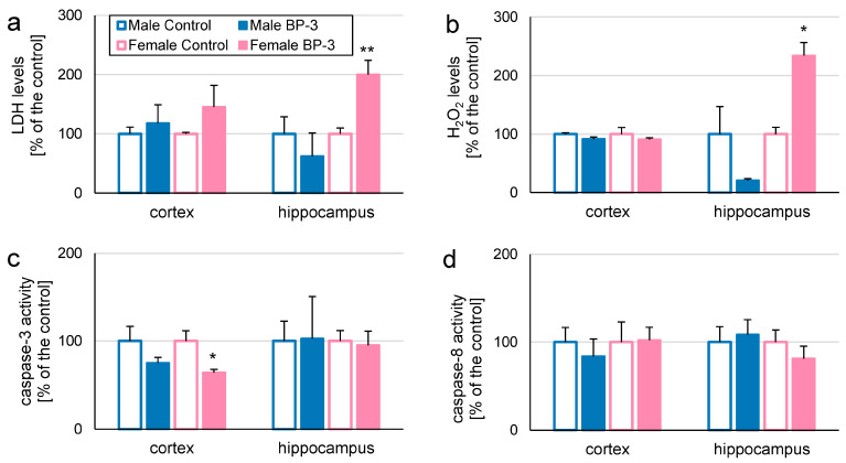
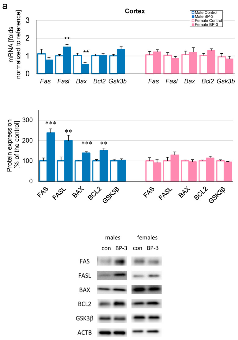
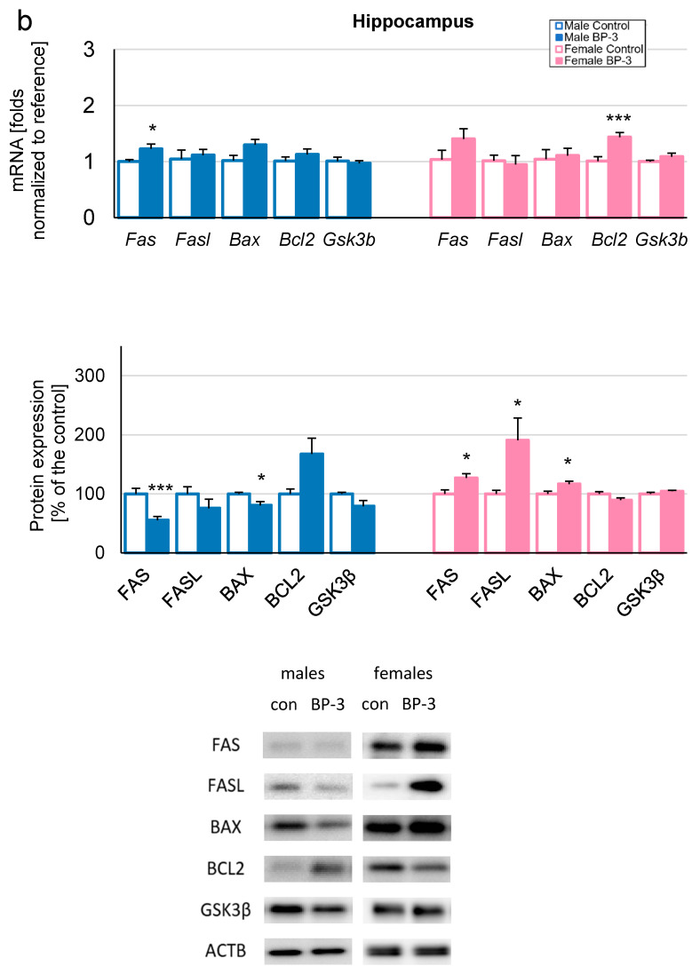
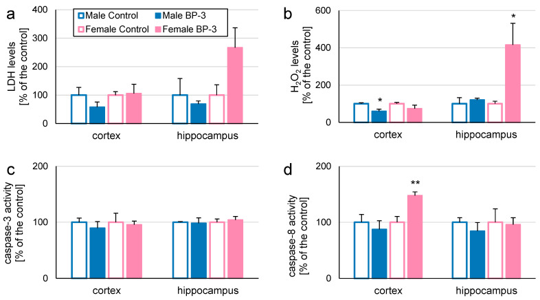
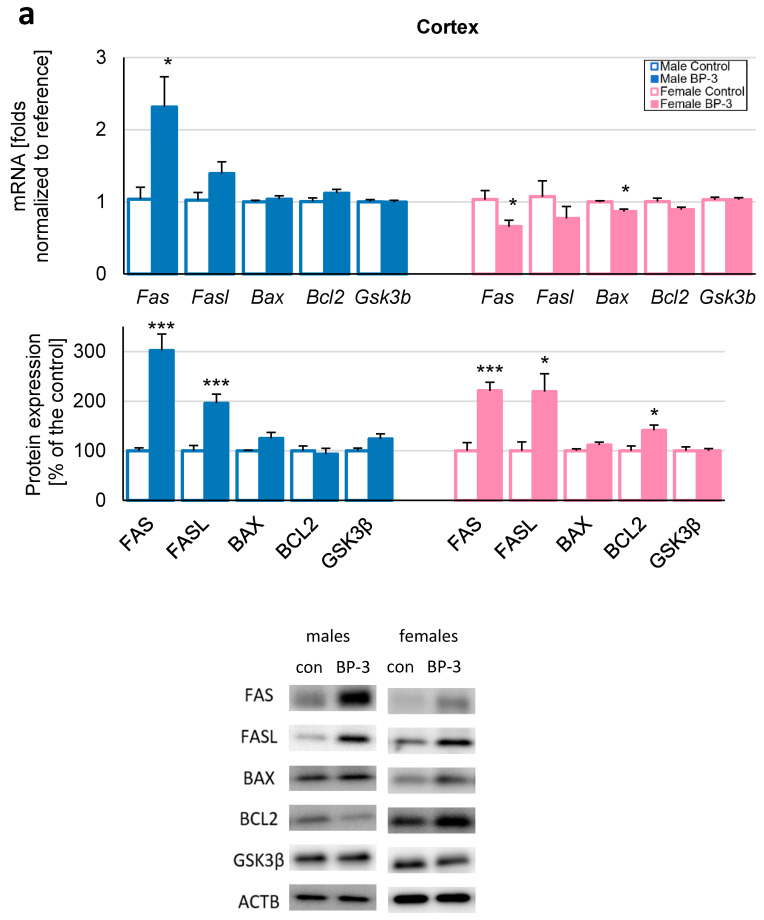
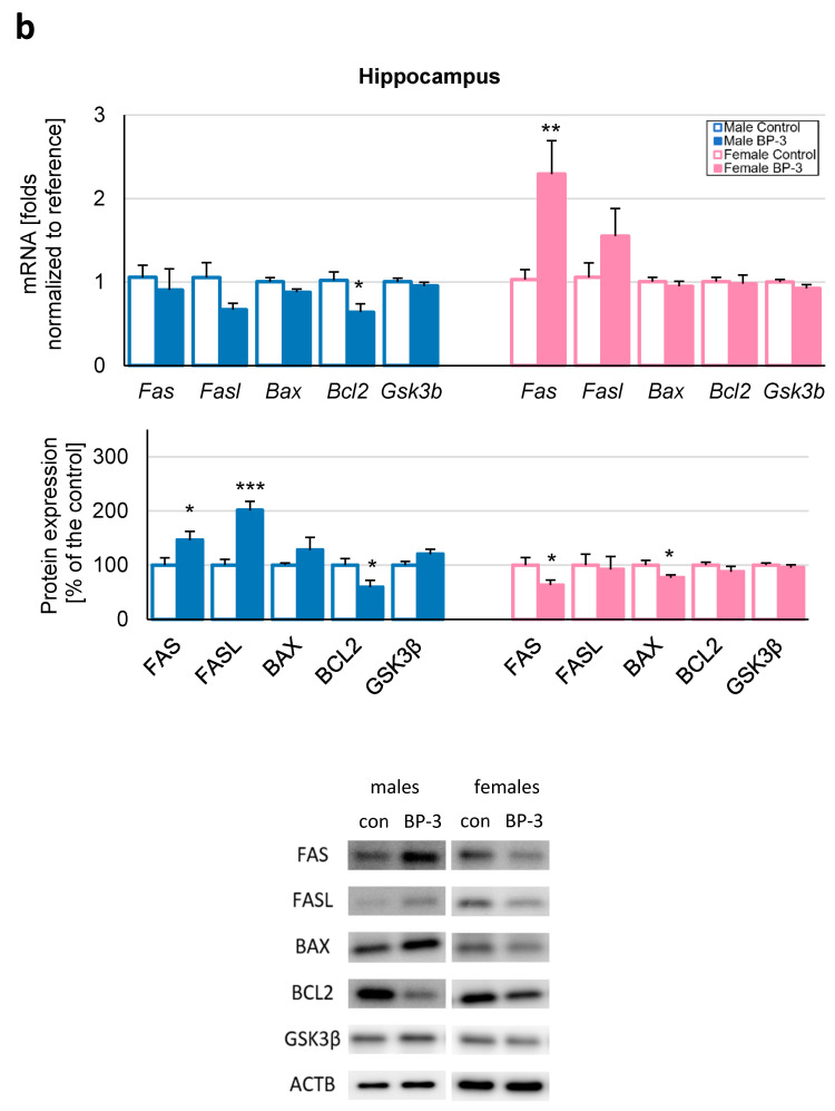
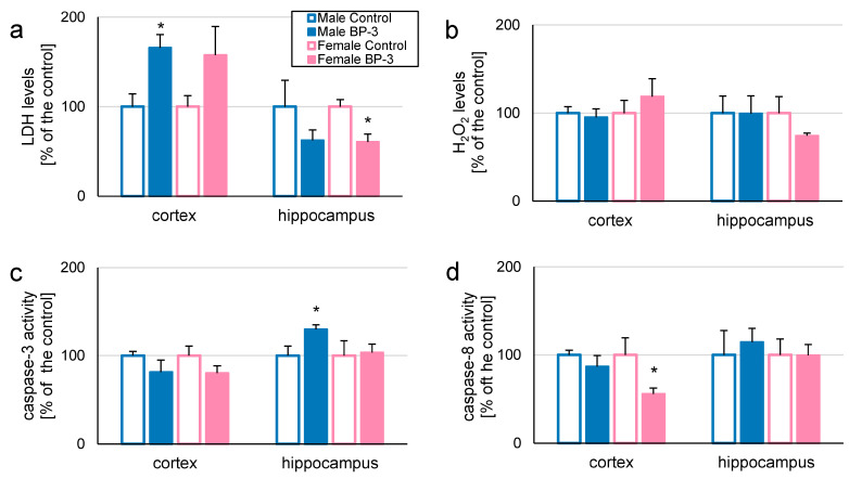
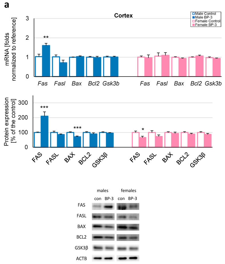
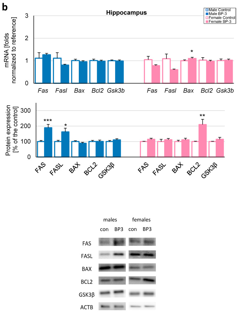
Benzophenone-3 (BP-3), commonly used as a UV filter in personal care products and as a stabilizer, is an alleged endocrine disruptor with potential neurodevelopmental impacts. Despite its abundance in the environment, the studies on its effect on brain development are scarce, especially in terms of multigenerational impact. In this work, for the first time, we examined neurotoxic and pro-apoptotic effects of BP-3 on mouse brain regions (cerebral cortex and hippocampus) in both the first (F1) and second (F2) generations after maternal exposure to environmentally relevant BP-3 levels. We found disregulated markers of cell damage (LDH, H2O2, caspase-3 and -8) and observed increased expression of pro-apoptotic Fas/FAS or Fasl/FASL. BP-3 exposure disrupted the BAX/BCL2 pathway, showing stronger effects in the F1 than in the F2 generation, with a dominance of extrinsic pathway (FAS, FASL, caspase-8) over intrinsic one (BAX, BCL2), suggesting that BP-3-induced apoptosis primarily operates via the extrinsic pathway and could impair brain homeostasis across generations. This study underscores the potential of BP-3 to increase multigenerational risks associated with disrupted neurodevelopment and highlights the importance of understanding its long-term neurotoxic effects.

Keywords: apoptosis; benzophenone-3; environmentally pervasive chemicals; multigenerational changes; neurotoxicity; prenatal exposure.


Conflict of interest statement

The authors have no relevant financial or non-financial interests to disclose.

Figures

Scheme 1
Scheme 1
Illustration of experimental paradigm–prenatal exposure to BP-3 and analyses performed on the F1 and F2 generations to determine the multigenerational neurotoxicity and apoptosis induced by BP-3.
Figure 1
Figure 1
The effects of prenatal exposure to BP-3 in 3-month-old mice from the F1 generation on levels of LDH, H2O2, and activity of caspase-3 and -8. Prenatal exposure to BP-3 increased LDH (a) and H2O2 levels (b) in female hippocampi and decreased caspase-3 activity in female cerebral cortices (c) in 3-month-old F1 mice. In the case of caspase-8, there were no significant changes in its activity in female and male cerebral cortices or hippocampi (d). Each control or experimental group consisted of animals from different litters, and the samples were collected from 5 to 6 animals per group. All results are expressed as percentages of the control ± SEM. * p < 0.05 and ** p < 0.01 versus the control animals.
Figure 2
Figure 2
The effects of prenatal exposure to BP-3 in 3-month-old mice from the F1 generation on the gene and protein expression of apoptosis-related factors. Prenatal exposure to BP-3 diminished the mRNA of Bax, but enhanced levels of FAS, Fasl/FASL, BAX, and BCL2 in 3-month-old F1 males cortices; however, there were no expression changes in apoptosis-related factors in the cerebral cortices of 3-month-old F1 females (a). For the hippocampi, prenatal exposure to BP-3 increased the mRNA expression of Fas, but decreased FAS and BAX in 3-month-old F1 males. Prenatal exposure of 3-month-old F1 females to BP-3 resulted in an increase in Bcl2 mRNA, as well as FAS, FASL, BAX protein levels in hippocampi (b). Each control or experimental group consisted of animals from different litters, and the samples were collected from 5 to 6 animals per group. All results are expressed as a fold change in the case of qPCR and a percentage of the control ± SEM in the case of Western blotting. * p < 0.05, ** p < 0.01, and *** p < 0.001 versus the control animals.
Figure 2
Figure 2
The effects of prenatal exposure to BP-3 in 3-month-old mice from the F1 generation on the gene and protein expression of apoptosis-related factors. Prenatal exposure to BP-3 diminished the mRNA of Bax, but enhanced levels of FAS, Fasl/FASL, BAX, and BCL2 in 3-month-old F1 males cortices; however, there were no expression changes in apoptosis-related factors in the cerebral cortices of 3-month-old F1 females (a). For the hippocampi, prenatal exposure to BP-3 increased the mRNA expression of Fas, but decreased FAS and BAX in 3-month-old F1 males. Prenatal exposure of 3-month-old F1 females to BP-3 resulted in an increase in Bcl2 mRNA, as well as FAS, FASL, BAX protein levels in hippocampi (b). Each control or experimental group consisted of animals from different litters, and the samples were collected from 5 to 6 animals per group. All results are expressed as a fold change in the case of qPCR and a percentage of the control ± SEM in the case of Western blotting. * p < 0.05, ** p < 0.01, and *** p < 0.001 versus the control animals.
Figure 3
Figure 3
The effects of prenatal exposure to BP-3 in 5-month-old mice from the F1 generation on levels of LDH, H2O2, and the activity of caspase-3 and -8. Prenatal exposure of 5-month-old F1 males reduced H2O2 levels in the cerebral cortex (b); while for females, significant changes concerned only an increase in caspase-8 activity in the cerebral cortex (d) and increases in H2O2 levels in the hippocampus (b). No changes were noticed in parameters determining LDH levels (a) and caspase-3 activity (c) in 5-month-old F1 males and females. Each control or experimental group consisted of animals from different litters, and the samples were collected from 5 to 6 animals per group. All results are expressed as a percentage of the control ± SEM. * p < 0.05 and ** p < 0.01 versus the control animals.
Figure 4
Figure 4
The effects of prenatal exposure to BP-3 in 5-month-old mice from the F1 generation on the gene and protein expression of apoptosis-related factors. Prenatal exposure to BP-3 increased Fas/FAS and FASL mRNA or protein levels in the cerebral cortices of 5-month-old males, while in females, there were decreases in mRNA Fas and Bax expression and enhancements in FAS, FASL, and BCL2 protein levels (a). Within the hippocampus, significant changes included a Bcl2/BCL2 decrease and FAS, FASL increases in 5-month-old males; in females, significant changes included an enhancement in Fas mRNA expression and diminishment in FAS and BAX protein levels (b). Each control or experimental group consisted of animals from different litters, and the samples were collected from 5 to 6 animals per group. All results are expressed as a fold change in the case of qPCR and a percentage of the control ± SEM in the case of Western blotting. * p < 0.05, ** p < 0.01, and *** p < 0.001 versus the control animals.
Figure 4
Figure 4
The effects of prenatal exposure to BP-3 in 5-month-old mice from the F1 generation on the gene and protein expression of apoptosis-related factors. Prenatal exposure to BP-3 increased Fas/FAS and FASL mRNA or protein levels in the cerebral cortices of 5-month-old males, while in females, there were decreases in mRNA Fas and Bax expression and enhancements in FAS, FASL, and BCL2 protein levels (a). Within the hippocampus, significant changes included a Bcl2/BCL2 decrease and FAS, FASL increases in 5-month-old males; in females, significant changes included an enhancement in Fas mRNA expression and diminishment in FAS and BAX protein levels (b). Each control or experimental group consisted of animals from different litters, and the samples were collected from 5 to 6 animals per group. All results are expressed as a fold change in the case of qPCR and a percentage of the control ± SEM in the case of Western blotting. * p < 0.05, ** p < 0.01, and *** p < 0.001 versus the control animals.
Figure 5
Figure 5
The effects of BP-3 in 1-month-old mice from the F2 generation on levels of LDH, H2O2, and the activity of caspase-3 and -8. Ancestral exposure of gestating mice to BP-3 resulted in increases in LDH levels in the cerebral cortices (a) and caspase-3 activity in the hippocampi (c) of 1-month-old F2 males. In females, the significant changes included decreases in caspase-8 activity in the cerebral cortex (d) and LDH levels in the hippocampus (a). The levels of H2O2 were not influenced (b). Each control or experimental group consisted of animals from different litters, and the samples were collected from 5 to 6 animals per group. All results are expressed as a percentage of the control ± SEM. * p < 0.05 versus the control animals.
Figure 6
Figure 6
The effects of BP-3 in 1-month-old mice from the F2 generation on the gene and protein expression of apoptosis-related factors. Ancestral exposure of gestating mice to BP-3 resulted in the enhancement of Fas/FAS and the diminishment of BAX expression in cerebral cortices of 1-month-old F2 males, while in females, only a decrease in the FAS level (a) was observed. Within the hippocampus, significant changes included an increase in FAS and FASL protein levels in 1-month-old F2 males; in females, the significant changes included the increase in the Bax mRNA expression level and in the BCL2 protein level (b). Each control or experimental group consisted of animals from different litters, and the samples were collected from 5 to 6 animals per group. All results are expressed as a fold change in the case of qPCR and as a percentage of the control ± SEM in the case of Western blotting. * p < 0.05, ** p < 0.01, and *** p < 0.001 versus the control animals.
Figure 6
Figure 6
The effects of BP-3 in 1-month-old mice from the F2 generation on the gene and protein expression of apoptosis-related factors. Ancestral exposure of gestating mice to BP-3 resulted in the enhancement of Fas/FAS and the diminishment of BAX expression in cerebral cortices of 1-month-old F2 males, while in females, only a decrease in the FAS level (a) was observed. Within the hippocampus, significant changes included an increase in FAS and FASL protein levels in 1-month-old F2 males; in females, the significant changes included the increase in the Bax mRNA expression level and in the BCL2 protein level (b). Each control or experimental group consisted of animals from different litters, and the samples were collected from 5 to 6 animals per group. All results are expressed as a fold change in the case of qPCR and as a percentage of the control ± SEM in the case of Western blotting. * p < 0.05, ** p < 0.01, and *** p < 0.001 versus the control animals.

Similar articles

References

    1. Watson M., Holman D.M., Maguire-Eisen M. Ultraviolet Radiation Exposure and Its Impact on Skin Cancer Risk. Seminars Oncol. Nurs. 2016;32:241–254. doi: 10.1016/j.soncn.2016.05.005. - DOI - PMC - PubMed
    1. Hasbani D.J., Ghaoui N., Haddad F., Shahla W.A., Saade S., Bandali T., Menhem Z., Saade D. Sun Protection Knowledge, Attitudes and Practices: A Lebanese-Based Survey. JEADV Clin. Pract. 2024;3:565–574. doi: 10.1002/jvc2.363. - DOI
    1. Afiouni R., Helou J., Bou-Orm I. Knowledge of the Risks of Ultraviolet Radiation, Sun Exposure Attitudes and Practices among Lebanese University Students. Prev. Med. Rep. 2024;47:102900. doi: 10.1016/j.pmedr.2024.102900. - DOI
    1. Tang X., Yang T., Yu D., Xiong H., Zhang S. Current Insights and Future Perspectives of Ultraviolet Radiation (UV) Exposure: Friends and Foes to the Skin and Beyond the Skin. Environ. Int. 2024;185:108535. doi: 10.1016/j.envint.2024.108535. - DOI - PubMed
    1. Sani M.A., Khezerlou A., Tavassoli M., Abedini A.H., McClements D.J. Development of Sustainable UV-Screening Food Packaging Materials: A Review of Recent Advances. Trends Food Sci. Technol. 2024;145:104366. doi: 10.1016/j.tifs.2024.104366. - DOI