

Development of Roselle (Hibiscus sabdariffa L.) Transcriptome-Based Simple Sequence Repeat Markers and Their Application in Roselle
- PMID: 39771215
- PMCID: PMC11679260
- DOI: 10.3390/plants13243517
Development of Roselle (Hibiscus sabdariffa L.) Transcriptome-Based Simple Sequence Repeat Markers and Their Application in Roselle
Abstract
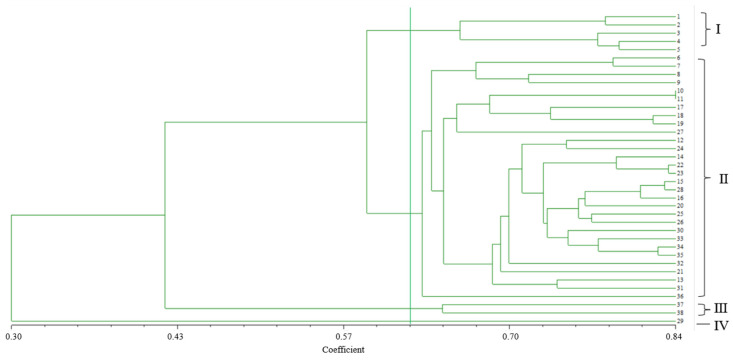
Roselle (Hibiscus sabdariffa L.) simple sequence repeat (SSR) markers were developed using RNA sequencing technology, providing a foundation for genetic analysis and the identification of roselle varieties. In this study, 10 785 unigenes containing 12 994 SSR loci with an average of one SSR locus per 6.87 Kb were identified, and the occurrence frequency of the SSR loci was 11.36%. Trinucleotide repeat motifs were the most abundant, followed by dinucleotide repeats, with AAG/CTT and AT/AT being the predominant types, respectively. After screening 100 primer pairs with a polymorphic ratio of 32.0%, we obtained 32 primer pairs, resulting in clear and stable polymorphic bands. Twenty-seven primer pairs were highly or moderately polymorphic, and seven primer pairs were highly polymorphic. Genetic relationship analysis based on the selected SSR primers showed that 38 roselle accessions were classified into different clades, with those from the same regions clustered into the same subgroups. In contrast, individuals with unique morphological traits were separated. DNA fingerprints of 38 roselle varieties were constructed using five SSR primers, providing an effective method for identifying roselle varieties at a molecular level. Our data provide novel insights into the genetics of H. sabdariffa and may be used in SSR-assisted roselle breeding.
Keywords: Hibiscus sabdariffa L.; application; simple sequence repeat markers; transcriptome-based.
Conflict of interest statement
The authors declare no conflicts of interest.
Figures


Similar articles
-
Development of EST-SSR markers for genetic diversity analysis in coconut (Cocos nucifera L.).Mol Biol Rep. 2020 Dec;47(12):9385-9397. doi: 10.1007/s11033-020-05981-8. Epub 2020 Nov 19. Mol Biol Rep. 2020. PMID: 33215363
-
[Analysis on SSR information in transcriptome of Astragalus membranaceus var. mongholicus and its polymorphism].Zhongguo Zhong Yao Za Zhi. 2018 May;43(9):1838-1843. doi: 10.19540/j.cnki.cjcmm.20180307.002. Zhongguo Zhong Yao Za Zhi. 2018. PMID: 29902894 Chinese.
-
Diversity analysis in Cannabis sativa based on large-scale development of expressed sequence tag-derived simple sequence repeat markers.PLoS One. 2014 Oct 20;9(10):e110638. doi: 10.1371/journal.pone.0110638. eCollection 2014. PLoS One. 2014. PMID: 25329551 Free PMC article.
-
Use of roselle extracted from Hibiscus sabdariffa for histological staining: a critical review and rational stain formulation.Biotech Histochem. 2021 Feb;96(2):94-101. doi: 10.1080/10520295.2020.1769864. Epub 2020 Jun 1. Biotech Histochem. 2021. PMID: 32476481 Review.
-
A review on phytochemistry and therapeutic uses of Hibiscus sabdariffa L.Biomed Pharmacother. 2018 Jun;102:575-586. doi: 10.1016/j.biopha.2018.03.023. Epub 2018 Apr 5. Biomed Pharmacother. 2018. PMID: 29597091 Review.
References
-
- Naim A.M.E., Ibrahim E.B., Abdalla A.W.H., Ibrahim A.E. Variability in some roselle (Hibiscus sabdariffa L.) genotypes for yield and its attributes. Int. J. Agric. For. 2013;3:261–266.
-
- Sharma H.K., Sarkar M., Choudhary S.B., Kumar A.A., Maruthi R.T., Mitra J., Karmakar P.G. Diversity analysis based on agro-morphological traits and microsatellite based markers in global germplasm collections of roselle (Hibiscus sabdariffa L.) Ind. Crops Prod. 2016;89:303–315. doi: 10.1016/j.indcrop.2016.05.027. - DOI
-
- Othman S.N., Yong S.Y.C., Karjiban R.A., Shakri A. Cloning and comparative protein modelling of two MADS-box genes, HsMADS1 and HsMADS2 isolated from Hibiscus sabdariffa L. var. UMKL (roselle) Aust. J. Crop Sci. 2016;10:207–215.
-
- Hashemi A., Shahani A. Effects of salt stress on the morphological characteristics, total phenol and total anthocyanin contents of Roselle (Hibiscus sabdariffa L.) Plant Physiol. Rep. 2019;24:210–214. doi: 10.1007/s40502-019-00446-y. - DOI
LinkOut - more resources
Full Text Sources
