Toxicological Assessment of 2-Hydroxychalcone-Mediated Photodynamic Therapy: Comparative In Vitro and In Vivo Approaches
- PMID: 39771502
- PMCID: PMC11728496
- DOI: 10.3390/pharmaceutics16121523
Toxicological Assessment of 2-Hydroxychalcone-Mediated Photodynamic Therapy: Comparative In Vitro and In Vivo Approaches
Abstract
Background: Photodynamic therapy (PDT) is a treatment modality that uses light to activate a photosensitizing agent, destroying target cells. The growing awareness of the necessity to reduce or eliminate the use of mammals in research has prompted the search for safer toxicity testing models aligned with the new global guidelines and compliant with the relevant regulations.
Objective: The objective of this study was to assess the impact of PDT on alternative models to mammals, including in vitro three-dimensional (3D) cultures and in vivo, in invertebrate animals, utilizing a potent photosensitizer, 2-hydroxychalcone.
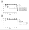
Methods: Cytotoxicity was assessed in two cellular models: monolayer (2D) and 3D. For this purpose, spheroids of two cell lines, primary dermal fibroblasts (HDFa) and adult human epidermal cell keratinocytes (HaCat), were developed and characterized following criteria on cell viability, shape, diameter, and number of cells. The survival percentages of Caenorhabditis elegans and Galleria mellonella were evaluated at 1 and 7 days, respectively.
Results: The findings indicated that all the assessed platforms are appropriate for investigating PDT toxicity. Furthermore, 2-hydroxychalcone demonstrated low toxicity in the absence of light and when mediated by PDT across a range of in vitro (2D and 3D cultures) and in vivo (invertebrate animal models, including G. mellonella and C. elegans) models.
Conclusion: There was a strong correlation between the in vitro and in vivo tests, with similar toxicity results, particularly in the 3D models and C. elegans, where the concentration for 50% viability was approximately 100 µg/mL.
Keywords: 2-hydroxychalcone; Caenorhabditis elegans; Galleria mellonella; monolayers; photodynamic therapy; three-dimensional tissue model; toxicological evaluation of compounds.
Conflict of interest statement
The authors declare no conflicts of interest.
Figures


Similar articles
-
Evaluation of the antineoplastic properties of the photosensitizer biscyanine in 2D and 3D tumor cell models and artificial skin models.J Photochem Photobiol B. 2025 Jan;262:113078. doi: 10.1016/j.jphotobiol.2024.113078. Epub 2024 Dec 9. J Photochem Photobiol B. 2025. PMID: 39671777
-
Antimicrobial and immunomodulatory responses of photodynamic therapy in Galleria mellonella model.BMC Microbiol. 2020 Jul 6;20(1):196. doi: 10.1186/s12866-020-01882-9. BMC Microbiol. 2020. PMID: 32631295 Free PMC article.
-
Comparative study of FosPeg® photodynamic effect on nasopharyngeal carcinoma cells in 2D and 3D models.J Photochem Photobiol B. 2020 Sep;210:111987. doi: 10.1016/j.jphotobiol.2020.111987. Epub 2020 Aug 7. J Photochem Photobiol B. 2020. PMID: 32801063
-
Alternative Non-Mammalian Animal and Cellular Methods for the Study of Host-Fungal Interactions.J Fungi (Basel). 2023 Sep 19;9(9):943. doi: 10.3390/jof9090943. J Fungi (Basel). 2023. PMID: 37755051 Free PMC article. Review.
-
Review of three-dimensional spheroid culture models of gynecological cancers for photodynamic therapy research.Photodiagnosis Photodyn Ther. 2024 Feb;45:103975. doi: 10.1016/j.pdpdt.2024.103975. Epub 2024 Jan 17. Photodiagnosis Photodyn Ther. 2024. PMID: 38237651 Review.
References
Grants and funding
- 2019/22188-8, 2020/15586-4, 2021/03805-6, 2017/18388-6, 2018/02785-9/Fundação de Amparo à Pesquisa do Estado de São Paulo
- 001; 88887.500765/2020-00/Coordenação de Aperfeiçoamento de Pessoal de Nível Superior (CAPES)
- 134559/2018-5, 142049/2019- 0/Conselho Nacional de Desenvolvimento Científico e Tecnológico (CNPq)
- 00/Programa de Apoio ao Desenvolvimento Científico (PADC) da Faculdade de Ciências Farma-cêuticas da UNESP
- PDI-UNESP-00/Pró-Reitoria de Pós-Graduação (PROPG)
LinkOut - more resources
Full Text Sources

