

Biomedical Application Prospects of Gadolinium Oxide Nanoparticles for Regenerative Medicine
- PMID: 39771605
- PMCID: PMC11676666
- DOI: 10.3390/pharmaceutics16121627
Biomedical Application Prospects of Gadolinium Oxide Nanoparticles for Regenerative Medicine
Abstract
Background/objectives: The aim was to study the possibilities of biomedical application of gadolinium oxide nanoparticles (Gd2O3 NPs) synthesized under industrial conditions, and evaluate their physicochemical properties, redox activity, biological activity, and safety using different human cell lines.
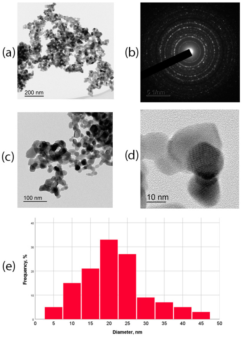
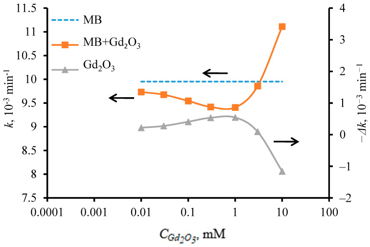
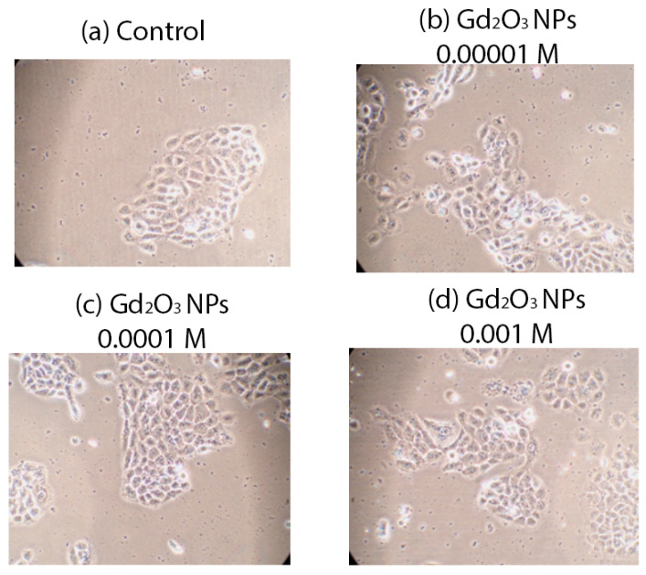
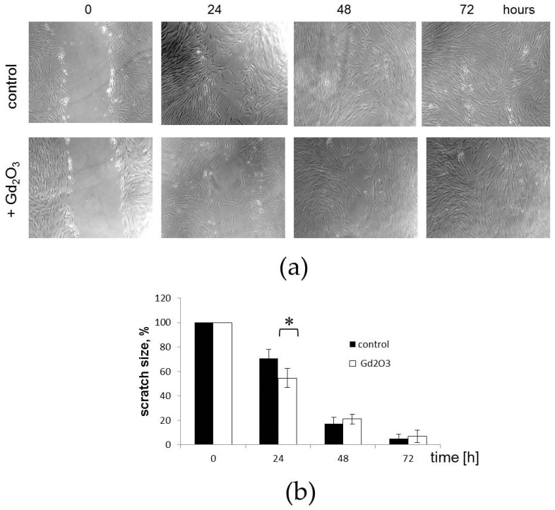
Methods: The powder of Gd2O3 NPs was obtained by a process of thermal decomposition of gadolinium carbonate precipitated from nitrate solution, and was studied using transmission electron microscopy (TEM), X-ray diffraction (XRD), Raman spectroscopy, mass spectrometry, and scanning electron microscopy (SEM) with energy dispersive X-ray analyzer (EDX). The redox activity of different concentrations of Gd2O3 NPs was studied by the optical spectroscopy (OS) method in the photochemical degradation process of methylene blue dye upon irradiation with an optical source. Biological activity was studied on different human cell lines (keratinocytes, fibroblasts, mesenchymal stem cells (MSCs)) with evaluation of the effect of a wide range of Gd2O3 NP concentrations on metabolic and proliferative cellular activity (MTT test, direct cell counting, dead cell assessment, and visual assessment of cytoarchitectonics). The test of migration activity assessment on a model wound was performed on MSC culture.
Results: According to TEM data, the size of the NPs was in the range of 2-43 nm, with an average of 20 nm. XRD analysis revealed that the f Gd2O3 nanoparticles had a cubic structure (C-form) of Gd2O3 (Ia3)¯ with lattice parameter a = 10.79(9) Å. Raman spectroscopy showed that the f Gd2O3 nanoparticles had a high degree of crystallinity. By investigating the photooxidative degradation of methylene blue dye in the presence of f Gd2O3 NPs under red light irradiation, it was found that f Gd2O3 nanoparticles showed weak antioxidant activity, which depended on the particle content in the solution. At a concentration of 10-3 M, the highest antioxidant activity of f Gd2O3 nanoparticles was observed when the reaction rate constant of dye photodegradation decreased by 5.5% to 9.4 × 10⁻³ min-1. When the concentration of f Gd2O3 NPs in solution was increased to 10-2 M upon irradiation with a red light source, their antioxidant activity changed to pro-oxidant activity, accompanied by a 15% increase in the reaction rate of methylene blue degradation. Studies on cell lines showed a high level of safety and regenerative potential of Gd2O3 NPs, which stimulated fibroblast metabolism at a concentration of 10-3 M (27% enhancement), stimulated keratinocyte metabolism at concentrations of 10-3 M-10-5 M, and enhanced keratinocyte proliferation by an average of 35% at concentrations of 10-4 M. Furthermore, it accelerated the migration of MSCs, enhancing their proliferation, and promoting the healing of the model wound.
Conclusions: The results of the study demonstrated the safety and regenerative potential of redox-active Gd2O3 NPs towards different cell lines. This may be the basis for further research to develop nanomaterials based on Gd2O3 NPs for skin wound healing and in regenerative medicine generally.
Keywords: biomedicine; cytotoxicity; drug development; gadolinium oxide; nanogadolinium; nanomaterials; nanoparticles; regeneration.
Conflict of interest statement
The authors declare no conflicts of interest. The funder (Sechenov University) was not involved in the study’s design, and did not affect its results. The «LANHIT» company did not sponsor this study and did not participate in the further instrumental analysis of the obtained materials that were not performed on the basis of LANHIT. LLC «LANHIT» company has been a manufacturer of high and special purity inorganic compounds since 1991, including rare earth metal compounds. E.L.C, A.A.G., and O.I.A. are affiliated with “LANHIT” Company. E.L.C. was responsible for study of the literature on the synthesis of gadolinium oxide nanoparticles, scientific substantiation of the choice of synthesis methodology, data curation and control over the obtained nanoparticles, and participation in writing the article in the part describing the synthesis. A.A.G., the chief technologist of the company, was responsible for the selection of synthesis methods, the technical side of the syntheses, model development of the technology and the transfer of the technology to an industrial basis, and writing the chemical part of the article. O.I.A. conducted the synthesis and assessed the physical characteristics of the obtained nanoparticles, and helped write the chemical part of the article. Besides, all instrumental studies were conducted within the framework of non-financial agreements (cooperation agreement) between the institutions indicated in the affiliation of the article independently of each other. In particular, there is a scientific cooperation agreement between I.M. Sechenov First Moscow State Medical University (Sechenov University), and LANHIT Company.
Figures


Similar articles
-
Development of Technology for the Synthesis of Nanocrystalline Cerium Oxide Under Production Conditions with the Best Regenerative Activity and Biocompatibility for Further Creation of Wound-Healing Agents.Pharmaceutics. 2024 Oct 25;16(11):1365. doi: 10.3390/pharmaceutics16111365. Pharmaceutics. 2024. PMID: 39598490 Free PMC article.
-
Biosynthesis of zinc oxide nanoparticles using Albizia lebbeck stem bark, and evaluation of its antimicrobial, antioxidant, and cytotoxic activities on human breast cancer cell lines.Int J Nanomedicine. 2018 Dec 20;14:87-100. doi: 10.2147/IJN.S186888. eCollection 2019. Int J Nanomedicine. 2018. PMID: 30587987 Free PMC article.
-
Effect of annealing on down-conversion properties of monoclinic Gd2O3:Er3+ nanophosphors.Luminescence. 2015 Sep;30(6):812-7. doi: 10.1002/bio.2824. Epub 2014 Dec 21. Luminescence. 2015. PMID: 25529921
-
Europium-doped Gd2O3 nanotubes cause the necrosis of primary mouse bone marrow stromal cells through lysosome and mitochondrion damage.J Inorg Biochem. 2015 May;146:28-36. doi: 10.1016/j.jinorgbio.2015.02.006. Epub 2015 Feb 19. J Inorg Biochem. 2015. PMID: 25725393
-
Raman spectroscopy and regenerative medicine: a review.NPJ Regen Med. 2017 May 15;2:12. doi: 10.1038/s41536-017-0014-3. eCollection 2017. NPJ Regen Med. 2017. PMID: 29302348 Free PMC article. Review.
Cited by
-
Fabrication of Radiopaque, Drug-Loaded Resorbable Polymer for Medical Device Development.Polymers (Basel). 2025 Mar 7;17(6):716. doi: 10.3390/polym17060716. Polymers (Basel). 2025. PMID: 40292576 Free PMC article.
-
Europium-doped cerium oxide nanoparticle-impregnated Hyalgan® (CartiOxgel): an intra-articular contrast agent for X-ray CT and optical imaging.RSC Adv. 2025 Jul 7;15(29):23374-23395. doi: 10.1039/d5ra01830g. eCollection 2025 Jul 4. RSC Adv. 2025. PMID: 40626062 Free PMC article.
-
Potential Applications of Rare Earth Metal Nanoparticles in Biomedicine.Pharmaceuticals (Basel). 2025 Jan 24;18(2):154. doi: 10.3390/ph18020154. Pharmaceuticals (Basel). 2025. PMID: 40005968 Free PMC article. Review.
References
-
- Wen J., Moloney E.B., Canning A., Donohoe E., Ritter T., Wang J., Xiang D., Wu J., Li Y. Synthesized nanoparticles, biomimetic nanoparticles and extracellular vesicles for treatment of autoimmune disease: Comparison and prospect. Pharmacol. Res. 2021;172:105833. doi: 10.1016/j.phrs.2021.105833. - DOI - PubMed
Grants and funding
LinkOut - more resources
Full Text Sources
Miscellaneous
