. 2024 Dec 11;24(24):7899.
doi: 10.3390/s24247899.

Affinity Peptide-Based Circularly Permuted Fluorescent Protein Biosensors for Non-Small Cell Lung Cancer Diagnosis

Affiliations

Affinity Peptide-Based Circularly Permuted Fluorescent Protein Biosensors for Non-Small Cell Lung Cancer Diagnosis

Dengyue Xu et al. Sensors (Basel). .

Abstract

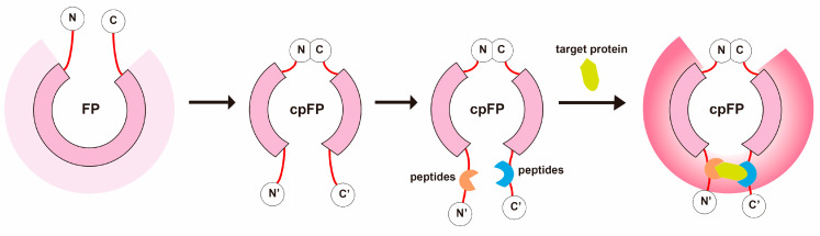
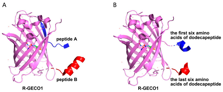
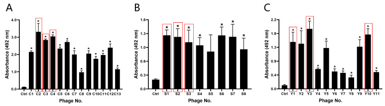
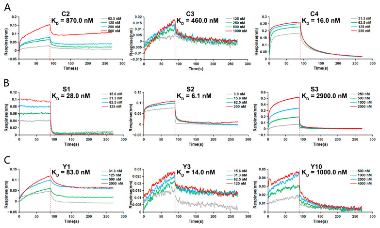
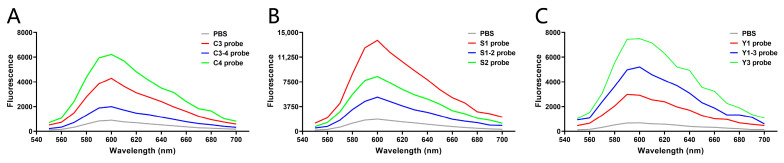
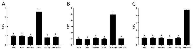
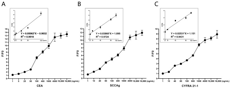
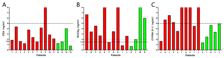
Non-small cell lung cancer (NSCLC) is the predominant form of lung cancer and poses a significant public health challenge. Early detection is crucial for improving patient outcomes, with serum biomarkers such as carcinoembryonic antigen (CEA), squamous cell carcinoma antigen (SCCAg), and cytokeratin fragment 19 (CYFRA 21-1) playing a critical role in early screening and pathological classification of NSCLC. However, due to being mainly based on corresponding antibody binding reactions, existing detection technologies for these serum biomarkers have shortcomings such as complex operations, high false positive rates, and high costs. This study aimed to develop new methods for detecting CEA, SCCAg, and CYFRA 21-1 to assist in the diagnosis of NSCLC. Affinity peptides of CEA, SCCAg, and CYFRA 21-1, respectively, were screened by phage display technology, and the peptides' binding affinities were determined by enzyme-linked immunosorbent assay and biolayer interferometry. Peptides with high affinity were then integrated as binding domains into biosensors by fusing them with circularly permuted fluorescent proteins (cpFPs) through genetic coding. The resulting biosensors, C4 biosensor for CEA, S1 biosensor for SCCAg, and Y3 biosensor for CYFRA 21-1, demonstrated robust sensitivity and specificity even at concentrations as low as 1 ng/mL for their respective tumor markers. When applied to clinical samples and recalibrated for the upper limit of normal concentrations, the biosensors exhibited enhanced sensitivity and specificity for NSCLC diagnosis. This study introduced innovative biosensors for the detection of CEA, SCCAg, and CYFRA 21-1, providing a highly sensitive, specific, rapid, and cost-effective diagnostic alternative that could significantly improve NSCLC screening rates.

Keywords: carcinoembryonic antigen; circularly permuted fluorescent protein; cytokeratin fragment 19; non-small cell lung cancer; phage display; squamous cell carcinoma antigen.


Conflict of interest statement

Author B.W. was employed by China Certification and Inspection Group Liaoning Co., Ltd. The remaining authors declare that the research was conducted in the absence of any commercial or financial relationships that could be construed as a potential conflict of interest.

Figures

Figure 1
Figure 1
Construct Design of the Circularly Permuted Fluorescent Protein (cpFP) Biosensor. This schematic illustrates the assembly of the cpFP biosensor. The native N- and C-termini of the cpFP are joined, creating new termini at alternative tolerance sites. The affinity peptides, identified through screening, are then attached to these new termini, serving as the binding domain proteins. Upon binding to the target protein, the microenvironment surrounding the chromophore of the rearranged fluorescent protein alters, leading to a significant fluorescence response.
Figure 2
Figure 2
The strategy for the construction of the cpFP biosensor. (A): Using R-GECO1 as a template, the cpFP biosensor was constructed with two selected dodecapeptides as the N-terminus and C-terminus, respectively. (B): Using R-GECO1 as a template, the cpFP biosensor was constructed with two hexapeptides separated from the selected dodecapeptide as the N-terminus and C-terminus.
Figure 3
Figure 3
The binding affinity of the screened binding phages. Absorbance values measured at 450 nm from ELISA assays for CEA (A), SCCAg (B), and CYFRA 21-1 (C). The sequences in the red box were the phage sequences selected for each marker for subsequent experiments. * p ˂ 0.05 vs. Control group. (n = 3).
Figure 4
Figure 4
Affinity analysis of screened peptides. The binding affinities of the screened peptides to corresponding serum tumor markers was detected by BLI assay. (A) Determination of the KD for the interaction between C2, C3, C4, and CEA, respectively. (B) Determination of the KD for the interaction between S1, S2, S3, and SCCAg, respectively. (C) Determination of the KD for the interaction between Y1, Y3, Y10, and CYFRA 21-1, respectively.
Figure 5
Figure 5
Sensitivity detection of the biosensors. Based on R-GECO1, the biosensors were constructed and synthesized according to the strategies, and the sensitivity of the reaction between each biosensor and corresponding tumor markers was detected. (A) Fluorescence values of PBS, C3 biosensor, C3-4 biosensor, and C4 biosensor mixed with CEA, respectively, tested by a microplate reader. (B) Fluorescence values of PBS, S1 biosensor, S1-2 biosensor, and S2 biosensor mixed with SCCAg, respectively, tested by a microplate reader. (C) Fluorescence values of PBS, Y1 biosensor, Y1-3 biosensor, and Y3 biosensor mixed with CYFRA 21-1, respectively, tested by a microplate reader.
Figure 6
Figure 6
Specificity detection of the biosensors. (A) Changes in fluorescence of C4 biosensor upon reacting with BSA, NSE, proGRP, CEA, SCCAg, and CYFRA 21-1, respectively. $ p ˂ 0.05 vs. CEA group. (B) Changes in fluorescence of S1 biosensor upon reacting with BSA, NSE, proGRP, CEA, SCCAg, and CYFRA 21-1, respectively. # p ˂ 0.05 vs. SCCAg group. (C) Changes in fluorescence of Y3 biosensor upon reacting with BSA, NSE, proGRP, CEA, SCCAg, and CYFRA 21-1, respectively. & p ˂ 0.05 vs. CYFRA 21-1 group. (n = 5).
Figure 7
Figure 7
Linearity test for the biosensors. The reaction system consists of 180 μL/well biosensor (4 μg/mL) and 20 μL/well serum tumor marker. (A) Obvious fluorescence change was observed when C4 biosensor reacted with CEA, and F/F0 increased with the increase of concentration of CEA (1–16,000 ng/mL). The linear regression analysis of C4 biosensor was performed using 5 increasing concentrations of CEA: 1, 5, 25, 50, 100 ng/mL. (B) Obvious fluorescence change was observed when S1 biosensor reacted with SCCAg, and F/F0 increased with the increase of concentration of SCCAg (1–16,000 ng/mL). The linear regression analysis of S1 biosensor was performed using 5 increasing concentrations of SCCAg: 1, 10, 40, 80, 100 ng/mL. (C) Obvious fluorescence change was observed when Y3 biosensor reacted with CYFRA 21-1, and F/F0 increased with the increase of concentration of CYFRA 21-1 (1–16,000 ng/mL). The linear regression analysis of Y3 biosensor was performed using 5 increasing concentrations of CYFRA 21-1: 1, 10, 40, 80, 100. (n = 5).
Figure 8
Figure 8
Evaluation of assay performance against patient samples. (A) The C4 biosensor was used to detect CEA in 11 NSCLC patients (red) and 4 non-NSCLC patients (green). (B) The S1 biosensor was used to detect SCCAg in 10 NSCLC patients (red) and 4 non-NSCLC patients (green). (C) The Y3 biosensor was used to detect CYFRA 21-1 in 10 NSCLC patients (red) and 5 non-NSCLC patients (green). The thick dashed lines represented the upper limit of normal concentrations currently used by the hospital. The thin dashed lines represented the adjusted upper limit of normal concentration for better diagnostic sensitivity and specificity.

Similar articles

References

    1. Leiter A., Veluswamy R.R., Wisnivesky J.P. The global burden of lung cancer: Current status and future trends. Nat. Rev. Clin. Oncol. 2023;20:624–639. doi: 10.1038/s41571-023-00798-3. - DOI - PubMed
    1. Nooreldeen R., Bach H. Current and Future Development in Lung Cancer Diagnosis. Int. J. Mol. Sci. 2021;22:8661. doi: 10.3390/ijms22168661. - DOI - PMC - PubMed
    1. China NHCotPsRo Guidelines for the Diagnosis and Treatment of Primary Lung Cancer. Chin. J. Ration. Drug Use. 2022;9:1–28.
    1. Adams S.J., Stone E., Baldwin D.R., Vliegenthart R., Lee P., Fintelmann F.J. Lung cancer screening. Lancet. 2023;401:390–408. doi: 10.1016/S0140-6736(22)01694-4. - DOI - PubMed
    1. Wang C., Long Y., Li W., Dai W., Xie S., Liu Y., Zhang Y., Liu M., Tian Y., Li Q., et al. Exploratory study on classification of lung cancer subtypes through a combined K-nearest neighbor classifier in breathomics. Sci. Rep. 2020;10:5880. doi: 10.1038/s41598-020-62803-4. - DOI - PMC - PubMed