. 2024 Nov 24;12(12):1316.
doi: 10.3390/vaccines12121316.

Rapid Development of Modified Vaccinia Virus Ankara (MVA)-Based Vaccine Candidates Against Marburg Virus Suitable for Clinical Use in Humans

Affiliations

Rapid Development of Modified Vaccinia Virus Ankara (MVA)-Based Vaccine Candidates Against Marburg Virus Suitable for Clinical Use in Humans

Alina Tscherne et al. Vaccines (Basel). .

Abstract

Background/objectives: Marburg virus (MARV) is the etiological agent of Marburg Virus Disease (MVD), a rare but severe hemorrhagic fever disease with high case fatality rates in humans. Smaller outbreaks have frequently been reported in countries in Africa over the last few years, and confirmed human cases outside Africa are, so far, exclusively imported by returning travelers. Over the previous years, MARV has also spread to non-endemic African countries, demonstrating its potential to cause epidemics. Although MARV-specific vaccines are evaluated in preclinical and clinical research, none have been approved for human use. Modified Vaccinia virus Ankara (MVA), a well-established viral vector used to generate vaccines against emerging pathogens, can deliver multiple antigens and has a remarkable clinical safety and immunogenicity record, further supporting its evaluation as a vaccine against MARV. The rapid availability of safe and effective MVA-MARV vaccine candidates would expand the possibilities of multi-factored intervention strategies in endemic countries.

Methods: We have used an optimized methodology to rapidly generate and characterize recombinant MVA candidate vaccines that meet the quality requirements to proceed to human clinical trials. As a proof-of-concept for the optimized methodology, we generated two recombinant MVAs that deliver either the MARV glycoprotein (MVA-MARV-GP) or the MARV nucleoprotein (MVA-MARV-NP).

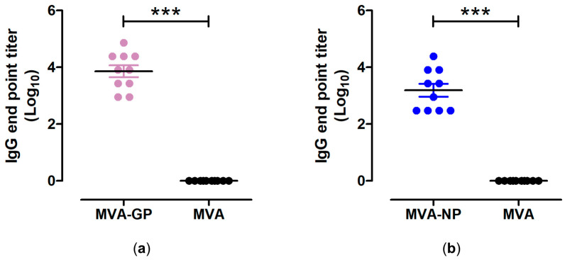
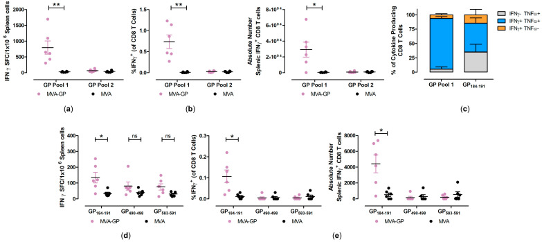
Results: Infections of human cell cultures with recombinant MVA-MARV-GP and MVA-MARV-NP confirmed the efficient synthesis of MARV-GP and MARV-NP proteins in mammalian cells, which are non-permissive for MVA replication. Prime-boost immunizations in C57BL/6J mice readily induced circulating serum antibodies binding to recombinant MARV-GP and MARV-NP proteins. Moreover, the MVA-MARV-candidate vaccines elicited MARV-specific T-cell responses in C57BL/6J mice.

Conclusions: We confirmed the suitability of our two backbone viruses MVA-mCherry and MVA-GFP in a proof-of-concept study to rapidly generate candidate vaccines against MARV. However, further studies are warranted to characterize the protective efficacy of these recombinant MVA-MARV vaccines in other preclinical models and to evaluate them as vaccine candidates in humans.

Keywords: Marburg virus; Modified Vaccinia virus Ankara (MVA); emerging viruses; rapid vaccine development; viral vector vaccine.


Conflict of interest statement

The authors declare no conflicts of interest.

Figures

Figure 1
Figure 1
Design, construction, and virological in vitro characterization of sfMVA-mCherry (MVA-mCherry) and sfMVA-GFP (MVA-GFP). (a,b) Schematic representation of the MVA genome. The six major deletion sites are indicated with I-VI. The encoding sequences of mCherry (a) and GFP (b) were introduced into non-recombinant MVA by homologous recombination. mCherry was inserted into the genomic region between the two MVA genes, MVA069R and MVA070L, within the MVA genome. GFP was inserted into deletion III within the MVA genome. PCR analysis was conducted by using a specific oligonucleotide primer targeting the respective insertion site. (c) Fluorescent microscopy of recombinant MVA-mCherry and MVA-GFP after serial round of plaque purification. Scale bar: 50 µm. (d) Genetic integrity of MVA-mCherry. PCR analysis of viral DNA with oligonucleotide primers specific for the target site confirmed the insertion of full-length mCherry gene sequence. (e) Viral growth profile of MVA-mCherry and non-recombinant MVA. (f) Genetic integrity of MVA-GFP. PCR analysis of viral DNA with oligonucleotide primers specific for the target site confirmed the insertion of a full-length GFP gene sequence. (g) Viral growth profile of MVA-GFP and non-recombinant MVA (MVA). hpi: hours post-infection.
Figure 1
Figure 1
Design, construction, and virological in vitro characterization of sfMVA-mCherry (MVA-mCherry) and sfMVA-GFP (MVA-GFP). (a,b) Schematic representation of the MVA genome. The six major deletion sites are indicated with I-VI. The encoding sequences of mCherry (a) and GFP (b) were introduced into non-recombinant MVA by homologous recombination. mCherry was inserted into the genomic region between the two MVA genes, MVA069R and MVA070L, within the MVA genome. GFP was inserted into deletion III within the MVA genome. PCR analysis was conducted by using a specific oligonucleotide primer targeting the respective insertion site. (c) Fluorescent microscopy of recombinant MVA-mCherry and MVA-GFP after serial round of plaque purification. Scale bar: 50 µm. (d) Genetic integrity of MVA-mCherry. PCR analysis of viral DNA with oligonucleotide primers specific for the target site confirmed the insertion of full-length mCherry gene sequence. (e) Viral growth profile of MVA-mCherry and non-recombinant MVA. (f) Genetic integrity of MVA-GFP. PCR analysis of viral DNA with oligonucleotide primers specific for the target site confirmed the insertion of a full-length GFP gene sequence. (g) Viral growth profile of MVA-GFP and non-recombinant MVA (MVA). hpi: hours post-infection.
Figure 2
Figure 2
Design, construction, and virological in vitro characterization of MVA-MARV-GP (MVA-GP) vaccine candidate. (a) Schematic representation of sfMVA-GFP (MVA-GFP) genome. The codon-optimized full-length MARV-GP gene sequence was inserted into MVA-GFP by homologous recombination. Removal of marker gene mCherry occurred by intragenomic recombination during plaque purification. (b) The genetic integrity of MVA-GP was determined by PCR analysis using an oligonucleotide primer specific for the targeted insertion site (deletion site III). (c) Viral growth profile of MVA-GP and non-recombinant MVA (MVA). (df) Expression of full-length MARV-GP in MVA-GP infected VeroE6 cells, analyzed by (d) immunofluorescence staining and (e,f) Western blot. (d) Permeabilized VeroE6 cells were probed with a primary antibody targeting MARV-GP and a polyclonal goat-anti-mouse secondary antibody for GP-specific staining (red). DAPI served to counterstain cell nuclei (blue). Scale bar: 50 µm. (e,f) Proteins in cell lysates of MVA-GP-infected VeroE6 cells were separated by SDS-PAGE and subsequently probed with a primary antibody targeting MARV-GP (GP2 subunit). (f) The blot was incubated with a primary antibody targeting GAPDH to confirm the loading of equal amounts of proteins (e) Deglycosylation of cell lysates was performed with PNGase F before Western blot analysis. Uninfected cells (mock) and cells infected with non-recombinant MVA (MVA) served as controls. hpi: hours post-infection.
Figure 3
Figure 3
Design, construction, and virological in vitro characterization of MVA-MARV-NP (MVA-NP). (a) Schematic representation of the sfMVA-mCherry genome. The codon-optimized full-length MARV-NP gene sequence was inserted into MVA-mCherry by homologous recombination. Removal of the marker gene GFP occurred by intragenomic recombination during plaque purification. (b) Genetic integrity of MVA-NP was determined by PCR analysis using an oligonucleotide primer specific for the targeted insertion site (intragenomic region between MVA069R and MVA070L). (c) Viral growth profile of MVA-NP and non-recombinant MVA (MVA). (d,e) Expression of full-length MARV-NP in MVA-NP infected VeroE6 cells, analyzed by (d) immunofluorescence staining and (e) Western blot. Permeabilized Vero cells were probed with a primary antibody targeting MARV-NP and a polyclonal goat-anti-rabbit secondary antibody for NP-specific staining (green). DAPI served for counterstaining cell nuclei (blue). Scale bar: 50 µm. (e) Proteins in cell lysates of MVA-NP infected Vero cells were separated by SDS-PAGE and subsequently probed with a primary antibody targeting MARV-NP. Uninfected cells (mock) and cells infected with non-recombinant MVA (MVA) served as controls. hpi: hours post infection.
Figure 4
Figure 4
Antigen-specific humoral immunity induced by MVA-MARV-GP (MVA-GP) and MVA-MARV-NP (MVA-NP). Groups of C57BL/6J mice (n = 10) were i.m. immunized twice with 107 PFU MVA- GP or MVA- NP. Serum samples were collected at day 35 after prime immunization and analyzed for MARV-GP- (a) or MARV-NP- (b) specific IgG binding titers by ELISA. Geometric means were calculated and data were log transformed and analyzed by unpaired two-tailed t test. Bars shows the mean + standard error of the mean (SEM). *** p < 0.001.
Figure 5
Figure 5
Activation of GP-specific cellular immune responses after prime-boost immunization with MVA-MARV-GP (MVA-GP). Groups of C57BL/6J mice (n = 6) were i.m. immunized twice with 107 PFU MVA-GP. Mice immunized with non-recombinant MVA served as controls. Splenocytes were collected and prepared on day 35 after prime immunization. (ac) Splenocytes were stimulated with pools containing H2-Db- and H2-Kb-restricted peptides and analyzed using IFN-γ ELISPOT assays and IFN-γ/TNF-α ICS plus FACS analysis. (d,e) Splenocytes were stimulated with the individual peptides GP184-191, GP490-498, GP583-591, and were tested using IFN-γ ELISPOT assays and IFN-γ/TNF-α ICS plus FACS analysis. (b,e) IFN-γ producing CD8+ T cells measured by FACS analysis. Graphs show the frequency and absolute number of IFN-γ+ CD8+ T cells. (c) Cytokine profile of MARV-GP-specific CD8 T cells. Graphs show the mean frequency of IFN-γ-TNF-α+, IFN-γ+TNF-α+, and IFN-γ+TNF-α- cells within the cytokine positive CD8 T cell compartment and the error bars represent the standard error of the mean (SEM). Bars in scatter plots represent the mean + SEM. For each peptide or peptide pools tested by IFN-γ ELISPOT and ICS, the MVA-GP and MVA groups were analyzed by two-tailed Mann–Whitney U test. ns not significant, * p < 0.05, ** p < 0.01.
Figure 6
Figure 6
Activation of NP-specific cellular immune response after prime-boost immunization with MVA-MARV-NP (MVA-NP). Groups of C57BL/6 mice (n = 6) were i.m. immunized twice with 107 PFU MVA-MARV-NP. Mice immunized with non-recombinant MVA served as controls. Splenocytes were collected and prepared on day 35 after prime immunization. (a,b) Splenocytes were stimulated with pools containing H2-Db- and H2-Kb-restricted peptides and tested using IFN-γ ELISPOT assay. (c,d) Splenocytes were stimulated with the individual peptides NP32-42, NP218-228, NP463-473, and were tested using IFN-γ ELISPOT assays and IFN-γ/TNF-α ICS plus FACS analysis. Bars in scatter plots represent the mean + the standard error of the mean (SEM). For each peptide or peptide pools tested by IFN-γ ELISPOT and ICS, the MVA-NP and MVA groups were analyzed by two-tailed Mann–Whitney U test. ns not significant, * p < 0.05, ** p < 0.01.

Similar articles

References

    1. Eneh S.C., Okonji O.C., Chiburoma A.G., Francisca Ogochukwu O., Tuwleh L., Gideon I., Okonji E.F., Bushabu F.N., Mgbere O. Marburg virus disease amid COVID-19 in West Africa: An emerging and re-emerging zoonotic epidemic threat, future implications and way forward. Ther. Adv. Infect. Dis. 2023;10:20499361231168520. doi: 10.1177/20499361231168520. - DOI - PMC - PubMed
    1. Mane Manohar M.P., Lee V.J., Chinedum Odunukwe E.U., Singh P.K., Mpofu B.S., Oxley M.C. Advancements in Marburg (MARV) Virus Vaccine Research With Its Recent Reemergence in Equatorial Guinea and Tanzania: A Scoping Review. Cureus. 2023;15:e42014. doi: 10.7759/cureus.42014. - DOI - PMC - PubMed
    1. Okonji O.C., Okonji E.F., Mohanan P., Babar M.S., Saleem A., Khawaja U.A., Essar M.Y., Hasan M.M. Marburg virus disease outbreak amidst COVID-19 in the Republic of Guinea: A point of contention for the fragile health system? Clin. Epidemiol. Glob. Health. 2021;13:100920. doi: 10.1016/j.cegh.2021.100920. - DOI - PMC - PubMed
    1. Kissling R.E., Murphy F.A., Henderson B.E. Marburg virus. Ann. N. Y. Acad. Sci. 1970;174:932–945. doi: 10.1111/j.1749-6632.1970.tb45614.x. - DOI - PubMed
    1. Crozier I., Kuhn J.H. A Forgotten Episode of Marburg Virus Disease: Belgrade, Yugoslavia, 1967. Microbiol. Mol. Biol. Rev. 2020;84:10–1128. doi: 10.1128/MMBR.00095-19. - DOI - PMC - PubMed

LinkOut - more resources