. 2024 Dec 11;12(12):1392.
doi: 10.3390/vaccines12121392.

Cell-Cultured Influenza Vaccine Enhances IFN-γ+ T Cell and Memory T Cell Responses Following A/Victoria/2570/2019 IVR-215 (A/H1N1) Infection

Affiliations

Cell-Cultured Influenza Vaccine Enhances IFN-γ+ T Cell and Memory T Cell Responses Following A/Victoria/2570/2019 IVR-215 (A/H1N1) Infection

Kyu-Ri Kang et al. Vaccines (Basel). .

Abstract

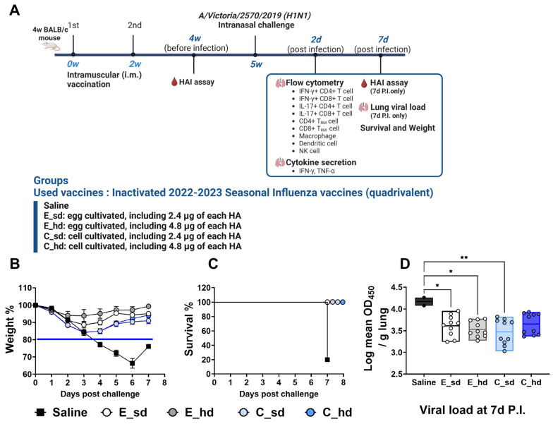
Background: Influenza remains a significant public health challenge, with vaccination being a substantial way to prevent it. Cell-cultured influenza vaccines have emerged to improve on the drawbacks of egg-based vaccines, but there are few studies focusing on T cell immunity with both types of vaccines. Therefore, we studied the following 2022-2023 seasonal influenza vaccines with a standard dose and high dose: cell-based (C_sd and C_hd) and egg-based (E_sd and E_hd) vaccines.

Methods: Along with a saline control group, C_sd, C_hd, E_sd, and E_hd vaccines were administered to BALB/c mice, followed by a challenge with the A/Victoria/2570/2019 (H1N1) strain.

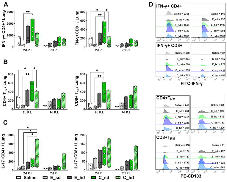
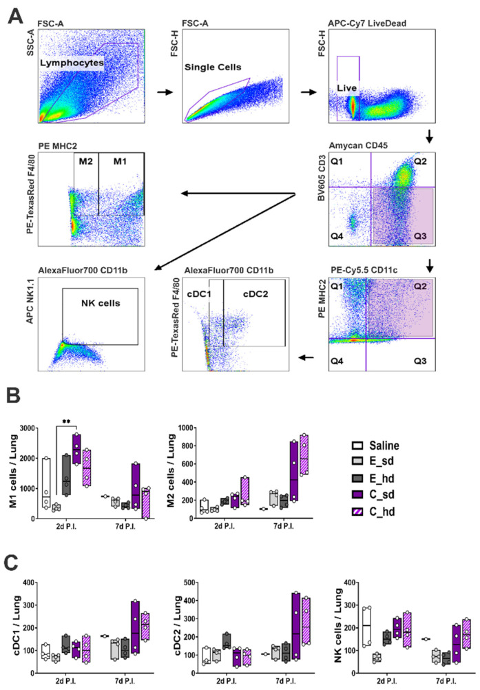
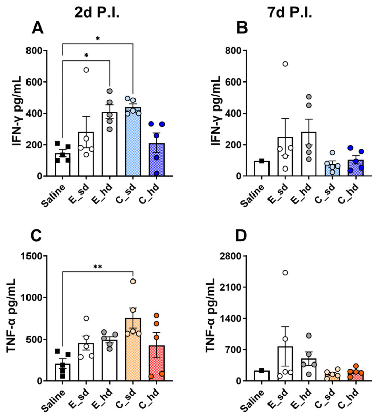
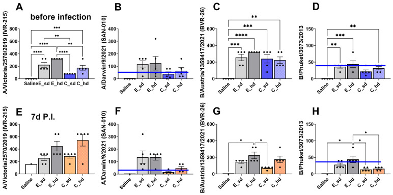
Results: After the challenge, four out of five mice in the saline group died by day 7 post-infection (P.I.). None of the vaccinated groups experienced over 20% weight loss or any deaths. On day 7 P.I., the lung viral load in the saline group (mean log value of 4.17) was higher than that in the vaccinated groups, with the C_sd group showing the lowest viral load (mean log value of 3.47). The C_sd group showed a significantly high response in macrophage 1 (M1), IFN-γ+ T cells, and tissue-resident memory (TRM) T cells compared with the E_sd group on day 2 P.I. These M1, IFN-γ+ T cells, and TRM cells showed similar trends (p < 0.01). In terms of humoral immunity, only the E_hd group showed HAI titers above 40 for all four strains before and after the challenge.

Conclusions: The high levels of T cells in the cell-cultured vaccines suggest, pending further real-world research, that these vaccines may offer advantages.

Keywords: T cells in influenza vaccine immunity; cell-cultured influenza vaccine; egg-based influenza vaccine; immune cells in influenza vaccine immunity; influenza vaccine efficacy.


Conflict of interest statement

P.-K.K., K.-M.J. and J.-Y.J. are employees of SK Bioscience. While every effort has been made to ensure the integrity and objectivity of the research, the potential for perceived or actual conflicts of interest exists.

Figures

Figure 1
Figure 1
Weight loss and viral titer in lungs. Three weeks after the second vaccination, mice were challenged intranasally with 50 µL of A/Victoria/2570/2019 IVR-215 (A/H1N1) virus (768 HA titer). Five mice were tested per group. (A) The scheme of the study is depicted using the BioRender program (Toronto, ON, Canada). (B) Body weight was monitored daily, and changes were expressed as a percentage relative to the pre-vaccination weight. The blue line indicates a humane endpoint of 20%. (C) Survival and symptoms were observed daily for 7 days post-infection. Survival significance thresholds were calculated using the log-rank (Mantel–Cox) test. (D) On day 7 post-infection, the lung viral titer was evaluated with the influenza NP antibody, and OD450 values were log-transformed and presented as mean ± SEM (standard error of the mean). Each group had 10 pieces of data, since a dilution factor of 2 (1:100, 1:1000) was used in all groups except for the saline group at 7 days P.I. (n = 1), when 2 pieces of data were used. Differences between groups in the lung viral titer were determined by one-way ANOVA with Tukey’s multiple comparison tests, with p-values indicated as * p < 0.05, ** p < 0.01.
Figure 2
Figure 2
IFN-γ+ T cell, CD4+ TRM cell, CD8+ TRM cell, and IL-17+ T cell response. The absolute numbers of T cells expressing (A) IFN-γ+ CD8+ and IFN-γ+ CD4+ are shown with box plots. (B) CD8+ TRM cell, CD4+ TRM cell, and (C) IL-17+ T cell counts are also shown with box plots (mean cell count depicted by line in min to max box). (D) IFN-γ+ T cell and T RM cell counts are depicted as histograms for the groups (n = 5, with 1 mouse in the saline group on day 7 P.I.). Differences between groups were determined by one-way ANOVA with Tukey’s multiple comparison test, with p-values indicated as * p < 0.05, ** p < 0.01.
Figure 3
Figure 3
Macrophage, dendritic cell, and NK cell responses. (A) Gating strategies for macrophages, dendritic cells, and NK cells. Live single cells were gated based on size and granularity by FSC-A and FSC-H and by live cell marker. Macrophages, dendritic cells, and NK cells were gated with CD45+ CD3-. Then, dendritic cells were gated with CD11c + MHC2+. Conventional DC1 was gated with CD11b- F4 80+, and cDC2 was gated with CD11b+ F4 80+. NK cells were defined with CD11b+ NK1.1+. Macrophage 1 and 2 were differentiated by MHC2 + F4 80+ for M1 and by MHC2-F4 80+ for M2. The absolute cell counts in lung tissues for (B) M1, M2, (C) cDC1, cDC2, and NK cells were plotted by box; the line in the box represents the median cell amount (n = 4, except for the saline group on day 7 P.I., where n = 1 was used). Differences between groups were determined by a non-parametric comparison test, with p-values indicated as ** p < 0.01.
Figure 4
Figure 4
Cytokine response. Single lung cells were cultured for 72 h at 2 days and 7 days post-infection. After culturing, the supernatant was collected, and cytokine ELISA kits (Proteintech) were used to measure the levels of IFN-γ and TNF-α for each group (n = 5, with n = 1 for the saline group on day 7 P.I.). (A) IFN-γ secretion on day 2 P.I.; (B) IFN-γ secretion on day 7 P.I.; (C) TNF-α secretion on day 2 P.I.; (D) TNF-α secretion on day 7 P.I. All results are presented as the mean ± standard error of the mean (SEM). Statistical differences were analyzed using one-way ANOVA. p-values are indicated as follows: * p < 0.05, ** p < 0.01.
Figure 5
Figure 5
HAI titers. The HAI titer was measured at 2 weeks after the last of 2 intramuscular injections of the 2022–2023 Northern Hemisphere quadrivalent influenza vaccine, before infection. The HAI titer against (A) A/Victoria/2570/2019 (IVR-215) (A/H1N1), (B) A/Darwin/9/2021 (SAN-010) antigen (A/H3N2), (C) B/Austria/1359417/2021 (BVR-26) (B Victoria lineage), and (D) B/Phuket/3073/2013 (B Yamagata lineage) are shown with bar graphs. One week after an intranasal challenge with the A/Victoria/2570/2019 (IVR-215) (A/H1N1) strain, which occurred 3 weeks after the last vaccination (P.I. 7d), HAI titers were measured again. The HAI titer against (E) A/Victoria/2570/2019 (IVR-215) (H1N1), (F) A/Darwin/9/2021 (SAN-010) antigen (A/H3N2), (G) B/Austria/1359417/2021 (BVR-26) (B Victoria lineage), and (H) B/Phuket/3073/2013 (B Yamagata lineage) are shown with bar graphs. The blue line indicates the HAI titer threshold of 40. Serum from 5 mice per group was used, and the results were presented as mean HAI titer ± SEM (standard error of the mean) (for the saline group at P.I. 7d, n = 1 was used). Differences between groups were determined by one-way ANOVA, with p-values indicated as * p < 0.05, ** p < 0.01, *** p < 0.001, and **** p < 0.0001.

Similar articles

References

    1. Dawood F.S., Iuliano A.D., Reed C., Meltzer M.I., Shay D.K., Cheng P.-Y., Bandaranayake D., Breiman R.F., Brooks W.A., Buchy P., et al. Estimated global mortality associated with the first 12 months of 2009 pandemic influenza A H1N1 virus circulation: A modelling study. Lancet Infect. Dis. 2012;12:687–695. doi: 10.1016/S1473-3099(12)70121-4. - DOI - PubMed
    1. Morens D.M., Taubenberger J.K., Harvey H.A., Memoli M.J. The 1918 influenza pandemic: Lessons for 2009 and the future. Crit. Care Med. 2010;38((Suppl. S4)):e10–e20. doi: 10.1097/CCM.0b013e3181ceb25b. - DOI - PMC - PubMed
    1. Carter R.W., Sanford J.C. A new look at an old virus: Patterns of mutation accumulation in the human H1N1 influenza virus since 1918. Theor. Biol. Med. Model. 2012;9:42. doi: 10.1186/1742-4682-9-42. - DOI - PMC - PubMed
    1. Conrad A., Valour F., Vanhems P. Burden of influenza in the elderly: A narrative review. Curr. Opin. Infect. Dis. 2023;36:296–302. doi: 10.1097/QCO.0000000000000931. - DOI - PubMed
    1. Molinari N.-A.M., Ortega-Sanchez I.R., Messonnier M.L., Thompson W.W., Wortley P.M., Weintraub E., Bridges C.B. The annual impact of seasonal influenza in the US: Measuring disease burden and costs. Vaccine. 2007;25:5086–5096. doi: 10.1016/j.vaccine.2007.03.046. - DOI - PubMed

LinkOut - more resources