. 2024 Dec 18;12(12):1428.
doi: 10.3390/vaccines12121428.

Pichia pastoris-Derived β-Glucan Capsules as a Delivery System for DNA Vaccines

Affiliations

Pichia pastoris-Derived β-Glucan Capsules as a Delivery System for DNA Vaccines

Samara Sousa de Pinho et al. Vaccines (Basel). .

Abstract

Background/objectives: DNA vaccines are rapidly produced and adaptable to different pathogens, but they face considerable challenges regarding stability and delivery to the cellular target. Thus, effective delivery methods are essential for the success of these vaccines. Here, we evaluated the efficacy of capsules derived from the cell wall of the yeast Pichia pastoris as a delivery system for DNA vaccines.

Methods: The capsules were extracted from the yeast Pichia pastoris strain GS115, previously grown in a YPD medium. pVAX1 expression vector was adopted to evaluate the DNA vaccine insertion and delivery. Three encapsulation protocols were tested to identify the most effective in internalizing the plasmid. The presence of plasmids inside the capsules was confirmed by fluorescence microscopy, and the encapsulation efficiency was calculated by the difference between the initial concentration of DNA used for insertion and the concentration of unencapsulated DNA contained in the supernatant. The capsules were subjected to different temperatures to evaluate their thermostability and were co-cultured with macrophages for phagocytosis analysis. HEK-293T cells were adopted to assess the cytotoxicity levels by MTT assay.

Results: The microscopy results indicated that the macrophages successfully phagocytosed the capsules. Among the protocols tested for encapsulation, the one with 2% polyethylenimine for internalization showed the highest efficiency, with an encapsulation rate above 80%. However, the vaccine capsules obtained with the protocol that used 5% NaCl showed better thermal stability and encapsulation efficiency above 63% without induction of cell viability loss in HEK 293T.

Conclusions: We successfully described a vaccine delivery system using yeast capsules derived from Pichia pastoris, demonstrating its potential for DNA vaccine delivery for the first time. Additional studies will be needed to characterize and improve this delivery strategy.

Keywords: glucan particle; nucleic acids; yeast cell wall.


Conflict of interest statement

The authors declare no conflicts of interest.

Figures

Figure 1
Figure 1
Schematic representation of the methodology used for capsule preparation. This figure outlines the process of yeast cell treatment and preparation. In (A), the whole yeast cells are cultured, providing the starting material for the yeast capsule procedure; after that, the cells undergo a chemical treatment (B), first with NaOH (1 M) at 60 °C for 1 h to disrupt cell components, followed by HCl (pH 4.5) at 55 °C for 1 h to further process and clean the cellular material. The treated material is subjected to multiple washes (C), including four rounds with isopropanol and two with acetone, to remove impurities and ensure purity of the capsules. The final capsules are dried at room temperature (RT) and stored at −20 °C to maintain their integrity and stability for further applications (D).
Figure 2
Figure 2
Methodologies for inserting plasmid DNA into the capsules. (A) Insertion methodology 1 (PI-1) using 2% PEI; (B) insertion methodology 2 (PI-2) using 5% NaCl. Grey arrows represents eletrostatic interactions between DNA and PEI/NaCl.
Figure 3
Figure 3
Optical microscopy of Pichia pastoris GS115 and yeast shells. Samples were stained with methylene blue and observed at 40× and 100× magnification. Microscopy with K55 OIT optical microscope (Kasvi, Pinhais, PR, Brazil) and image manual capture system.
Figure 4
Figure 4
Comparison of the plasmid DNA incorporation efficiency between the three protocols adopted. %EE = encapsulation efficiency percentage. Different DNA concentrations were adopted to verify the loading capacity using 50, 250, and 500 ng/µL (x-axis). Values correspond to mean ± standard deviation.
Figure 5
Figure 5
Fluorescence microscopy of capsules yeasts. YS: Empty capsules; YS + DNA + PEI 2%; and YS + DNA + NaCl 5%. Observation of the labeling of YS with DAPI (blue), indicating the presence of nucleic acid in the capsules. YS was used as a negative expression control and displayed only fluorescence background. Images captured through a Leica DMLB epi-fluorescence microscope (Leica, Wetzlar, Alemanha) at 100× magnification. Images were captured by the Leica DFC345 FX. Scale bar: 5 μm.
Figure 6
Figure 6
Evaluation of the maintenance of plasmid DNA inside the capsules under different heat treatments. (A) PEI 2% and (B) NaCl 5% were the compounds used to incorporate the DNA inside the capsules. The y-axis corresponds to the DNA concentration, and the x-axis corresponds to the temperatures that were tested. Bar = means ± SD. Asterisks represent statistical significance (* p < 0.05; ** p < 0.01; *** p < 0.001; **** p < 0.0001).
Figure 7
Figure 7
Phagocytosis verification by fluorescence microscopy. Groups: THP-1 cells only and THP-1 + YS (yeast shell), observed under bright-field and fluorescence microscopy. YS were labeled with 5′DTAF (green), and THP-1 cells were labeled with DAPI (blue). The white arrows highlight the YS phagocytosed by the macrophages. Images were captured through a fluorescence microscope (Motic AE31E) at 40× magnification. Images were captured by the Moticam S6 camera using Motic Images Plus 3.0 software. Scale bar: 5 μm.
Figure 8
Figure 8
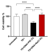
Cell viability evaluation by MTT assay. HEK 293-T cells were treated by incubation with yeast shells (capsules). Asterisks represent statistical significance (**** p < 0.0001), determined by the variance test (ANOVA). Values correspond to mean ± standard deviation.
Figure 9
Figure 9
Comparison of DNAse assay results. (A) Empty yeast capsules and capsules with two insertion protocols (PEI 2% and NaCl 5%) whose inserted DNA (pVAX1) was stained with DAPI (blue color) without being subjected to DNAse treatment. (B) Empty yeast capsules and capsules with two insertion protocols with DNA stained with DAPI but subjected to DNAse treatment. It was possible to observe that there was no degradation of the DNA inserted in the capsules, even in medium containing DNAse. Images captured through a fluorescence microscope (Motic AE31E) at 40× magnification. Images were captured by the Moticam S6 camera using Motic Images Plus 3.0 software.

References

    1. Plotkin S.A. Vaccines: Past, Present and Future. Nat. Med. 2005;11:S5–S11. doi: 10.1038/nm1209. - DOI - PMC - PubMed
    1. Pollard A.J., Bijker E.M. Publisher Correction: A Guide to Vaccinology: From Basic Principles to New Developments. Nat. Rev. Immunol. 2021;21:129. doi: 10.1038/s41577-020-00497-5. - DOI - PMC - PubMed
    1. Gebre M.S., Brito L.A., Tostanoski L.H., Edwards D.K., Carfi A., Barouch D.H. Novel Approaches for Vaccine Development. Cell. 2021;184:1589–1603. doi: 10.1016/j.cell.2021.02.030. - DOI - PMC - PubMed
    1. Wang C., Yuan F. A Comprehensive Comparison of DNA and RNA Vaccines. Adv. Drug Deliv. Rev. 2024;210:115340. doi: 10.1016/j.addr.2024.115340. - DOI - PMC - PubMed
    1. Yang J., Li Y., Jin S., Xu J., Wang P.C., Liang X.-J., Zhang X. Engineered Biomaterials for Development of Nucleic Acid Vaccines. Biomater. Res. 2015;19:5. doi: 10.1186/s40824-014-0025-8. - DOI - PMC - PubMed

LinkOut - more resources