. 2024 Nov 29;16(12):1862.
doi: 10.3390/v16121862.

Investigation of RNA Viruses in Culicoides Latreille, 1809 (Diptera: Ceratopogonidae) in a Mining Complex in the Southeastern Region of the Brazilian Amazon

Affiliations

Investigation of RNA Viruses in Culicoides Latreille, 1809 (Diptera: Ceratopogonidae) in a Mining Complex in the Southeastern Region of the Brazilian Amazon

Sâmia Luzia Sena da Silva et al. Viruses. .

Abstract

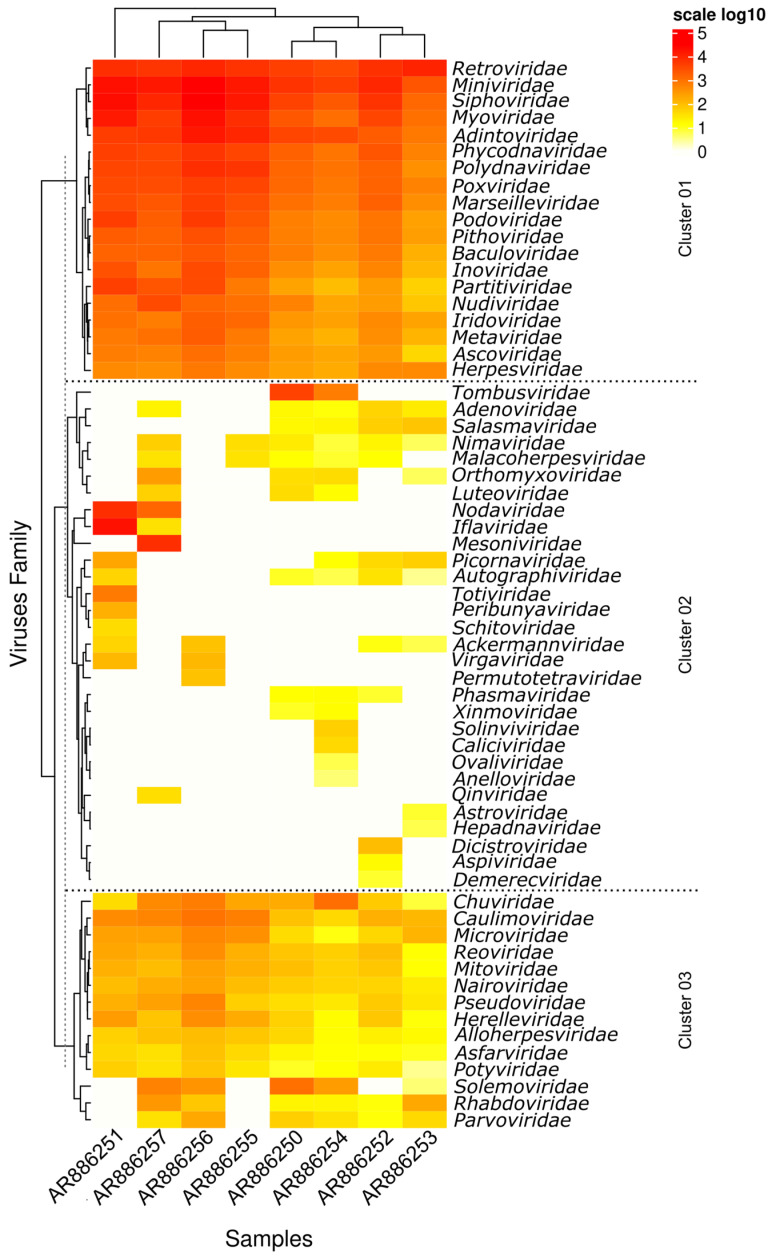
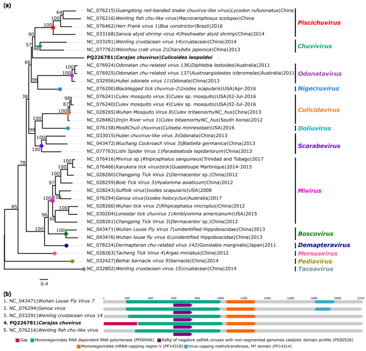
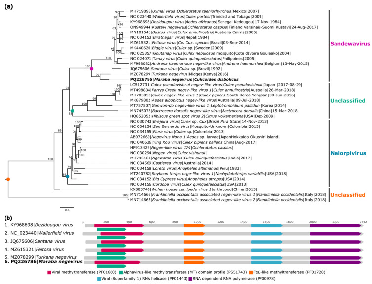
The biting midges Culicoides Latreille, 1809 (Diptera: Ceratopogonidae) is highly relevant to epidemiology and public health, as it includes species that are potential vectors of human and animal arboviruses. The aim of this study was to investigate the presence of RNA viruses in species of the genus Culicoides collected in the Carajás mining complex in the state of Pará. The biting midges were collected in the municipalities of Canaã dos Carajás, Curionópolis and Marabá and morphologically identified. A total of 1139 specimens of seven Culicoides species were grouped into eight pools and subjected to metagenomic analysis. Eight new insect-specific viruses (ISVs) were characterized and assigned to the order Tolivirales, the families Chuviridae, Nodaviridae, Iflaviridae, Mesoniviridae, and Flaviviridae, and the taxon Negevirus. All viruses identified were assigned to clades, families and taxa never reported in Culicoides in Brazil. This study demonstrated that biting midges harbor a significant diversity of RNA viruses, many of which are still unknown, highlighting the importance of studies aiming at virome of these insects.

Keywords: Culicoides; biting midges; insect specific-viruses; metagenomic; virome; viruses.


Conflict of interest statement

The authors declare no conflicts of interest.

Figures

Figure 1
Figure 1
Map showing the collection points of ceratopogonids in the municipalities of Canaã dos Carajás, Curionópolis, and Marabá, located in the state of Pará, Brazilian Amazonia.
Figure 2
Figure 2
Distribution of reads of viral families found in Culicoides samples. The side bar corresponds to the log10 values of the total reads for each sample.
Figure 3
Figure 3
Phylogenetic relationship and genomic organization of Ouro Verde tombus-like virus 1. (a) Phylogenetic inference was performed using the Maximum Likelihood (ML) method based on the amino acid sequences of the RNA-dependent RNA Polymerase (RdRp) protein, using the LG+F+R6 matrix as the best nucleotide substitution model, and measurement of the phylogenetic signal in the dataset, showing only 33% unresolved quartets and 67% resolved quartets. Different groups are identified by different colors. The samples identified in this study are highlighted in bold. The numbers at each main node of the tree correspond to bootstrap values in percentages (1000 replicates). The scale bar corresponds to amino acid divergence per site between sequences. (b) Domains are displayed as colored boxes, and the sequence size is shown as the number of nucleotides.
Figure 4
Figure 4
Phylogenetic relationship and genomic organization of Carajas chuvirus. (a) Phylogenetic inference was performed using the Maximum Likelihood (ML) method based on the amino acid sequences of the RNA-dependent RNA Polymerase (RdRp) protein, using the LG+F+R6 matrix as the best nucleotide substitution model, and measurement of the phylogenetic signal in the dataset, showing only 11.8% unresolved quartets and 88.2% resolved quartets. The genera are identified by different colors on the right side of the tree. The samples identified in this study are highlighted in bold. The numbers at each main node of the tree correspond to bootstrap values in percentages (1000 replicates). The scale bar corresponds to amino acid divergence per site between sequences. (b) Domains are displayed as colored boxes, and the sequence size is shown as the number of nucleotides.
Figure 5
Figure 5
Phylogenetic relationship and genomic organization of Ouro Verde nodavirus. (a) Phylogenetic inference was performed using the Maximum Likelihood (ML) method based on the amino acid sequences of the RNA-dependent RNA Polymerase (RdRp) protein, using the LG+F+I+G4 matrix as the best nucleotide substitution model, and measurement of the phylogenetic signal in the dataset, showing only 11.4% unresolved quartets and 88.6% resolved quartets. The genera are identified by different colors on the right side of the tree. The samples identified in this study are highlighted in bold. The numbers at each main node of the tree correspond to bootstrap values in percentages (1000 replicates). The scale bar corresponds to amino acid divergence per site between sequences. (b) Domains are displayed as colored boxes, and the sequence size is shown as the number of nucleotides.
Figure 6
Figure 6
Phylogenetic relationship and genomic organization of Carajas iflavirus. (a) Phylogenetic inference was performed using the Maximum Likelihood (ML) method based on the amino acid sequences of the polyprotein, using the VT+F+R7 matrix as the best nucleotide substitution model, and measurement of the phylogenetic signal in the dataset, showing only 18% unresolved quartets and 82% resolved quartets. The sample identified in this study is highlighted in bold. The numbers at each main node of the tree correspond to bootstrap values in percentages (1000 replicates). The scale bar corresponds to amino acid divergence per site between sequences. (b) Domains are displayed as colored boxes, and the sequence size is shown as the number of nucleotides.
Figure 7
Figure 7
Phylogenetic relationship and genomic organization of Maraba mesonivirus. (a) Phylogenetic inference was performed using the Maximum Likelihood (ML) method based on the amino acid sequences of polyprotein 1a, using the LG+F+R5 matrix as the best amino acid substitution model, and measurement of the phylogenetic signal in the dataset, showing only 8.2% unresolved quartets and 91.8% resolved quartets. The genera are identified by different colors on the right side of the tree. The sample identified in this study is highlighted in bold. The numbers at each main node of the tree correspond to bootstrap values in percentages (1000 replicates). The scale bar corresponds to amino acid divergence per site between sequences. (b) Domains are displayed as colored boxes, and the sequence size is shown as the number of nucleotides.
Figure 8
Figure 8
Phylogenetic relationship and genomic organization of Carajing-like virus 1 and Carajing-like virus 2. (a) Phylogenetic inference was performed using the Maximum Likelihood (ML) method based on the amino acid sequences of the NSP2 protein, with the LG+F+R4 matrix used as the best amino acid substitution model, and the measurement of the phylogenetic signal in the dataset, showing only 17% unresolved quartets and 82% resolved quartets. The classification of viruses according to their hosts is indicated by different background colors in the tree. The samples identified in this study are highlighted in bold. The numbers at each main node of the tree correspond to bootstrap values in percentages (1000 replicas). The scale bar corresponds to amino acid divergence per site among sequences. (b) Domains are displayed as colored boxes, and the sequence size is shown as the number of nucleotides.
Figure 9
Figure 9
Phylogenetic relationship and genomic organization of Turkana-like negevirus. (a) Phylogenetic inference was performed using the Maximum Likelihood (ML) method based on the amino acid sequences of hypothetical protein 1, with the LG+F+I+G4 matrix used as the best amino acid substitution model, and the measurement of the phylogenetic signal in the dataset, showing only 8.2% unresolved quartets and 79.9% resolved quartets. The classification of viruses according to their hosts is indicated by the different background colors in the tree. The samples identified in this study are highlighted in bold. The numbers at each main node of the tree correspond to bootstrap values in percentages (1000 replicas). The scale bar corresponds to amino acid divergence per site among sequences. (b) Domains are displayed as colored boxes, and the sequence size is shown as the number of nucleotides.

Similar articles

Cited by

References

    1. Silva A.D.S.D., Delatorre E.O., Leon L.A.A., Azevedo S.S.D.D., Leite T.C.N.F., Paula V.S.D. Propriedades Gerais Dos Vírus. In: Lemos E.R.S.D., Villar L.M., Leon L.A.A., Guimarães M.L., Teixeira S.L.M., Paula V.S.D., editors. Tópicos em Virologia. Editora Fiocruz; Rio de Janeiro, Brazil: 2023. pp. 29–45.
    1. Geoghegan J.L., Holmes E.C. Predicting Virus Emergence amid Evolutionary Noise. Open Biol. 2017;7:170189. doi: 10.1098/rsob.170189. - DOI - PMC - PubMed
    1. Silva A.D.S.D., Lemos A.S., Alves A.D.R., Marques B.C.L., Ribeiro C.R.D.A., Passaes C.P.B., Santos D.R.L.D., Caetano D.G., Costa E.V.D., Bottino F.D.O., et al. Diagnóstico de Infecções Virais. In: Lemos E.R.S.D., Villar L.M., Leon L.A.A., Guimarães M.L., Teixeira S.L.M., Paula V.S.D., editors. Tópicos Em Virologia. Editora Fiocruz; Rio de Janeiro, Brazil: 2023. pp. 47–86.
    1. Zuo K., Gao W., Wu Z., Zhang L., Wang J., Yuan X., Li C., Xiang Q., Lu L., Liu H. Evolution of Virology: Science History through Milestones and Technological Advancements. Viruses. 2024;16:374. doi: 10.3390/v16030374. - DOI - PMC - PubMed
    1. Zhou S., Liu B., Han Y., Wang Y., Chen L., Wu Z., Yang J. ZOVER: The Database of Zoonotic and Vector-Borne Viruses. Nucleic Acids Res. 2022;50:D943–D949. doi: 10.1093/nar/gkab862. - DOI - PMC - PubMed

Publication types

LinkOut - more resources