. 2024 Dec 11;16(12):1908.
doi: 10.3390/v16121908.

The Pestivirus RNase Erns Tames the Interferon Response of the Respiratory Epithelium

Affiliations

The Pestivirus RNase Erns Tames the Interferon Response of the Respiratory Epithelium

Guillaume Beilleau et al. Viruses. .

Abstract

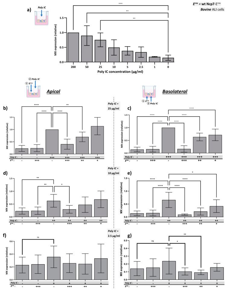
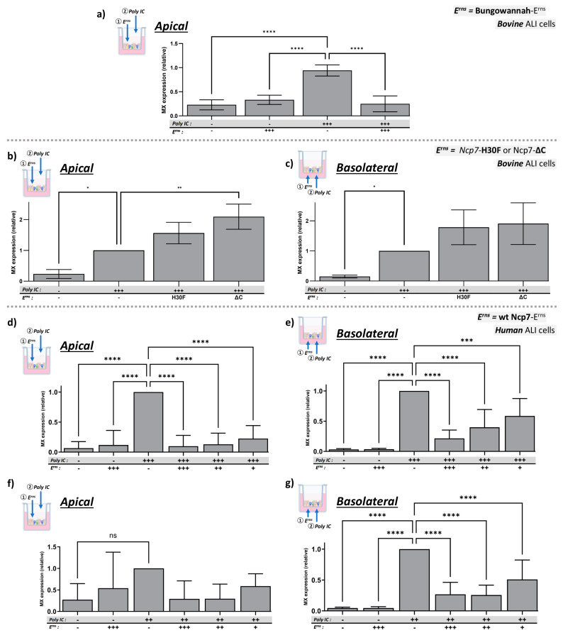
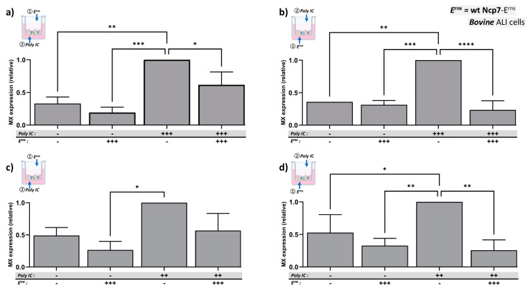
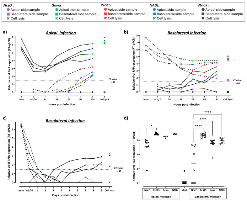
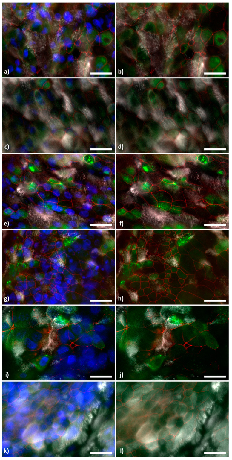
Bovine viral diarrhea virus (BVDV), a pestivirus in the family Flaviviridae, is a major livestock pathogen. Horizontal transmission leads to acute transient infections via the oronasal route, whereas vertical transmission might lead to the birth of immunotolerant, persistently infected animals. In both cases, BVDV exerts an immunosuppressive effect, predisposing infected animals to secondary infections. Erns, an immunomodulatory viral protein, is present on the envelope of the virus and is released as a soluble protein. In this form, it is taken up by cells and, with its RNase activity, degrades single- and double-stranded (ds) RNA, thus preventing activation of the host's interferon system. Here, we show that Erns of the pestiviruses BVDV and Bungowannah virus effectively inhibit dsRNA-induced IFN synthesis in well-differentiated airway epithelial cells cultured at the air-liquid interface. This activity was observed independently of the side of entry, apical or basolateral, of the pseudostratified, polarized cell layer. Virus infection was successful from both surfaces but was inefficient, requiring several days of incubation. Virus release was almost exclusively restricted to the apical side. This confirms that primary, well-differentiated respiratory epithelial cells cultured at the air-liquid interface are an appropriate model to study viral infection and innate immunotolerance in the bovine respiratory tract. Furthermore, evidence is presented that Erns might contribute to the immunosuppressive effect observed after BVDV infections, especially in persistently infected animals.

Keywords: IFN antagonist; air–liquid interface (ALI); bovine viral diarrhea virus (BVDV); glycosaminoglycan (GAG)-binding site; innate immune evasion; interferon type-I; pestivirus; respiratory epithelium; viral RNase; viral endonuclease.


Conflict of interest statement

The authors declare no conflicts of interest.

Figures

Figure 1
Figure 1
Inhibition of dsRNA-induced Mx expression by Erns in bBAEC cultured in an ALI system. (a) Poly IC was applied to the apical side of well-differentiated bBAEC cultures. After 20 h of incubation, cytosolic extracts were assayed for Mx expression by Western blot using β-actin as loading control. The relative expression of Mx at the highest concentration of Poly IC was set to 1 (100%). (bg) Strep-tag purified wild-type Erns from the BVDV strain Ncp7 was pre-incubated for 90 min on bBAEC cultures prior to the addition of Poly IC, both to the apical (b,d,f) and basolateral sides (c,e,g). The signal for Poly IC-induced Mx expression was quantified with the one in the absence of Erns present on each gel being set to 1 (100%), and the means of each column was compared to this reference value (mean ± SD, n = 8–9 with cells from 3 different donors). Concentrations indicated in the figure: (+++): 25 μg/mL Poly IC or 25 ng/mL Erns; (++): 10 μg/mL Poly IC or 10 ng/mL Erns; (+): 2.5 μg/mL Poly IC or 2.5 ng/mL Erns; (-): no Poly IC or Erns. Ordinary one-way analysis of variance (ANOVA) for multiple comparisons was performed using GraphPad Prism 10.4.0 software with significant differences indicated as follows: **** (p < 0.0001), *** (p < 0.001), ** (p < 0.01), * (p < 0.1), or ns (not significant). For clarity of the figure, most of the non-significant comparisons to the reference columns were omitted.
Figure 2
Figure 2
Inhibition of dsRNA-induced Mx expression by wt- or mutant- Erns in bBAEC and hBAEC cultured in an ALI system. (a) Strep-tag purified wild-type Erns from the porcine pestivirus Bungo wannah virus (25 ng/mL) was pre-incubated or not for 90 min on the apical side of bBAEC cultures prior to the addition of Poly IC (25 μg/mL) at the same side for another 20 h. (b,c) RNase-inactive Erns (H30F) or mutant Erns lacking the C-terminal amphipathic helix (ΔC) from the BVDV strain Ncp7 was pre-incubated on the apical (b) or basolateral (c) side of bBAEC as described in the methods section prior to the addition of Poly IC at 25 μg/mL. (dg) Strep-tag purified wild-type Erns from the BVDV strain Ncp7 was pre-incubated for 90 min at the indicated concentrations on human hBAEC cultures prior to the addition of Poly IC at 10 (f,g) or 25 μg/mL (d,e), both at the apical (d,f) or basolateral side (e,g). In all panels, the signal for Poly IC-induced Mx expression in the absence of Erns, present on every gel, was set to 100% with the mean of each column being compared to this reference column (mean ± SD, n = 3 to 8 with cells from 3 different donors). Ordinary one-way analysis of variance (ANOVA) for multiple comparisons was performed using GraphPad Prism 10.4.0 software with significant differences indicated as follows: **** (p < 0.0001), *** (p < 0.001), ** (p < 0.01), * (p < 0.1), or ns (not significant). For clarity of the figures, most of the non-significant comparisons to the reference columns were omitted. Concentrations indicated in the figure: (+++): 25 μg/mL Poly IC or 25 ng/mL Erns; (++): 10 μg/mL Poly IC or 10 ng/mL Erns; (+): 2.5 ng/mL Erns; (-): no Poly IC or Erns.
Figure 3
Figure 3
Inhibition of dsRNA induced Mx expression by Erns added to opposite sides of bBAEC cultures. Strep-tag purified wild-type Erns from BVDV strain Ncp7 at 25 ng/mL (+++) was pre-incubated on the apical side of the bBAEC cultures for 90 min followed by its removal and further incubation with the apical side exposed to air for a total of 24 h (a,c). On the basolateral side (b,d), this Erns was incubated with the bBAEC cultures for the complete incubation time of 24 h. Thereafter, Poly IC was added at 25 μg/mL (+++) (a,b) or 10 μg/mL (++) (c,d) to the opposite side compared to Erns. On the basolateral side, the dsRNA was incubated for 20 h, whereas on the apical side, Poly IC was removed after 2 h followed by 18 h of incubation with the apical side exposed to air. Thereafter, cytosolic extracts were assayed for Mx expression by Western blotting using β-actin as the loading control. The signal for Poly IC-induced Mx expression in the absence of Erns present on each gel was set to 1 (100%), and the mean of each column was compared to this reference column (mean ± SD, n = 3 to 6 with cells from 3 different donors). Ordinary one-way analysis of variance (ANOVA) for multiple comparisons was performed using GraphPad Prism 10.4.0 software with significant differences indicated as follows: **** (p < 0.0001), *** (p < 0.001), ** (p < 0.01), * (p < 0.1). For clarity of the figure, the non-significant comparisons to the reference columns were omitted.
Figure 4
Figure 4
Detection of viral RNA of different BVDV strains after infection of bBAEC cultured in an ALI system. (ac) Two different bovine donors were infected with four different BVDV strains (Ncp7, Pe515, Suwa, NADL) apically and basolaterally for 5 days (a,b) or basolaterally for 9 days (c) (n = 3). Infection was monitored with a sample obtained every 24 h on each side (apical, continuous line; basolateral, dashed line). (d) Cells from an additional, freshly isolated bovine donor were included. The bBAEC from the three different donors were infected with the same BVDV strains apically and basolaterally as before. After 10 days of incubation (without intermittent sampling in order not to stress the pseudo-stratified layer), cells were lysed and analyzed (n = 4–9). Total RNA from all samples (ad) was extracted, purified, and analyzed by RT-qPCR for the quantification of pestiviral RNA. The relative amount of viral RNA based on the Ct value was obtained as described in the Materials and Methods section. Statistical analysis was as described in Figure 3.
Figure 5
Figure 5
Detection of infectious BVD viruses after infection of bBAEC cultured in an ALI system. (a,b) Two bovine donors were infected with four different BVDV strains (Ncp7, Pe515, Suwa, NADL) apically (a) and basolaterally (b) for 10 days (n = 6). Infection was monitored with a sample obtained every 24 h on each side (apical, continuous line; basolateral, dashed line). (c) Cells from an additional, freshly isolated bovine donor were included. The bBAEC from the three different donors were infected with the same BVDV strains apically and basolaterally. After 10 days of incubation (without intermittent sampling), cells were lysed and analyzed (n = 4–8). The virus titer of all samples was determined by titration on BT cells as described in the Materials and Methods section. Ordinary one-way analysis of variance (ANOVA) for multiple comparisons was performed using GraphPad Prism 10.4.0 software with significant differences indicated as follows: **** (p < 0.0001), ** (p < 0.01), * (p < 0.1). For clarity of (c), the significant differences of the virus titers to uninfected cells (Mock) (p < 0.001 for Ncp7; p < 0.0001 for Pe515, NADL, and Suwa) were omitted.
Figure 6
Figure 6
Detection of BVDV after infection of bBAEC cultured in an ALI system by immunofluorescence microscopy. Well-differentiated bBAEC were infected with the BVDV strains Ncp7 (a,b), Pe515 (c,d), Suwa (e,f), or NADL (g,h), with Bungowannah pestivirus (i,j) or left uninfected (k,l) for 10 days (n = 3). Thereafter, the cells were stained as described for the nuclei (blue), tight junctions (red), cilia (white), and dsRNA (green). Scale bar = 20 μm. For better visibility of the virus-infected cells (green channel), each picture is presented with all color channels (a,c,e,g,i,k) or without the nuclei staining (blue channel) (b,d,f,h,j,l).

Similar articles

References

    1. Smith D.B., Meyers G., Bukh J., Gould E.A., Monath T., Muerhoff A.S., Pletnev A., Rico-Hesse R., Stapleton J.T., Simmonds P., et al. Proposed revision to the taxonomy of the genus Pestivirus, family Flaviviridae. J. Gen. Virol. 2017;98:2106–2112. doi: 10.1099/jgv.0.000873. - DOI - PMC - PubMed
    1. Postler T.S., Beer M., Blitvich B.J., Bukh J., de Lamballerie X., Drexler J.F., Imrie A., Kapoor A., Karganova G.G., Lemey P., et al. Renaming of the genus Flavivirus to Orthoflavivirus and extension of binomial species names within the family Flaviviridae. Arch. Virol. 2023;168:224. doi: 10.1007/s00705-023-05835-1. - DOI - PubMed
    1. Childs T. X Disease of cattle—Saskatchewan. Can. J. Comp. Med. Vet. Sci. 1946;10:316–319. - PMC - PubMed
    1. Olafson P., MacCallum A.D., Fox F.H. An apparently new transmissible disease of cattle. Cornell Vet. 1946;36:205–213. - PubMed
    1. Evermann J.F., Barrington G.M. Clinical features. In: Goyal S.M., Ridpath J.F., editors. Bovine Viral Diarrhea Virus: Diagnosis, Management, and Control. Blackwell Publishing; Ames, IA, USA: 2005. pp. 105–119.

Publication types