. 2024 Dec 13;16(12):1913.
doi: 10.3390/v16121913.

DHX15 and Rig-I Coordinate Apoptosis and Innate Immune Signaling by Antiviral RNase L

Affiliations

DHX15 and Rig-I Coordinate Apoptosis and Innate Immune Signaling by Antiviral RNase L

Barkha Ramnani et al. Viruses. .

Abstract

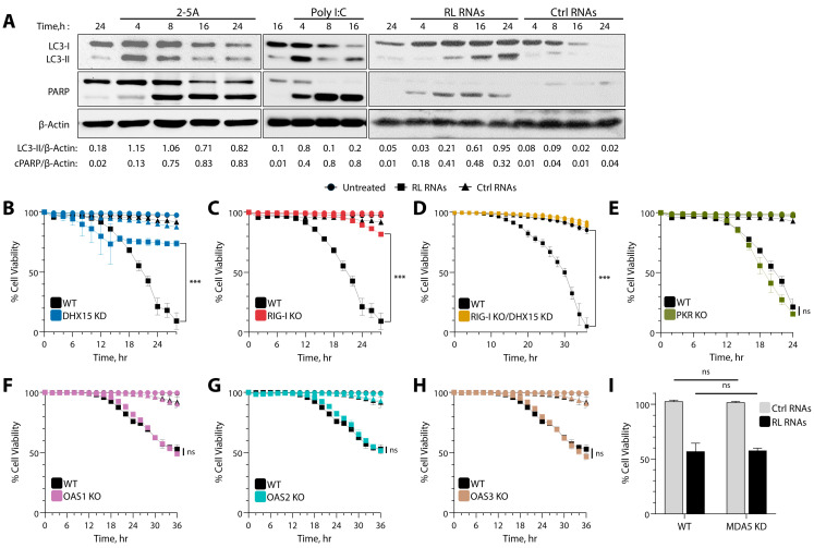
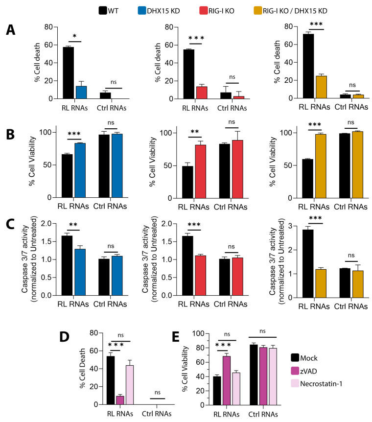
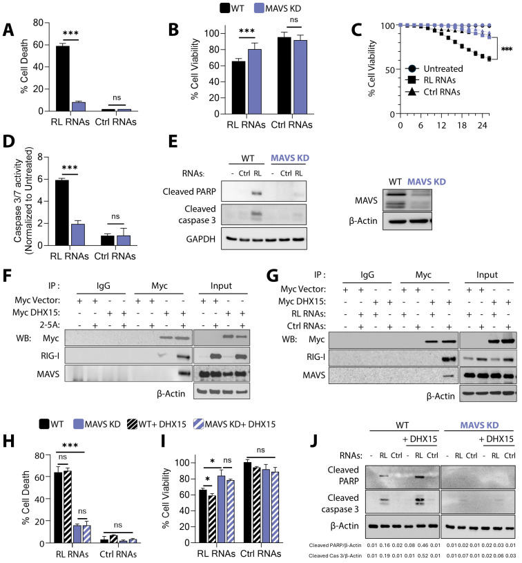
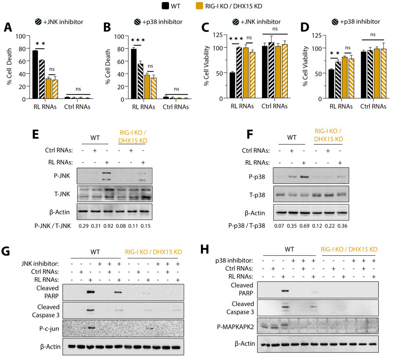
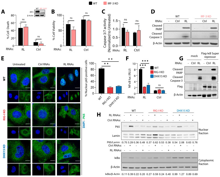
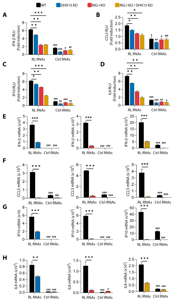
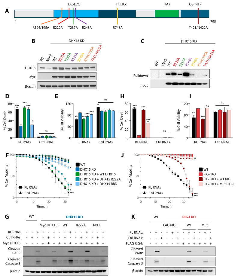
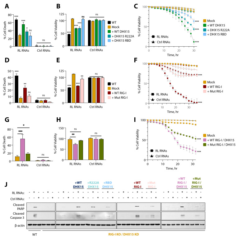
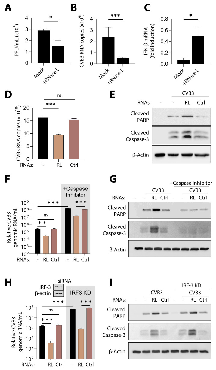
During virus infection, the activation of the antiviral endoribonuclease, ribonuclease L (RNase L), by a unique ligand 2'-5'-oilgoadenylate (2-5A) causes the cleavage of single-stranded viral and cellular RNA targets, restricting protein synthesis, activating stress response pathways, and promoting cell death to establish broad antiviral effects. The immunostimulatory dsRNA cleavage products of RNase L activity (RL RNAs) recruit diverse dsRNA sensors to activate signaling pathways to amplify interferon (IFN) production and activate inflammasome, but the sensors that promote cell death are not known. In this study, we found that DEAH-box polypeptide 15 (DHX15) and retinoic acid-inducible gene I (Rig-I) are essential for apoptosis induced by RL RNAs and require mitochondrial antiviral signaling (MAVS), c-Jun amino terminal kinase (JNK), and p38 mitogen-activated protein kinase (p38 MAPK) for caspase-3-mediated intrinsic apoptosis. In RNase L-activated cells, DHX15 interacts with Rig-I and MAVS, and cells lacking MAVS expression were resistant to apoptosis. RL RNAs induced the transcription of genes for IFN and proinflammatory cytokines by interferon regulatory factor 3 (IRF-3) and nuclear factor kB (NF-kB), while cells lacking both DHX15 and Rig-I showed a reduced induction of cytokines. However, apoptotic cell death is independent of both IRF-3 and NF-kB, suggesting that cytokine and cell death induction by RL RNAs are uncoupled. The RNA binding of both DHX15 and Rig-I is required for apoptosis induction, and the expression of both single proteins in cells lacking both DHX15 and Rig-I is insufficient to promote cell death by RL RNAs. Cell death induced by RL RNAs suppressed Coxsackievirus B3 (CVB3) replication, and inhibiting caspase-3 activity or cells lacking IRF-3 showed that the induction of apoptosis directly resulted in the CVB3 antiviral effect, and the effects were independent of the role of IRF-3.

Keywords: DHX15; RNase L; Rig-I; apoptosis; innate immune signaling.


Conflict of interest statement

The authors declare no conflicts of interest. The funders had no role in the design of the study; in the collection, analyses, or interpretation of data; in the writing of the manuscript, or in the decision to publish the results.

Figures

Figure 1
Figure 1
Role of dsRNA sensors in cell death induced by RNase L. (A) HT1080 cells were transfected with 2-5A (10 µM), Poly I:C (2 µg/mL), RL RNAs, or Ctrl RNAs (2 µg/mL) for indicated time points and the conversion of LC3-I to LC3-II. the cleavage of PARP was normalized to β-actin by immunoblotting, and band intensities were plotted as ratios. Kinetics of cell death induced by RL RNAs (2 µg/mL) were compared to Ctrl RNAs (2 µg/mL), and the percentage of the cell viability was determined by real-time imaging using dual dyes in WT HT1080 cells and compared to (B) DHX15 KD, (C) Rig-I KO, (D) Rig-I KO/DHX15 KD, (E) PKR KO, (F) OAS1 KO, (G) OAS2 KO, and (H) OAS3 KO cells. The percentage of cell survival in each well was determined by quantitating dead cells and normalized to the total number of cells at each time point. Data are representative of four values per well performed in triplicate and shown as mean ± SD. (I) HT1080 cells were transfected with control or MDA5 siRNA (5 nM) and, after 24 h, treated with RL RNAs or Ctrl RNAs (2 µg/mL)thepercentage of the cell viability was determined by MTT assay. The results are representative of three independent experiments performed in triplicate ± SD. WT: wild-type; RL RNAs: RNase L-cleaved small RNAs; Ctrl RNAs: control small RNAs; *** p < 0.001; ns: not significant.
Figure 2
Figure 2
RL RNAs signal through DHX15 and Rig-I to induce apoptotic cell death. HT1080 WT, DHX15 KD, Rig-I KO, or Rig-I KO/DHX15 KD cells were transfected with RL RNAs or Ctrl RNAs (2 µg/mL). (A) The percentage of cell death by trypan blue exclusion, (B) the percentage of cell viability by MTT assay, and (C) caspase-3/7 activity were determined. WT cells were mock-treated, pretreated with zVAD-FMK (20 μΜ) or necrostatin-1 (20 μΜ), and transfected after 1 h with RL RNAs or Ctrl RNAs (2 µg/mL). (D) The percentage of cell death by trypan blue exclusion and (E) the percentage of cell viability by MTT assay were determined. The results are representative of three independent experiments performed in triplicate ± SD. KO: knockout; KD: knockdown; WT: wild-type; RL RNAs: RNase L-cleaved small RNAs; Ctrl RNAs: control small RNAs; * p < 0.05; ** p < 0.01; *** p < 0.001; ns: not significant.
Figure 3
Figure 3
MAVS is required for RL RNA-mediated apoptosis. HT1080 WT and MAVS KD cells were transfected with RL RNAs or Ctrl RNAs (2 µg/mL). (A) The percentage of cell death by trypan blue exclusion, (B) the percentage of cell viability by MTT assay, (C) Kinetics of cell death in real time, and the percentage of cell viability were estimated. (D) Caspase-3/7 activity was determined. (E) Cell lysates were analyzed for cleaved PARP, cleaved caspase-3, and MAVS by immunoblotting. HT1080 cells expressing Myc vector or Myc-DHX15 were transfected with (F) 2-5A (10 µM) or (G) RL RNAs and Ctrl RNAs (2 µg/mL). Cell lysates were immunoprecipitated using anti-Myc or isotype control IgG, and the presence of Rig-I and MAVS analyzed by immunoblotting. The expression of the proteins in total cell lysates was analyzed using specific antibodies and normalized to β-actin levels. WT and MAVS KD cells overexpressing DHX15 or not were transfected with RL RNAs or Ctrl RNAs (2 µg/mL), and (H) the percentage of cell death by trypan blue exclusion, (I) the percentage of cell viability by MTT assay, and (J) the cleaved PARP and cleaved caspase-3 on immunoblots were determined. The band intensities were plotted as ratios. The representative immunoblots of experiments performed in duplicate are shown. The results are representative of three independent experiments performed in triplicate ± SD. KO: knockout; KD: knockdown; WT: wild-type; RL RNAs: RNase L-cleaved small RNAs; Ctrl RNAs: control small RNAs; * p < 0.05; *** p < 0.001; ns: not significant.
Figure 4
Figure 4
RL RNAs induce JNK and p38 phosphorylation and cell death: HT1080 WT and Rig-I KO/DHX15 KD cells were transfected with RL RNAs or Ctrl RNAs (2 µg/mL) in the absence or presence of the JNK inhibitor or p38 inhibitor as indicated. (A,B) The percentage of cell death by trypan blue exclusion and (C,D) cell viability by MTT assay were determined. Cell lysates were analyzed for the phosphorylation of JNK (E) and p38 (F) and normalized to the level of total JNK and p38, respectively, on immunoblots. Cell lysates from WT and Rig-I KO/DHX15 KD cells treated as above were analyzed on immunoblots for cleaved PARP, cleaved caspase-3, and phospho-c-Jun (G) or phospho-MAPKAPK2 (H) and normalized to β-actin levels. The representative immunoblots of experiments performed in duplicate are shown. The results are representative of three independent experiments performed in triplicate ± SD. KO: knockout; KD: knockdown; WT: wild-type; RL RNAs: RNase L-cleaved small RNAs; Ctrl RNAs: control small RNAs; ** p < 0.01; *** p < 0.001; ns: not significant.
Figure 5
Figure 5
RL RNA-mediated apoptosis is independent of IRF-3 and NF-kB signaling. HT1080 WT and IRF-3 KO cells were transfected with RL RNAs or Ctrl RNAs (2 µg/mL). (A) The percentage of cell death by trypan blue exclusion, (B) the percentage of cell viability by MTT assay, and (C) caspase-3/7 activity were determined. The inset in (A) shows immunoblot of WT and IRF-3 KO cells for IRF-3 levels. (D) Cell lysates were analyzed for cleaved PARP and cleaved caspase-3 on immunoblots and normalized to β-actin levels. WT, Rig-I KO, and DHX15KD were (E) transfected with RL RNAs or Ctrl RNAs (2 µg/mL) for 8 h, and the nuclear translocation of the NF-kB p65 subunit (green) was determined by immunofluorescence; nuclei were stained with DAPI. The representative images of cells were visualized under a confocal microscope at 60×. The scale bar represents 50 μm. The percentage of cells showing nuclear p65 cells were quantified in 3 random fields. At least 100 cells were quantified. The data represent mean ± SD; (F) co-transfected with NF-kB-luc reporter plasmids and Renilla-luc plasmid. After 24 h, the cells were treated with RL RNAs or Ctrl RNAs (2 µg/mL), and 8 h later, luciferase activity was measured and normalized to the levels of Renilla luciferase and shown as fold induction. Results are representative of three independent experiments performed in triplicate ± SD. (G) HT1080 WT cells were transfected with FLAG-IκB super-repressor and, 24 h later, with RL RNAs or Ctrl RNAs (2 µg/mL). Cell lysates were analyzed for cleaved PARP, cleaved caspase-3, and FLAG-IkB on immunoblots and normalized to β-actin levels. (H) WT, DHX15 KD, and RIG I KO cells were transfected with RL RNAs or Ctrl RNAs (2 µg/mL) for 8 h. Cell lysates were fractionated into nuclear and cytosol fractions and analyzed by immunoblotting using indicated antibodies. The levels of NF-κB p65 subunit in the nuclear extract normalized to Lamin or the degradation of IκBα in cytosol extract normalized to β-actin levels was determined by Image J analysis. The representative immunoblots of experiments performed in duplicate are shown. KO: knockout; WT: wild-type; RL RNAs: RNase L-cleaved small RNAs; Ctrl RNAs: control small RNAs; ** p < 0.01; *** p < 0.001; ns: not significant.
Figure 6
Figure 6
IFN and cytokine induction by RL RNA is mediated by DHX15 and Rig-I. HT1080 WT, Rig-I KO, DHX15KD, and Rig-I KO/DHX15KD cells were transfected with (A) IFN-β-luc, (B) CCL5-luc, (C) IP-10-luc, and (D) IL-8-luc reporter plasmids along with Renilla luc or β-galactosidase plasmids. After 24 h, the cells were treated with RL RNAs or Ctrl RNAs (2 µg/mL), and 8 h later, luciferase activity was measured and normalized to levels of Renilla luciferase or β-galactosidase levels and shown as fold induction. HT1080 WT, Rig-I KO, DHX15KD, and Rig-I KO/DHX15KD cells were transfected with RL RNAs or Ctrl RNAs (2 µg/mL), and 8 h later, (E) IFN-β, (F) CCL5, (G) IP-10, and (H) IL-8 mRNA levels were measured by qRT-PCR and normalized to GAPDH mRNA levels. Induction by RL RNAs in the various genotypes was compared to WT and significance shown as *. Induction by RL RNAs compared to Ctrl RNAs within a cell type is indicated by #. The data represent mean ± SD performed in triplicates. WT: wild-type, KO: knockout; KD: knockdown; RL RNAs: RNase L-cleaved small RNAs; Ctrl RNAs: control small RNAs; * p < 0.05, # p < 0.05; ** p < 0.01, ## p < 0.01; *** p < 0.001, ### p < 0.001; ns: not significant.
Figure 7
Figure 7
Effect of DHX15 and Rig-I mutations on apoptosis. (A) Schematic presentation of the domain structure of DHX15 and sites of point mutations. DExD/C: DEAD-like helicase superfamily domain; HELICc: helicase superfamily C-terminal domain: HA2: C-terminal helicase-associated domain; OB_NTP: oligonucleotide binding fold domain. (B) The expression of DHX15 WT and mutant plasmids in DHX15KD cells was analyzed by immunoblotting using indicated antibodies. ℜ-Actin was used as the loading control. (C) DHX15KD cells were transfected with empty vector (mock) or the indicated Myc-DHX15 plasmid constructs, and cell lysates were incubated with polyI:C-agarose beads and pulled-down DHX15 proteins detected by immunoblotting. Immunoblots without pulldown show the input levels of the Myc-DHX15 proteins. HT1080 WT and DHX15 KD cells expressing the indicated DHX15 plasmids were transfected with RL RNAs or Ctrl RNAs (2 µg/mL). (D) The percentage of cell death by trypan blue exclusion, (E) the percentage of cell viability by MTT assay, (F) Kinetics of cell death in real time, and the percentage of cell viability were estimated. The data represent mean ± SD performed in triplicates. (G) Cell lysates were analyzed for cleaved PARP and cleaved caspase-3 on immunoblots and normalized to b-actin levels. HT1080 WT and Rig-I KO cells expressing the indicated Rig-I plasmids were transfected with RL RNAs or Ctrl RNAs (2 µg/mL). (H) The percentage of cell death by trypan blue exclusion, (I) the percentage of cell viability by MTT assay, (J) Kinetics of cell death in real time, and the percentage of cell viability were estimated. The data represent mean ± SD performed in triplicates. (K) Cell lysates were analyzed for cleaved PARP and cleaved caspase-3 on immunoblots and normalized to β-actin levels. The representative immunoblots of experiments performed in duplicate are shown. WT: wild-type, Mut: mutant, KO: knockout; KD: knockdown; RL RNAs: RNase L-cleaved small RNAs; Ctrl RNAs: control small RNAs; * p < 0.05; ** p < 0.01; *** p < 0.001; ns: not significant.
Figure 8
Figure 8
RNA binding defective mutants of DHX15 and Rig-I promote cell viability in response to RL RNAs. Rig-I KO/DHX15 KD cells expressing the indicated DHX15, Rig-I, or both DHX15 and Rig-I plasmids were transfected with RL RNAs or Ctrl RNAs (2 µg/mL). (A,D,G) The percentage of cell death by trypan blue exclusion, (B,E,H) the percentage of cell viability by MTT assay, (C,F,I) the kinetics of cell death in real time, and the percentage of cell viability were estimated. The data represent mean ± SD performed in triplicates. (J) Cell lysates were analyzed for cleaved PARP and leaved caspase-3 on immunoblots and normalized to β-actin levels. The representative immunoblots of experiments performed in duplicate are shown. Mut: RNA binding mutant, WT: wild-type, KO: knockout; KD: knockdown; RL RNAs: RNase L-cleaved small RNAs; Ctrl RNAs: control small RNAs; * p < 0.05; ** p < 0.01; *** p < 0.001; ns: not significant.
Figure 9
Figure 9
Apoptosis by RL RNAs enhances RNase L antiviral effect during CVB3 infection. HeLa-M cells (mock) or stably expressing WT RNase L were infected with CVB3 (MOI = 0.1) for 24 h. (A) Viral yields were determined by plaque assay, (B) the copy numbers of CVB3 genomic RNA were determined by real-time RT-PCR as described under methods, and (C) IFN-β mRNA levels in CVB3-infected cells were measured by qRT-PCR, normalized to GAPDH mRNA levels, and shown as fold induction. The data represent mean ± SD performed in triplicates. HeLa-M cells were transfected with RL RNAs or Ctrl RNAs (2 µg/mL) and infected with CVB3 (MOI 0.1), and after 24 h, (D) the copy numbers of CVB3 genomic RNA were determined by real-time RT-PCR. The data represent mean ± SD performed in triplicates. (E) Cell lysates were analyzed for cleaved PARP and cleaved caspase-3 on immunoblots and normalized to β-actin levels. (F) HeLa-M cells were transfected with RL RNAs or Ctrl RNAs (2 µg/mL) and infected with CVB3 (MOI = 1.0) with or without Ac-DEVD-CHO (20 mM), and after 24 h, the copy numbers of CVB3 genomic RNA were determined by real-time RT-PCR. The data represent mean ± SD performed in triplicates, and (G) cell lysates were analyzed for cleaved PARP and cleaved caspase-3 on immunoblots and normalized to β-actin levels. HeLa-M cells were transfected with control or siRNA specific for IRF-3 (50 mM) and, after 36 h, transfected with RL RNAs or Ctrl RNAs (2 µg/mL) and infected with CVB3 (MOI = 1.0). (H) The copy numbers of CVB3 genomic RNA were determined by real-time RT-PCR. The inset shows the immunoblot of WT and siIRF-3 cells for IRF-3 levels. The data represent mean ± SD performed in triplicates, and (I) cell lysates were analyzed for cleaved PARP and cleaved caspase-3 on immunoblots and normalized to β-actin levels. The representative immunoblots of experiments performed in duplicate are shown. PFU: plaque forming units; MOI: multiplicity of infection; RL RNAs: RNase L-cleaved small RNAs; Ctrl RNAs: control small RNAs; KD: knockdown * p < 0.05; ** p < 0.01; *** p < 0.001; ns: not significant.

Similar articles

References

    1. Takeuchi O., Akira S. Innate immunity to virus infection. Immunol. Rev. 2009;227:75–86. doi: 10.1111/j.1600-065X.2008.00737.x. - DOI - PMC - PubMed
    1. Takeuchi O., Akira S. Recognition of viruses by innate immunity. Immunol. Rev. 2007;220:214–224. doi: 10.1111/j.1600-065X.2007.00562.x. - DOI - PubMed
    1. Akira S., Uematsu S., Takeuchi O. Pathogen recognition and innate immunity. Cell. 2006;124:783–801. doi: 10.1016/j.cell.2006.02.015. - DOI - PubMed
    1. Takeuchi O., Akira S. Pattern recognition receptors and inflammation. Cell. 2010;140:805–820. doi: 10.1016/j.cell.2010.01.022. - DOI - PubMed
    1. Takeda K., Akira S. Toll-like receptors in innate immunity. Int. Immunol. 2005;17:1–14. doi: 10.1093/intimm/dxh186. - DOI - PubMed

Publication types

MeSH terms

LinkOut - more resources