. 2024 Dec 13;16(12):1915.
doi: 10.3390/v16121915.

Phenotypic Differences Between the Epidemic Strains of Vesicular Stomatitis Virus Serotype Indiana 98COE and IN0919WYB2 Using an In-Vivo Pig (Sus scrofa) Model

Affiliations

Phenotypic Differences Between the Epidemic Strains of Vesicular Stomatitis Virus Serotype Indiana 98COE and IN0919WYB2 Using an In-Vivo Pig (Sus scrofa) Model

Kate Hole et al. Viruses. .

Abstract

During the past 25 years, vesicular stomatitis virus (VSV) has produced multiple outbreaks in the US, resulting in the emergence of different viral lineages. Currently, very little is known about the pathogenesis of many of these lineages, thus limiting our understanding of the potential biological factors favoring each lineage in these outbreaks. In this study, we aimed to determine the potential phenotypic differences between two VSV Indiana (VSIV) serotype epidemic strains using a pig model. These strains are representative of the epidemic lineages that affected the US between 1997 and 1998 (IN98COE) and between 2019 and 2020 (IN0919WYB2), the latter responsible for one of the most extensive outbreaks in the US. Our initial genome analysis revealed the existence of 121 distinct mutations between both strains, including the presence of a 14-nucleotide insertion in the intergenic region between the G and L genes observed in IN0919WYB2. The levels of viral RNA in clinical samples between pigs infected with IN98COE or IN0919WYB2 were compared. Overall, higher and prolonged expression of viral RNA in pigs infected with IN98COE was observed. However, clinically, IN0919WYB2 was slightly more virulent than IN98COE, as well as more efficient at producing infection through contact transmission. Additionally, infectious virus was recovered from more samples when the pigs were infected with IN0919WYB2, as revealed by virus isolation in cell culture, indicating the increased ability of this virus to replicate in pigs. Sequence analyses conducted from isolates recovered from both experimental groups showed that IN0919WYB2 produced more variability during the infection, denoting the potential of this strain to evolve rapidly after a single infection-contact transmission event in pigs. Collectively, the results showed that epidemic strains of VSIV may represent disparate phenotypes in terms of virulence/transmissibility for livestock, a situation that may impact the intensity of an epidemic outbreak. This study also highlights the relevance of pathogenesis studies in pigs to characterize phenotypic differences in VSV strains affecting livestock in the field.

Keywords: epidemics; evolution; pathogenesis; transmissibility; vesicular stomatitis virus; virulence.


Conflict of interest statement

The authors declare that the research was conducted without any commercial or financial relationships that could potentially create a conflict of interest.

Figures

Figure 1
Figure 1
Summary of the epidemiological dynamics of two distinct epidemic VSIV lineages affecting the US during the 1997–1998 and 2019–2020 outbreaks. (A) Geographical distribution of the VSIV 1997–1998 and VSIV 2019–2020 lineages associated with the strains 98COE and IN0919WYB2, respectively. States are abbreviated as Arizona (AZ), Arkansas (AR), Colorado (CO), Kansas (KS), Missouri (MO), Nebraska (NE), New Mexico (NM), Oklahoma (OK), Texas (TX), Utah (UT), and Wyoming (WY). (B) Differences between the number of affected premises during the 1997–1998 and 2019–2020 VSIV outbreaks in the US.
Figure 2
Figure 2
General overview of the experiment design. Two groups of pigs were used to evaluate the pathogenesis of the strains 98COE and IN0919WYB2, representative of the epidemic lineages responsible for outbreaks of VSIV in the US during 1997–1998 and 2019–2020. Each experimental group comprised six pigs, with three of the pigs in each group being inoculated in the snout. In comparison, the remaining three pigs evaluated the ability of each strain to be transmitted by direct contact. Contact pigs in each room co-mingled with inoculated pigs after 24 h post-inoculation (hpi). Pig# reflects diverse pigs’ roles during the experiment (inoculated or contact). This figure was created using BioRender.com under the agreement number YH27EC6GAL.
Figure 3
Figure 3
Genomic characterization between the VSIV epidemic strains 98COE and IN0919WYB2. (A) Phylogenetic analysis showing the genetic relationship between 98COE and IN0919WYB2. The analysis was conducted using representative VSIV strains from genetic origins in North America (NA), Central America (CA), and South America (SA). The phylogenetic tree was produced using MEGA version 10.2.5. (B,C) Pairwise distance analysis between 98COE and IN019WYB showing the genetic distance produced between both strains through the accumulation of synonymous and nonsynonymous mutations at different genes. Bars in the graphics represent pairwise distance using a window of 50 nucleotides, with a step of 25. The graph was produced using SSE version 1.2. (D) Summary of the distinctive number of synonymous and nonsynonymous mutations between 98COE and IN019WYB within the different genes of VSIV. The selection class reflects a summary of the evolutionary dynamics at different codon sites in each gene where distinctive mutations were predicted. The selection class at different codons was obtained from a previous study assessing the evolution in natural populations of VSIV [3]. (E) Location of mutations and insertion at non-coding genome regions of VSIV between 98COE and IN019WYB strains. To enhance the analysis, the original sequences of these viruses previously published in the GenBank database were included (AF473864_98COE and T4337283.2_IN0919WYB2). Nucleotide (nt) boundaries between non-coding regions are based on sequence T4337283.2. Analysis was conducted using the software Jalview version 2.11.1.4.
Figure 4
Figure 4
Comparison of clinical outcomes produced by infection with 98COE and IN0919WYB2. Rectal temperatures were monitored on specific days to detect fever in inoculated (A) and in contact pigs (B) after infection with different viruses. In inoculated pigs, red circles, squares, and triangles denote measurements for pigs 300, 301, and 302, respectively. The same shapes are used for the contact pigs to identify pigs 303, 304, and 305 from the IN0919WYB2 group. A similar pattern is followed for blue shapes (98COE group), identifying pigs 306, 307, and 308 (inoculated) and 309, 310, and 311 (contact), respectively. Final clinical scores were recorded from inoculated (C) and contact pigs (D) at each group. Scoring key: Pig #300: vesicular lesion in the inoculation site (1 point). Pig #301: vesicular lesions in the hind coronary bands (2 points each one). Pig #302: vesicular lesion in the inoculation site (1 point) and vesicular lesions in the hind coronary bands (2 points each one). Pig #307: vesicular lesion in the inoculation site (1 point). Pig #307: vesicular lesion in the inoculation site (1 point). Graphics were produced using the software GraphPad version 9.5.0.
Figure 5
Figure 5
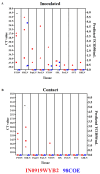
Shedding dynamics produced by infection with 98COE and IN0919WYB2. RT-qPCR was used to evaluate the shedding of viral RNA during infection in nasal, oral, and rectal swabs collected from inoculated ((A), (C), and (E), respectively) and contact ((B), (D), and (F), respectively) animals infected with different viruses. The right Y-axis of each graph represents the predicted TCID50/mL based on the CT value. Predicted levels of infectious virus were considered based on the results of a previous validation [20]. For these graphs, inoculated pigs (IN0919WYB2 group) are represented by red circles, squares, and triangles for pigs 300, 301, and 302, respectively, and to identify contact pigs 303, 304, and 305 (IN0919WYB2 group). Similarly, the same pattern in blue is followed for the 98COE group, identifying pigs 306, 307, and 308 (inoculated) and 309, 310, and 311 (contact), respectively. Asterisks represent time points where means were significantly different between groups (p-value < 0.05) by the Welch’s t-test.
Figure 6
Figure 6
Analysis of the recovery of infectious viral particles from nasal and oral swabs during infection with 98COE and IN0919WYB2. (A) Differences in the recovery of infectious virus from nasal swabs between pigs infected with 98COE and IN0919WYB2. (B) CT values obtained from the same nasal swabs subjected to viral isolation. (C) Differences in the recovery of infectious virus from oral swabs between pigs infected with 98COE and IN0919WYB2. (D) CT values obtained from the same oral swabs subjected to viral isolation. Specific days and pigs where viral isolations were obtained are highlighted in yellow in all cases. (E) Proportion of positive viral isolates using nasal and oral swabs at specific CT value ranges. The number of isolates obtained for each CT group is denoted by the numbers (in red) on the top of the bars.
Figure 7
Figure 7
Postmortem evaluation between 98COE and IN0919WYB2. At 21 dpi, necropsies were conducted in inoculated (A) and contact (B) pigs from each group. Tissues were evaluated by RT-qPCR. The right Y-axis reflects the predicted TCID50/mL based on the CT value. Abbreviations indicate tonsil of the soft palate (PTON), submandibular lymph node (SMLN), popliteal lymph node (R-PopLN), parotid lymph node (ParLN), nasopharyngeal tonsil (NTON), spleen (SPL), prescapular lymph node (PreLN), and gastrohepatic lymph node (GHLN). IN0919WYB2 group pigs identified by red circles, squares, and triangles denote measurements of pigs 300, 301, and 302, respectively. Identical shapes are used for contact pigs to identify pigs 303, 304, and 305. A similar pattern is used to denote the 98COE group, with blue shapes identifying pigs 306, 307, and 308 (inoculated) and 309, 310, and 311 (contact), respectively. Asterisks represent tissues where means were significantly different between groups (p < 0.05) by the Welch’s t-test.
Figure 8
Figure 8
Differences in the adaptive immune response between 98COE and IN0919WYB2 groups during the infection in pigs. Graphics show the titer of neutralizing antibodies against VSIV at different times in inoculated (A) and contact (B) pigs infected with 98COE or IN0919WYB2. In inoculated pigs, red circles, squares, and triangles denote measurements from pigs 300, 301, and 302, respectively. The same shapes are used for contact pigs to identify pigs 303, 304, and 305 from the IN0919W9YB2 group. Similarly, the blue shapes (98COE group) identify pigs 306, 307, and 308 (inoculated) and 309, 310, and 311 (contact), respectively.
Figure 9
Figure 9
Genetic characteristics of the viral progeny produced during the infection of IN0919WYB2 and 98COE in pigs. To assess the viral progeny diversity produced during the infection with strains IN0919WYB2 (A) and 98COE (B), viral isolates recovered from pigs infected with each of the strains were subjected to next-generation sequencing (NGS). Mutations at different codons were identified by gene/codon position and mutation type (dS = synonymous, dN = nonsynonymous substitutions). Gray highlights denote substitutions at specific codon positions compared with parental phenotype in the viral stock. Letters in parentheses reflect the amino acid encoded by different codons.

Similar articles

Cited by

References

    1. Rodriguez L.L. Emergence and re-emergence of vesicular stomatitis in the United States. Virus Res. 2002;85:211–219. doi: 10.1016/S0168-1702(02)00026-6. - DOI - PubMed
    1. Pelzel-McCluskey A., Christensen B., Humphreys J., Bertram M., Keener R., Ewing R., Cohnstaedt L.W., Tell R., Peters D.P.C., Rodriguez L. Review of Vesicular Stomatitis in the United States with Focus on 2019 and 2020 Outbreaks. Pathogens. 2021;10:993. doi: 10.3390/pathogens10080993. - DOI - PMC - PubMed
    1. Velazquez-Salinas L., Pauszek S.J., Zarate S., Basurto-Alcantara F.J., Verdugo-Rodriguez A., Perez A.M., Rodriguez L.L. Phylogeographic characteristics of vesicular stomatitis New Jersey viruses circulating in Mexico from 2005 to 2011 and their relationship to epidemics in the United States. Virology. 2014;449:17–24. doi: 10.1016/j.virol.2013.10.025. - DOI - PubMed
    1. Zarate S., Bertram M., Rodgers C., Reed K., Pelzel-McCluskey A., Gomez-Romero N., Rodriguez L.L., Mayo C., Mire C., Pond S.L.K., et al. Phylogenomic Signatures of a Lineage of Vesicular Stomatitis Indiana Virus Circulating During the 2019–2020 Epidemic in the United States. Viruses. 2024;16:1803. doi: 10.3390/v16111803. - DOI - PMC - PubMed
    1. Dietzgen R.G. Morphology. Genome Organization, Transcription and Replication of Rhabdoviruses. In: Dietzgen R.G., Kuzmin I.V., editors. Rhabdoviruses. Caister Academic Press; Norfolk, UK: 2012. pp. 5–11.

Publication types

LinkOut - more resources