. 2024 Dec 14;16(12):1918.
doi: 10.3390/v16121918.

Heterogeneous Ribonucleoprotein K Is a Host Regulatory Factor of Chikungunya Virus Replication in Astrocytes

Affiliations

Heterogeneous Ribonucleoprotein K Is a Host Regulatory Factor of Chikungunya Virus Replication in Astrocytes

Lisa Pieterse et al. Viruses. .

Abstract

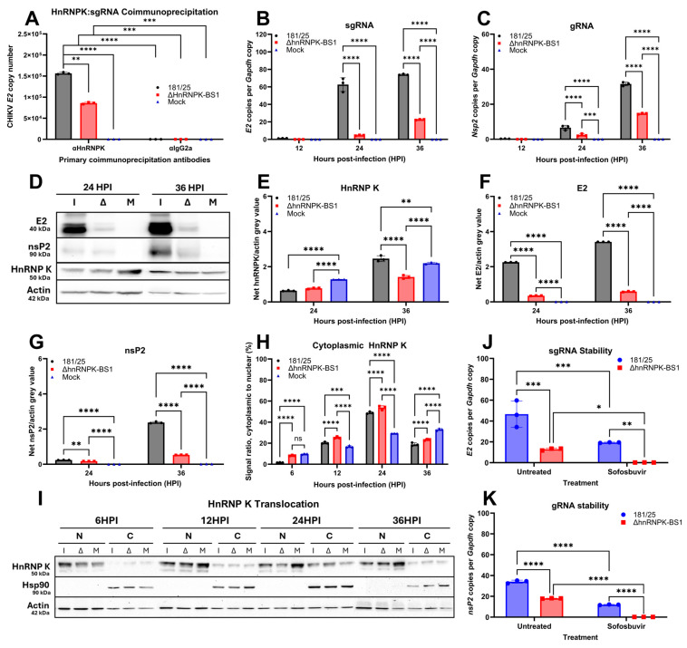
Chikungunya virus (CHIKV) is an emerging, mosquito-borne arthritic alphavirus increasingly associated with severe neurological sequelae and long-term morbidity. However, there is limited understanding of the crucial host components involved in CHIKV replicase assembly complex formation, and thus virus replication and virulence-determining factors, within the central nervous system (CNS). Furthermore, the majority of CHIKV CNS studies focus on neuronal infection, even though astrocytes represent the main cerebral target. Heterogeneous ribonucleoprotein K (hnRNP K), an RNA-binding protein involved in RNA splicing, trafficking, and translation, is a regulatory component of alphavirus replicase assembly complexes, but has yet to be thoroughly studied in the context of CHIKV infection. We identified the hnRNP K CHIKV viral RNA (vRNA) binding site via sequence alignment and performed site-directed mutagenesis to generate a mutant, ΔhnRNPK-BS1, with disrupted hnRNPK-vRNA binding, as verified through RNA coimmunoprecipitation and RT-qPCR. CHIKV ΔhnRNPK-BS1 demonstrated hampered replication in both NSC-34 neuronal and C8-D1A astrocytic cultures. In astrocytes, disruption of the hnRNPK-vRNA interaction curtailed viral RNA transcription and shut down subgenomic RNA translation. Our study demonstrates that hnRNP K serves as a crucial RNA-binding host factor that regulates CHIKV replication through the modulation of subgenomic RNA translation.

Keywords: RNA-binding protein; alphavirus; astrocytes; chikungunya virus; heterogeneous ribonucleoprotein K; host–virus interactions; neurons; neurovirulence; subgenomic RNA translation; viral replication.


Conflict of interest statement

Diane E. Griffin is a member of the advisory boards for GlaxoSmithKline, Academia Sinica, and the University of Vermont and has also received funding from Gilead, MeVox, Merck, and the US National Institutes of Health.

Figures

Figure 1
Figure 1
Mutagenesis of the heterogeneous ribonucleoprotein K (hnRNP K) chikungunya virus (CHIKV) viral RNA (vRNA)-binding site modifies the vRNA secondary structure. (A) RT-qPCR analysis of total RNA bound to coimmunoprecipitated hnRNP K protein collected from C8-D1A murine astrocytes infected with CHIKV 181/25 (WT) versus mock demonstrated hnRNP K interaction with the postulated hnRNPK–vRNA binding site originally identified via cross-sequence alignment with the Sindbis virus (SINV) hnRNPK–vRNA reference sequence. HnRNP K vRNA-binding site sequence annotations of attenuated CHIKV 181/25 versus (B) ΔhnRNPK-BS1 and (C) ΔhnRNPK-BS2 mutants with four or eight silent mutation substitutions, respectively. Site-directed silent mutations are highlighted in black and cross-aligned with the CHIKV 181/25 hnRNPK–vRNA-binding site consensus sequence. Translational products are delineated in a gray box below the alignments to illustrate the lack of change in amino acid translation. Predicted dot plot modeling of secondary structure of (D) CHIKV 181/25, (E) ΔhnRNPK-BS1, and (F) ΔhnRNPK-BS2 hnRNPK–vRNA-binding site sequences, as modeled using RNAComposer and RiboSketch software. Silent mutations resulted in RNA secondary structure modifications, as illustrated by the addition of an internal loop, an expanded hairpin loop, and disrupted overhang structures found in the vRNA of both ΔhnRNPK mutants. Three-dimensional ball-and-stick RNA secondary structure modeling schematics of the (G) CHIKV 181/25, (H) ΔhnRNPK-BS1, and (I) ΔhnRNPK-BS2 mutants, as generated by Geneious (Java Version 11.0.18+10) using the standard Corey-Pauling-Koltun color convention. Statistical significance was determined via two-way ANOVAs, assuming Gaussian distribution; * p < 0.05, mock versus 181/25.
Figure 2
Figure 2
Mutagenesis of the heterogeneous ribonucleoprotein K CHIKV viral RNA-binding site negatively regulates viral replication and disrupts hnRNPK–vRNA binding. Murine astrocytic C8-D1A and neuronal NSC-34 cell lines were infected with the CHIKV 181/25, ΔhnRNPK-BS1, or ΔhnRNPK-BS2 passage 1 (P1) mutant virus at a multiplicity of infection (MOI) of 5. Supernatant was collected at various timepoints and viral titers in (A) murine C8-D1A astrocytes and (B) NSC-34 neurons were determined via Vero plaque assays; #, statistical difference from 181/25; †, significant difference from ΔhnRNPK-BS1; ‡, significant difference from ΔhnRNPK-BS2. (C) Mutagenesis of hnRNPK–vRNA diminishes CHIKV infectious center formation in C8-D1A astrocytes. (D) RT-qPCR analysis of total RNA pulled-down from coimmunoprecipitated hnRNP K protein isolated from CHIKV 181/25 or ΔhnRNPK-BS1-infected C8-D1A cells illustrated significantly diminished hnRNPK–vRNA-binding site transcript quantities bound to hnRNP K in ΔhnRNPK-BS1-infected cells. The experimental design included the use of three biological replicates per condition. Individual replicate values are denoted by symbols delineated by the legend, while standard deviations of the mean are represented by error bars. Statistical significance was determined via two-way ANOVAs, assuming Gaussian distribution; * p < 0.05; ** p < 0.01; **** p < 0.0001.
Figure 3
Figure 3
Heterogeneous ribonucleoprotein K interacts with CHIKV subgenomic RNA (sgRNA) and hnRNPK–vRNA binding regulates sgRNA translation. (A) Coimmunoprecipitation was performed using protein lysates harvested from murine astrocytes infected with CHIKV 181/25, CHIKV ΔhnRNPK-BS1, or media (mock) only. Protein pull-down was performed using αHnRNPK or αIgG2a isotype control and agarose-conjugated beads, followed by RNA isolation of protein-linked RNA, cDNA synthesis, and RT-qPCR to quantify sgRNA expression. RT-qPCR analysis of total RNA isolated from CHIKV 181/25 or ΔhnRNPK-BS1-infected C8-D1A astrocytes was performed to quantify (B) sgRNA and (C) genomic RNA (sgRNA) transcript copies, as normalized to host Gapdh copy numbers. (D) Western blot analysis illustrating E2 glycoprotein and nsP2 expression in CHIKV 181/25, ΔhnRNPK-BS1, or mock-infected C8-D1A cells. Densitometric analyses to quantify signal intensities of (E) hnRNP K, (F) E2, and (G) nsP2 from the blot depicted in (D). (H) Densitometric analyses of hnRNP K signal intensity in (I) immunoblotting depicting nuclear (N) and cytoplasmic (C) fractions of CHIKV 181/25, CHIKV ΔhnRNPK-BS1 (Δ), or mock (M)-infected C8-D1A astrocytes at various hours post-infection (HPI). Hsp90 was utilized as a cytoplasmic-specific marker. (J) C8-D1A cells were infected with CHIKV 181/25 or ΔhnRNPK-BS1 for 1 h, followed by 50 µM sofosbuvir treatment. Lysate was collected at 12 HPI, and sgRNA and (K) gRNA were quantified via RT-qPCR. The experimental design included the use of three biological replicates per condition. Individual replicate values are denoted by symbols delineated by the legend, while standard deviations of the mean are represented by error bars. Statistical significance was determined via two-way ANOVAs, assuming Gaussian distribution; * p < 0.05; ** p < 0.01; *** p < 0.001; **** p < 0.0001.
Figure 4
Figure 4
Host hnRNP K availability regulates CHIKV replication in astrocytes. (A) Small interfering RNA (siRNA)-mediated Hnrnpk gene knockdown was performed in murine astrocytes in vitro for 48 h prior to infection with CHIKV 181/25 at an MOI of 5. Quantification of Hnrnpk gene expression, as normalized to Gapdh-VIC, was then performed via RT-qPCR using RNA lysates harvested at designated timepoints. #, statistical difference between Mock siControl versus Mock siHnRNPK; †, statistical difference between 181/25 siControl versus 181/25 siHnRNPK. Vero plaque assays were used to quantify viral titers in longitudinal supernatants collected from siHnRNPK or siControl-transfected C8-D1A cells infected with (B) CHIKV 181/25 or (C) ΔhnRNPK-BS1. (D) CHIKV 181/25 or ΔhnRNPK-BS1 titers were additionally compared in siHnRNPK-transfected cells. RT-qPCR analysis of RNA isolated from siHnRNPK versus siControl-transfected, CHIKV 181/25-, ΔhnRNPK-BS1, or mock-infected C8-D1A cells was performed to quantify (E) relative sgRNA or (F) gRNA quantities. (G) Infectious center assays of siHnRNPK or siControl-transfected, CHIKV 181/25-infected C8-D1A cells were performed to quantify replication initiation. HnRNP K was constitutively knocked down in C8-D1A cells via short hairpin RNA (shRNA) vectors generating small interfering RNAs (siRNAs) that mediate hnRNP K gene expression interference. (H) Knockdown was confirmed via RT-qPCR analysis. Vero plaque assays demonstrating (I) CHIKV 181/25 and (J) ΔhnRNPK-BS1 titers in C8-D1A shHnRNPK versus shControl. Comparisons of CHIKV 181/25 versus ΔhnRNPK-BS1 titers in (K) shHnRNPK and (L) shControl-transduced C8-D1A cells. RT-qPCR analysis of lysates collected 24 HPI demonstrated significantly diminished (M) sgRNA (E2) and (N) gRNA (Nsp2) transcript levels in both CHIKV 181/25 and ΔhnRNPK-BS1-infected C8-D1A shHnRNPK clones. Experimental design included the use of three biological replicates per condition. Individual replicate values are denoted by symbols delineated by the legend, while standard deviations of the mean are represented by error bars. Statistical significance was determined via two-way ANOVAs, assuming Gaussian distribution; * p < 0.05; ** p < 0.01; *** p < 0.001; **** p < 0.0001; ns (non-significant).
Figure 5
Figure 5
Altered CHIKV replication with respect to host hnRNP K availability regulates viral protein translation. (A) Western blot analysis of lysates collected from siHnRNPK-transfected astrocytes infected with CHIKV 181/25 versus ΔhnRNPK-BS1 virus illustrated dramatic differences in E2 glycoprotein expression. Densitometric analyses of the Western blot illustrated in (A) were conducted by quantifying the net mean pixelation intensity by determining mean gray values of (B) hnRNP K, (C) E2, and (D) nsP2 normalized to actin. (E) Protein lysates collected from CHIKV 181/25 or ΔhnRNPK-BS1-infected C8-D1A astrocytes with constituent hnRNP K knockdown via shRNA lentiviral construct transduction were collected at 24 HPI. Lysates were processed via Western blot analysis, and densitometry was performed to examine (F) hnRNP K, (G) E2, and (H) nsP2 expression. Experimental design included the use of three biological replicates per condition. Individual replicate values are denoted by symbols delineated by the legend, while standard deviations of the mean are represented by error bars. Statistical significance was determined via two-way ANOVAs, assuming Gaussian distribution; * p < 0.05; ** p < 0.01; *** p < 0.001; **** p < 0.0001.

Similar articles

References

    1. Barr K.L., Vaidhyanathan V. Chikungunya in Infants and Children: Is Pathogenesis Increasing? Viruses. 2019;11:294. doi: 10.3390/v11030294. - DOI - PMC - PubMed
    1. Silva M.M.O., Tauro L.B., Kikuti M., Anjos R.O., Santos V.C., Goncalves T.S.F., Paploski I.A.D., Moreira P.S.S., Nascimento L.C.J., Campos G.S., et al. Concomitant Transmission of Dengue, Chikungunya, and Zika Viruses in Brazil: Clinical and Epidemiological Findings From Surveillance for Acute Febrile Illness. Clin. Infect. Dis. 2019;69:1353–1359. doi: 10.1093/cid/ciy1083. - DOI - PMC - PubMed
    1. Mehta R., Soares C.N., Medialdea-Carrera R., Ellul M., da Silva M.T.T., Rosala-Hallas A., Jardim M.R., Burnside G., Pamplona L., Bhojak M., et al. The spectrum of neurological disease associated with Zika and chikungunya viruses in adults in Rio de Janeiro, Brazil: A case series. PLoS Negl. Trop. Dis. 2018;12:e0006212. doi: 10.1371/journal.pntd.0006212. - DOI - PMC - PubMed
    1. Balavoine S., Pircher M., Hoen B., Herrmann-Storck C., Najioullah F., Madeux B., Signate A., Valentino R., Lannuzel A., Saint Louis M., et al. Guillain-Barre Syndrome and Chikungunya: Description of All Cases Diagnosed during the 2014 Outbreak in the French West Indies. Am. J. Trop. Med. Hyg. 2017;97:356–360. doi: 10.4269/ajtmh.15-0753. - DOI - PMC - PubMed
    1. Bandeira A.C., Campos G.S., Sardi S.I., Rocha V.F., Rocha G.C. Neonatal encephalitis due to Chikungunya vertical transmission: First report in Brazil. IDCases. 2016;5:57–59. doi: 10.1016/j.idcr.2016.07.008. - DOI - PMC - PubMed

Publication types

Substances

LinkOut - more resources