. 2024 Dec 16;16(12):1923.
doi: 10.3390/v16121923.

Sequence Analysis of microRNAs Encoded by Simian Lymphocryptoviruses

Affiliations

Sequence Analysis of microRNAs Encoded by Simian Lymphocryptoviruses

Yan Chen et al. Viruses. .

Abstract

Lymphocryptoviruses (LCVs) are ubiquitous gamma-herpesviruses that establish life-long infections in both humans and non-human primates (NHPs). In immunocompromised hosts, LCV infections are commonly associated with B cell disorders and malignancies such as lymphoma. In this study, we evaluated simian LCV-encoded small microRNAs (miRNAs) present in lymphoblastoid cell lines (LCLs) derived from a Mauritian cynomolgus macaque (Macaca fascicularis) with cyLCV-associated post-transplant lymphoproliferative disease (PTLD) as well as the viral miRNAs expressed in a baboon (Papio hamadryas) LCL that harbors CeHV12. Via sequence comparisons, we further predicted viral miRNAs encoded by LCVs that infect two additional NHP species: stump-tailed macaques (Macaca arctoides) and bonobos (Pan paniscus). Together, these species represent two arms of the primate phylogeny: Hominoids (Pan) and Old-World monkeys (Macaca, Papio). Through our analysis, we defined sequences for >95 viral miRNAs encoded by these four NHP LCVs. Our study provides the most comprehensive annotation of NHP LCV miRNAs to date, yielding a resource for developing sequence-specific reagents to detect these molecules. Importantly, we further demonstrate that cyLCV miRNAs can be detected in circulation in vivo and have biomarker potential for LCV-related PTLD.

Keywords: biomarkers; herpesvirus; lymphocryptovirus; microRNAs.


Conflict of interest statement

The authors declare no conflicts of interest.

Figures

Figure 1
Figure 1
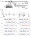
Identification of cyLCV miRNAs. (A) Distribution of reads aligned to cyLCV genome. (B) Positions of viral pre-miRNAs in the cyLCV genome. Red indicates experimental evidence of miRNA expression; gray indicates a predicted cyLCV pre-miRNA based on homology to the rLCV and EBV pre-miRNAs. (C) qRT-PCR analysis of three conserved viral miRNAs in primary cyLCLs established from tissue biopsies from MCM 35132. miRNA expression is shown relative to levels in Raji and rLCL 309-98 cells and normalized to miR-16. PBMCs = cyLCLs sequenced in (A); Ax LN = axillary lymph node; Ing LN = inguinal lymph node; Mes LN = mesenteric lymph node. (D) Sequence alignments of cyLCV, EBV, and rLCV miRNAs tested by qRT-PCR.
Figure 2
Figure 2
Pan paniscus LCV1 miRNAs. (A) Positions of predicted viral miRNAs encoded within the Pan paniscus LCV1 genome. (B) Putative hairpin structures for eight miRNA precursors encoded within the conserved BART Cluster I.
Figure 3
Figure 3
Predicted HVMA miRNAs. (A) Positions of predicted viral miRNAs encoded within the HVMA genome. (B) Putative pre-miRNA structures for the four BHRF1 miRNA homologs and miR-maL-25 encoded antisense to HVMA BALF5.
Figure 4
Figure 4
CeHV12 BHRF1 miRNA homologs expressed in S594 cells. (A) Distribution of small RNA reads aligned to the baboon genome (Panu3.0) and contigs of the CeVH12 genome. 4% of reads aligned to two CeHV12 contigs that encompass the homologous BHRF1 region (AF200364.1 and AH009709.2). (B) Genome plot of small RNA sequencing reads aligned to the CeHV12 BHRF1 region. Y-axis indicates read counts (RC) (C) Putative pre-miRNA folding structures and experimentally defined 5p (colored in red) and 3p (colored in blue) sequences of the CeHV12 BHRF1 miRNAs.
Figure 5
Figure 5
Identification of BART Cluster II miRNA homologs in CeHV12. (A) Schematic showing PCR primer positions and regions amplified from S594 genomic DNA. Primers correspond to miRNA sequences conserved with EBV (denoted with B) or rLCV (denoted with R). Insert: agarose gel electrophoresis following PCR amplification of CeHV12 BART regions. (B) Genome plot of reads aligned to the CeHV12 BART Cluster II region. (C) Predicted pre-miRNA folding structures with the highlighted experimentally defined 5p (colored in red) and 3p (colored in blue) sequences of the CeHV12 BART miRNAs.
Figure 6
Figure 6
Detection of cyLCV miRNAs in circulation. (A) Plasma was collected from MCM 35132 (recipient) at times indicated prior to or post hematopoetic stem cell transplant (HSCT). (B) Plasma was collected from MCM 35133 (donor) at three different time points starting ~6.5 months after graft collection. For both (A,B), miRNAs were extracted from plasma samples and tested by qRT-PCR. Values are normalized to miR-16 and reported relative to the respective EBV miRNA homolog in Raji cells. RNA isolated from rLCL 309-98 and from cyLCL 35132 (spontaneously cultured out of PBMCs) were included as controls. PCR was performed in duplicate. * Student’s t-test, p < 0.05.

Similar articles

References

    1. Muhe J., Wang F. Non-human Primate Lymphocryptoviruses: Past, Present, and Future. Curr. Top. Microbiol. Immunol. 2015;391:385–405. doi: 10.1007/978-3-319-22834-1_13. - DOI - PubMed
    1. Young L.S., Yap L.F., Murray P.G. Epstein-Barr virus: More than 50 years old and still providing surprises. Nat. Rev. Cancer. 2016;16:789–802. doi: 10.1038/nrc.2016.92. - DOI - PubMed
    1. Farrell P.J. Epstein-Barr Virus and Cancer. Annu. Rev. Pathol. 2019;14:29–53. doi: 10.1146/annurev-pathmechdis-012418-013023. - DOI - PubMed
    1. Escalante G.M., Mutsvunguma L.Z., Muniraju M., Rodriguez E., Ogembo J.G. Four Decades of Prophylactic EBV Vaccine Research: A Systematic Review and Historical Perspective. Front. Immunol. 2022;13:867918. doi: 10.3389/fimmu.2022.867918. - DOI - PMC - PubMed
    1. Fujiwara S., Nakamura H. Animal Models for Gammaherpesvirus Infections: Recent Development in the Analysis of Virus-Induced Pathogenesis. Pathogens. 2020;9:116. doi: 10.3390/pathogens9020116. - DOI - PMC - PubMed

Publication types

MeSH terms

LinkOut - more resources