Review
. 2024 Dec 17;16(12):1928.
doi: 10.3390/v16121928.

The 'Oma's of the Gammas-Cancerogenesis by γ-Herpesviruses

Affiliations
Review

The 'Oma's of the Gammas-Cancerogenesis by γ-Herpesviruses

Anwesha Banerjee et al. Viruses. .

Abstract

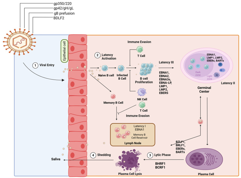
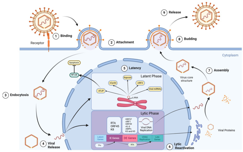
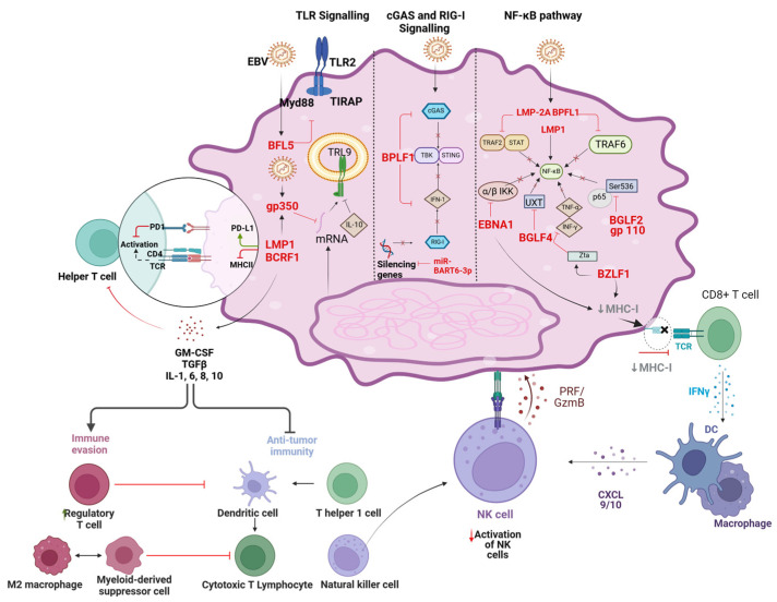
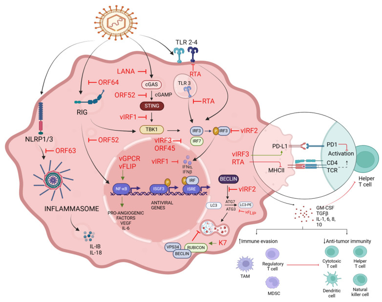
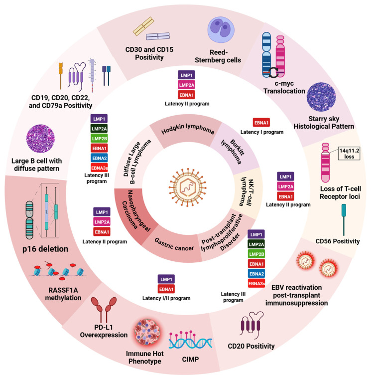
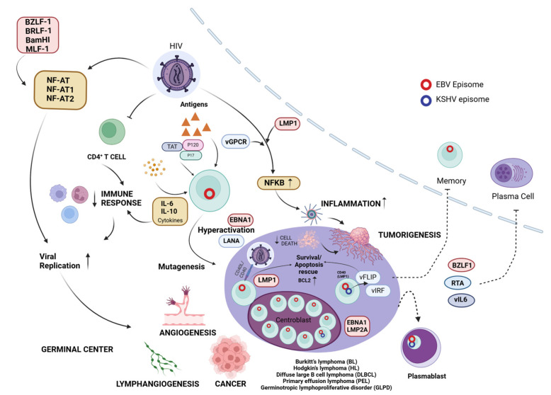
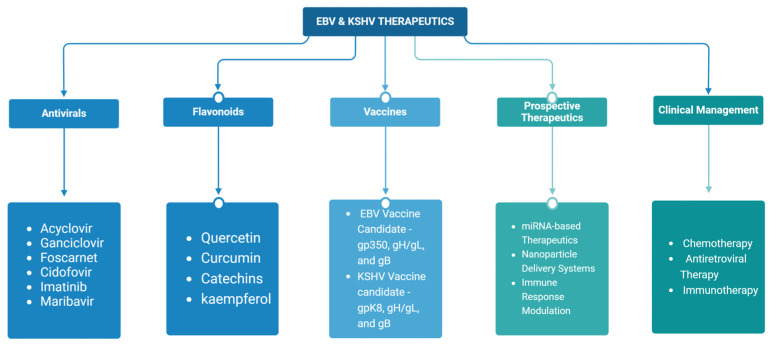
Epstein-Barr virus (EBV) and Kaposi's sarcoma-associated herpesvirus (KSHV), which are the only members of the gamma(γ) herpesviruses, are oncogenic viruses that significantly contribute to the development of various human cancers, such as Burkitt's lymphoma, nasopharyngeal carcinoma, Hodgkin's lymphoma, Kaposi's sarcoma, and primary effusion lymphoma. Oncogenesis triggered by γ-herpesviruses involves complex interactions between viral genetics, host cellular mechanisms, and immune evasion strategies. At the genetic level, crucial viral oncogenes participate in the disruption of cell signaling, leading to uncontrolled proliferation and inhibition of apoptosis. These viral proteins can modulate several cellular pathways, including the NF-κB and JAK/STAT pathways, which play essential roles in cell survival and inflammation. Epigenetic modifications further contribute to EBV- and KSHV-mediated cancerogenesis. Both EBV and KSHV manipulate host cell DNA methylation, histone modification, and chromatin remodeling, the interplay of which contribute to the elevation of oncogene expression and the silencing of the tumor suppressor genes. Immune factors also play a pivotal role in the development of cancer. The γ-herpesviruses have evolved intricate immune evasion strategies, including the manipulation of the major histocompatibility complex (MHC) and the release of cytokines, allowing infected cells to evade immune detection and destruction. In addition, a compromised immune system, such as in HIV/AIDS patients, significantly increases the risk of cancers associated with EBV and KSHV. This review aims to provide a comprehensive overview of the genetic, epigenetic, and immune mechanisms by which γ-herpesviruses drive cancerogenesis, highlighting key molecular pathways and potential therapeutic targets.

Keywords: Epstein–Barr; Kaposi’s sarcoma; antiviral; co-infection; epigenetic; gamma herpesviruses; immune evasion; lymphoma; oncogene; vaccine.


Conflict of interest statement

The authors declare no conflicts of interest.

Figures

Figure 1
Figure 1
Life cycle of Epstein–Barr virus (EBV). This figure depicts the infection and persistence mechanism of Epstein–Barr virus (EBV). The process is illustrated in several stages. (1) Viral entry—EBV enters the epithelial cells using viral glycoproteins (gp350/220, gp42/gH/gL, gB prefusion, BDLF2). (2) Latency activation—the virus infects B cells, leading to their proliferation and the establishment of latent infection. (3) Lytic phase—in some cells, EBV enters the lytic phase, resulting in the production of new viral particles and plasma cell lysis. (4) Shedding—viral particles are shed into saliva, facilitating transmission. The figure also illustrates different latency stages (I, II, III) and the associated viral proteins (EBNA1, EBNA2, EBNA3s, LMP1, LMP2). It shows how EBV evades the immune system by manipulating T cells and NK cells. The germinal center reaction and the establishment of a memory B cell reservoir in lymph nodes are also depicted, highlighting the lifecycle of EBV and its ability to persist in the host.
Figure 2
Figure 2
Life cycle of Kaposi’s sarcoma-associated herpesvirus (KSHV). This figure illustrates the life cycle of Kaposi’s sarcoma-associated herpesvirus (KSHV). The process is divided into several key stages: (1) Binding—KSHV attaches to a cell receptor. (2) Attachment—the virus adheres to the cell membrane. (3) Endocytosis—the virus enters the cell through endocytosis. (4) Viral release—the viral capsid is released into the cytoplasm. (5) Latency—KSHV enters a latent phase, where LANA plays a crucial role in maintaining the viral genome and regulating host cell processes. (6) Lytic reactivation—the virus switches to the lytic phase, where viral DNA replication and gene expression occur. (7) Assembly—new virus particles are assembled. (8) Budding—mature virions bud from the cell membrane. (9) Release—new viral particles are released to infect other cells. The image also shows key viral proteins and their functions in both latent (vFLIP, vCyclin, kaposin, vIRF3) and lytic (RTA, ORF45, K8) phases. The figure demonstrates how KSHV manipulates cellular processes like apoptosis and NF-κB signaling to ensure its survival and replication.
Figure 3
Figure 3
Immune evasion mechanisms of EBV. This figure illustrates the immune evasion strategies employed by Epstein–Barr virus (EBV) showing various cellular pathways and their manipulation by the virus. The figure depicts three main signaling pathways: TLR signaling, cGAS and RIG-I signaling, and the NF-κB pathway. EBV proteins (in red) such as LMP1, LMP2A, EBNA1, and BGLF4 interfere with these pathways to promote viral survival. The figure also shows how EBV modulates cell surface markers like PD-L1 and MHC-I to evade T-cell recognition. Key viral strategies include silencing genes, manipulating cytokine production, and interfering with antiviral responses. The bottom of the figure illustrates the broader immune landscape, showing how EBV influences various immune cells, including helper T cells, cytotoxic T cells, NK cells, dendritic cells, and macrophages. It highlights the balance between immune evasion and anti-tumor immunity, with EBV tipping the scales towards evasion through mechanisms like increasing regulatory T cells and myeloid-derived suppressor cells while inhibiting cytotoxic T lymphocytes and NK cell activation. Overall, the figure provides insights into how EBV subverts multiple aspects of the host immune response to establish persistent infection and potentially contribute to oncogenesis.
Figure 4
Figure 4
Immune evasion mechanisms of KSHV. This figure illustrates the immune evasion strategies employed by Kaposi’s sarcoma-associated herpesvirus (KSHV). It shows a KSHV-infected cell (red) interacting with immune cells, particularly a helper T cell. The figure depicts viral proteins (in red) interfering with host cell receptors, signaling pathways, and transcription factors. Key cellular components like the inflammasome, autophagy machinery, and antiviral gene expression are shown. The figure also highlights how KSHV modulates cell surface markers like PD-L1 and MHC II to evade T-cell recognition. Various immune cells in the tumor microenvironment are represented at the bottom, including regulatory T cells, cytotoxic T cells, and natural killer cells. Cytokines and growth factors involved in immune modulation and angiogenesis are also indicated. Overall, the figure demonstrates that KSHV is used to subvert the host immune response and promote its survival and potential oncogenesis.
Figure 5
Figure 5
Epstein–Barr virus (EBV)-associated malignancies and latency. Hodgkin’s lymphoma is characterized by CD30/CD15 positivity and the presence of Reed–Sternberg cells. Burkitt’s lymphoma is distinguished by C-myc translocation and a starry sky histological pattern. Diffuse large B-cell lymphoma presents with large B cells along with CD19, CD20, and CD22 positivity. Nasopharyngeal carcinoma is marked by p16 deletion and RASSF1A methylation, while gastric cancer exhibits PD-L1 overexpression and an immune host phenotype. NK/T-cell lymphoma features CD56 positivity alongside the loss of T-cell receptor loci. Post-transplant lymphoproliferative disorder involves EBV reactivation under conditions of immunosuppression. Latency I, II, and III programs are associated with these malignancies as shown.
Figure 6
Figure 6
Molecular interplay between HIV, EBV, and KSHV in the context of immune dysregulation and oncogenesis. This figure illustrates the interplay between HIV, EBV, and KSHV in immune dysregulation and oncogenesis. It depicts how HIV infection compromises the immune system, particularly CD4+ T cells, creating an environment conducive to EBV- and KSHV-associated malignancies. EBV and KSHV, represented by their respective episomes, are shown to exploit the weakened immune system. Key viral proteins, such as LMP1 (EBV) and vGPCR (KSHV), activate NF-κB, a central mediator of inflammation and cell survival. Additionally, the figure shows how HIV with EBV viral proteins leads to the activation of NF-AT, NF-AT1, and NF-AT2, which are important transcription factors in T-cell activation and viral replication. These activations, along with other viral factors like EBNA1 and LANA, promote cell proliferation, inhibit apoptosis, and induce angiogenesis and lymphangiogenesis. The figure highlights the germinal center reaction, where EBV-infected B cells undergo hyperactivation and mutagenesis. This process, coupled with impaired immune surveillance, can lead to the development of various lymphomas, including Burkitt’s lymphoma, Hodgkin’s lymphoma, diffuse large B-cell lymphoma, and primary effusion lymphoma. The figure also illustrates the inhibition from infected cells to memory B cells and plasma cells, emphasizing the role of these viruses in manipulating B-cell differentiation and survival. Overall, this figure highlights the interactions between HIV, EBV, and KSHV, demonstrating how these viruses cooperatively subvert host immune responses, induce chronic inflammation, and drive lymphomagenesis.
Figure 7
Figure 7
Antiviral mechanisms/therapeutic strategies for EBV and KSHV. This flowchart presents an overview of current and prospective therapeutic approaches for EBV- and KSHV-associated diseases. It outlines five main categories of interventions: antivirals, flavonoids, vaccines, prospective therapeutics, and clinical management strategies. The figure details specific examples within each category, such as acyclovir and ganciclovir as antivirals, quercetin and curcumin as flavonoids, and vaccine candidates targeting viral glycoproteins. It also highlights emerging therapies like miRNA-based therapeutics and nanoparticle delivery systems. The figure provides a comprehensive summary of the multifaceted approach to treating EBV and KSHV-related conditions, encompassing both established and innovative therapeutic strategies.

Similar articles

Cited by

References

    1. Barton E., Mandal P., Speck S.H. Pathogenesis and Host Control of Gammaherpesviruses: Lessons from the Mouse. Annu. Rev. Immunol. 2011;29:351–397. doi: 10.1146/annurev-immunol-072710-081639. - DOI - PubMed
    1. Britt W. Virus entry into host, establishment of infection, spread in host, mechanisms of tissue damage. In: Arvin A., Campadelli-Fiume G., Mocarski E., Moore P.S., Roizman B., Whitley R., Yamanishi K., editors. Human Herpesviruses: Biology, Therapy, and Immunoprophylaxis. Cambridge University Press; Cambridge, UK: 2007. - PubMed
    1. Dunmire S.K., Verghese P.S., Balfour H.H. Primary Epstein-Barr Virus Infection. J. Clin. Virol. 2018;102:84–92. doi: 10.1016/j.jcv.2018.03.001. - DOI - PubMed
    1. Corvalán A.H., Ruedlinger J., de Mayo T., Polakovicova I., Gonzalez-Hormazabal P., Aguayo F. The Phylogeographic Diversity of EBV and Admixed Ancestry in the Americas−Another Model of Disrupted Human-Pathogen Co-Evolution. Cancers. 2019;11:217. doi: 10.3390/cancers11020217. - DOI - PMC - PubMed
    1. Jemal A., Bray F., Center M.M., Ferlay J., Ward E., Forman D. Global Cancer Statistics. CA Cancer J. Clin. 2011;61:69–90. doi: 10.3322/caac.20107. - DOI - PubMed

MeSH terms

LinkOut - more resources