. 2024 Dec 18;16(12):1932.
doi: 10.3390/v16121932.

Development and Evaluation of a Newcastle Disease Virus-like Particle Vaccine Expressing SARS-CoV-2 Spike Protein with Protease-Resistant and Stability-Enhanced Modifications

Affiliations

Development and Evaluation of a Newcastle Disease Virus-like Particle Vaccine Expressing SARS-CoV-2 Spike Protein with Protease-Resistant and Stability-Enhanced Modifications

Yu Chen et al. Viruses. .

Abstract

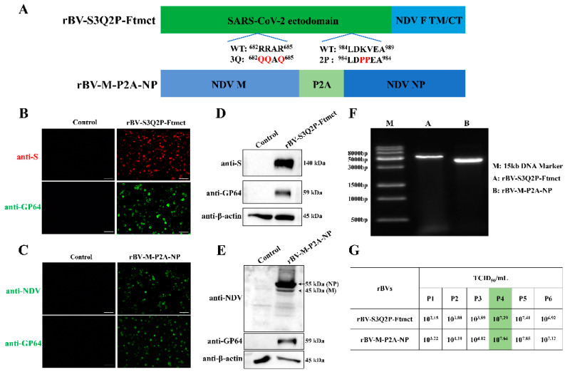
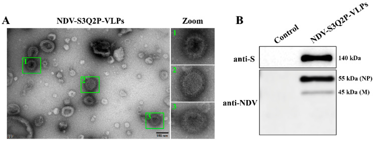
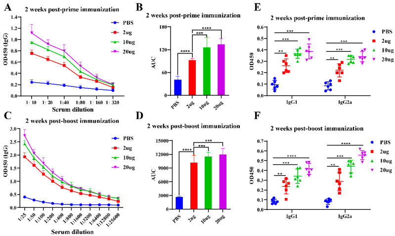
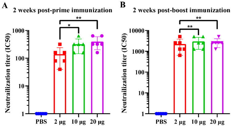
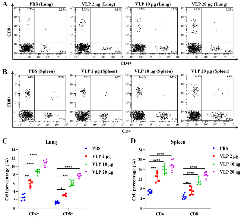
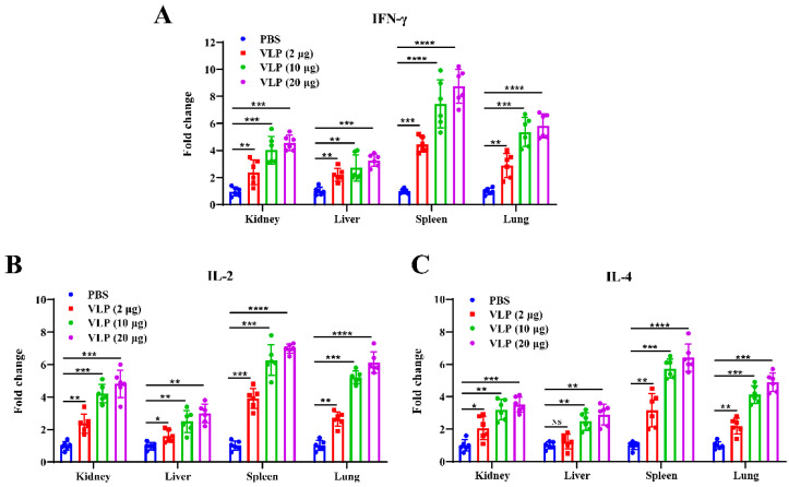
The ongoing global health crisis caused by severe acute respiratory syndrome coronavirus 2 (SARS-CoV-2) necessitates the continuous development of innovative vaccine strategies, especially in light of emerging viral variants that could undermine the effectiveness of existing vaccines. In this study, we developed a recombinant virus-like particle (VLP) vaccine based on the Newcastle Disease Virus (NDV) platform, displaying a stabilized prefusion form of the SARS-CoV-2 spike (S) protein. This engineered S protein includes two proline substitutions (K986P, V987P) and a mutation at the cleavage site (RRAR to QQAQ), aimed at enhancing both its stability and immunogenicity. Using a prime-boost regimen, we administered NDV-VLP-S-3Q2P intramuscularly at different doses (2, 10, and 20 µg) to BALB/c mice. Robust humoral responses were observed, with high titers of S-protein-specific IgG and neutralizing antibodies against SARS-CoV-2 pseudovirus, reaching titers of 1:2200-1:2560 post-boost. The vaccine also induced balanced Th1/Th2 immune responses, evidenced by significant upregulation of cytokines (IFN-γ, IL-2, and IL-4) and S-protein-specific IgG1 and IgG2a. Furthermore, strong activation of CD4+ and CD8+ T cells in the spleen and lungs confirmed the vaccine's ability to promote cellular immunity. These findings demonstrate that NDV-S3Q2P-VLP is a potent immunogen capable of eliciting robust humoral and cellular immune responses, highlighting its potential as a promising candidate for further clinical development in combating COVID-19.

Keywords: NDV; SARS-CoV-2; VLP; immune response; vaccine.


Conflict of interest statement

The authors declare no conflicts of interest.

Figures

Figure 1
Figure 1
Construction and characterization of recombinant baculoviruses (rBVs). (A) Schematic diagram of the rBV constructs. rBV-S3Q2P-Ftmct includes the ectodomain of the SARS-CoV-2 S protein fused to the transmembrane and cytoplasmic tail (TM/CT) domains of the NDV F protein, with the S1/S2 cleavage site (RRAR) mutated to QQAQ and two proline substitutions (K986P, V987P) for stabilization. rBV-M-P2A-NP includes the NDV M and NP proteins, linked via the P2A sequence. (B,C) Immunofluorescence assay (IFA) of Sf9 cells infected with rBV-S3Q2P-Ftmct (B) and rBV-M-P2A-NP (C). Cells were stained with anti-S (red) or anti-NDV (green) antibodies, along with anti-GP64 (green) as a baculovirus marker. Scale bars,100 μm. (D,E) Western blot analysis of Sf9 cells infected with rBV-S3Q2P-Ftmct (D) and rBV-M-P2A-NP (E). Proteins were detected using antibodies against SARS-CoV-2 S protein, NDV NP, M, and GP64, and β-actin was used as a control. (F) PCR confirmation of the rBV constructs. The presence of the expected gene fragments for rBV-S3Q2P-Ftmct (lane A) and rBV-M-P2A-NP (lane B) was confirmed. (G) Virus titration results (TCID50/mL) of the rBVs across passages (P1–P6).
Figure 2
Figure 2
Production and characterization of NDV-S3Q2P-VLPs. (A) Transmission electron microscopy (TEM) image of NDV-S3Q2P-VLPs. Sf9 cells were co-infected with rBV-S3Q2P-Ftmct and rBV-M-P2A-NP, and VLPs were purified at 72 hpi. VLPs were negatively stained and visualized under a transmission electron microscope. The zoomed-in sections (1, 2, and 3) provide magnified views of individual VLPs. Scale bars, 100 nm. (B) Western blot analysis of NDV-S3Q2P-VLPs. Purified VLPs were lysed and subjected to SDS-PAGE, followed by immunoblot with anti-SARS-CoV-2 S protein and anti-NDV antibodies. S protein (140 kDa), NDV NP (55 kDa), and M (45 kDa) were detected.
Figure 3
Figure 3
Validation of 293T-hACE2 cell line and SARS-CoV-2 pseudovirus infectivity: (A) Western blot analysis of ACE2 expression in 293T wild-type (WT) and 293T-hACE2 cells from passages 1 to 8: Cells were lysed and subjected to SDS-PAGE, followed by immunoblotting using anti-ACE2 and anti-β-actin antibodies. (B) Luciferase assay to assess pseudovirus infectivity: 293T-WT and 293T-hACE2 cells were infected with SARS-CoV-2 pseudovirus or VSV-G pseudovirus, and luciferase activity was measured after 48 h to evaluate infection efficiency. (C) Fluorescence microscopy to visualize pseudovirus infection: 293T-WT and 293T-hACE2 cells were infected with SARS-CoV-2 pseudovirus or VSV-G pseudovirus, and GFP fluorescence was observed at 48 hpi. Scale bars, 100 μm. (D) Pseudovirus infection dose–response experiment: 293T-hACE2 cells were infected with increasing volumes of SARS-CoV-2 pseudovirus (10 μL, 20 μL, 30 μL, 40 μL, and 50 μL), and luciferase activity was measured at 48hpi to determine the optimal pseudovirus dose. NS means not significant, **** p < 0.0001.
Figure 4
Figure 4
Immunization schedule and groups: BALB/c mice (n = 6 per group) were immunized with different doses of NDV-S3Q2P-VLPs (2 μg, 10 μg, and 20 μg) or PBS as a control. Mice were immunized intramuscularly (I.M.) with a prime dose at week 0 and a boost dose at week 2. Blood samples were collected at week 2 and week 4 to assess immune responses.
Figure 5
Figure 5
S-protein-specific IgG, IgG1, and IgG2a antibody responses induced by NDV-S3Q2P-VLPs immunization. (A) ELISA analysis of S-protein-specific IgG antibody titers at 2 weeks post-prime immunization. BALB/c mice were immunized with NDV-S3Q2P-VLPs at doses of 2 μg, 10 μg, or 20 μg, with PBS as the control. Serum samples were collected and serially diluted (1:10 to 1:320) to assess IgG titers. (B) Area under the curve (AUC) analysis of S-protein-specific IgG titers at 2 weeks post-prime immunization: AUC values were calculated to quantify the antibody responses across different doses. (C) ELISA analysis of S-protein-specific IgG antibody titers at 2 weeks post-boost immunization: serum samples were serially diluted (1:25 to 1:25,600) to measure the increase in IgG titers following the booster dose. (D) AUC analysis of S-protein-specific IgG titers at 2 weeks post-boost immunization: AUC values were calculated to evaluate the dose-dependent response after the boost. (E) ELISA analysis of S-protein-specific IgG1 and IgG2a antibody titers at 2 weeks post-prime immunization: serum samples were diluted at 1:40. (F) ELISA analysis of S-protein-specific IgG1 and IgG2a antibody titers at 2 weeks post-boost immunization: serum samples were diluted at 1:400. Statistical analyses were performed using one-way ANOVA for (B,D) and two-way ANOVA for (E,F). Data are presented as means ± standard deviation (SD). ** p < 0.01, *** p < 0.001, **** p < 0.0001.
Figure 6
Figure 6
Neutralizing-antibody titers against SARS-CoV-2 pseudovirus induced by NDV-S3Q2P-VLPs immunization: (A) Neutralizing-antibody titers (IC50) at 2 weeks post-prime immunization. BALB/c mice were immunized with NDV-S3Q2P-VLPs at doses of 2 μg, 10 μg, or 20 μg, with PBS as the control. Serum samples were collected and tested for neutralization of SARS-CoV-2 pseudovirus. IC50 values represent the dilution at which 50% neutralization was achieved. (B) Neutralizing antibody titers (IC50) at 2 weeks post-boost immunization: serum samples were collected and tested for neutralization activity against SARS-CoV-2 pseudovirus following the booster dose. Statistical analyses were performed using one-way ANOVA, and data are presented as means ± SD. * p < 0.05, ** p < 0.01.
Figure 7
Figure 7
NDV-S3Q2P-VLPs induced CD4+ and CD8+ T cell responses in lung and spleen two weeks after booster immunization: Lung and spleen single-cell suspensions were isolated from mice at 2 weeks post-boost immunization with different doses of NDV-S3Q2P-VLPs or PBS control. The cells were stained with mouse anti-CD4 and anti-CD8 antibodies to evaluate T cell populations. (A) Flow cytometry analysis of CD4+ and CD8+ T cell populations in the lung. (B) Flow cytometry analysis of CD4+ and CD8+ T cell populations in the spleen. (C) Proportional analysis of CD4+ and CD8+ T cells in the lung. (D) Proportional analysis of CD4+ and CD8+ T cells in the spleen. Data were analyzed with two-way ANOVA. * p < 0.05, ** p < 0.01, *** p < 0.001, **** p < 0.0001.
Figure 8
Figure 8
Cytokine expression levels in tissues from mice immunized with NDV-S3Q2P-VLPs. Total RNA was extracted from kidney, liver, spleen, and lung tissues of NDV-S3Q2P-VLP-immunized mice or PBS control at 2 weeks post-boost immunization. qRT-PCR was used to measure the expression levels of IFN-γ, IL-2, and IL-4 in these tissues. (A) Fold change in IFN-γ expression in kidney, liver, spleen, and lung tissues. (B) Fold change in IL-2 expression in kidney, liver, spleen, and lung tissues. (C) Fold change in IL-4 expression in kidney, liver, spleen, and lung tissues. Cytokine expression was normalized to β-actin, and relative expression was calculated using the 2−ΔΔCt method. Statistical analyses were performed using two-way ANOVA. NS means not significant, * p < 0.05, ** p < 0.01, *** p < 0.001, **** p < 0.0001.

Similar articles

Cited by

References

    1. Mansour H.M. The interference between SARS-COV-2 and Alzheimer’s disease: Potential immunological and neurobiological crosstalk from a kinase perspective reveals a delayed pandemic. Ageing Res. Rev. 2024;94:102195. doi: 10.1016/j.arr.2024.102195. - DOI - PubMed
    1. Yee R., Carranza D., Kim C., Trinidad J.P., Tobias J.L., Bhatkoti R., Kuwabara S. COVID-19 Vaccination Site Accessibility, United States, December 11, 2020-March 29, 2022. Emerg. Infect. Dis. 2024;30:947–955. doi: 10.3201/eid3005.230357. - DOI - PMC - PubMed
    1. Carabelli A.M., Peacock T.P., Thorne L.G., Harvey W.T., Hughes J., Peacock S.J., Barclay W.S., de Silva T.I., Towers G.J., Robertson D.L. SARS-CoV-2 variant biology: Immune escape, transmission and fitness. Nat. Rev. Microbiol. 2023;21:162–177. doi: 10.1038/s41579-022-00841-7. - DOI - PMC - PubMed
    1. Harrison A.G., Lin T., Wang P. Mechanisms of SARS-CoV-2 Transmission and Pathogenesis. Trends Immunol. 2020;41:1100–1115. doi: 10.1016/j.it.2020.10.004. - DOI - PMC - PubMed
    1. Lacek K.A., Rambo-Martin B.L., Batra D., Zheng X.Y., Hassell N., Sakaguchi H., Peacock T., Groves N., Keller M., Wilson M.M., et al. SARS-CoV-2 Delta-Omicron Recombinant Viruses, United States. Emerg. Infect. Dis. 2022;28:1442–1445. doi: 10.3201/eid2807.220526. - DOI - PMC - PubMed

Publication types

MeSH terms