. 2024 Dec 18;16(12):1935.
doi: 10.3390/v16121935.

MARCH8 Restricts RSV Replication by Promoting Cellular Apoptosis Through Ubiquitin-Mediated Proteolysis of Viral SH Protein

Affiliations

MARCH8 Restricts RSV Replication by Promoting Cellular Apoptosis Through Ubiquitin-Mediated Proteolysis of Viral SH Protein

Takashi Okura et al. Viruses. .

Abstract

Numerous host factors function as intrinsic antiviral effectors to attenuate viral replication. MARCH8 is an E3 ubiquitin ligase that has been identified as a host restriction factor that inhibits the replication of various viruses. This study elucidated the mechanism by which MARCH8 restricts respiratory syncytial virus (RSV) replication through selective degradation of the viral small hydrophobic (SH) protein. We demonstrated that MARCH8 directly interacts with RSV-SH and catalyzes its ubiquitination at lysine 13, leading to SH degradation via the ubiquitin-lysosomal pathway. Functionally, MARCH8 expression enhances RSV-induced apoptosis through SH degradation, ultimately reducing viral titers. Conversely, an RSV strain harboring the SH-K13R mutation exhibited prolonged SH protein stability and attenuated apoptosis in infected cells, even in the presence of MARCH8. Targeted depletion of MARCH8 enhances cellular survival and potentially increases viral persistence. These findings demonstrate that MARCH8 promotes the early elimination of virus-infected cells by abrogating the anti-apoptotic function of SH, thereby reducing viral transmission. Our study provides novel insights into the interplay between host restriction factors and viral evasion strategies, potentially providing new therapeutic approaches for RSV infections.

Keywords: MARCH8; apoptosis; respiratory syncytial virus; small hydrophobic protein; ubiquitination.


Conflict of interest statement

T.K. is currently an employee of Kanto Chemical Co., Inc. All authors have submitted the ICMJE Form for Disclosure of Potential Conflicts of Interest.

Figures

Figure 1
Figure 1
Reduction of RSV-SH and IAV-M2 by overexpression of MARCH1 and MARCH8. Schematic representation of the flow for transfection experiments using MARCH and RSV-SH fused with mStrawberry (mSB), influenza A virus (IAV) M2 fused with mSB, or mSB expression plasmids (A). 293T cells were co-transfected with MARCH1, 2, 4, 6, and 8 expression plasmids along with expression plasmids of RSV-SH-mSB, IAV-M2-mSB, and mSB. At 48 hpt, the cells were observed with fluorescence microscopy. All images were taken at the same magnification and exposure time (B). The fluorescence intensity acquired from at least five of each image was quantified using ImageJ. The relative fluorescence intensity of SH-mSB or M2-mSB to that of mSB is shown in the graph. Asterisks represent statistically significant differences using the Student’s t-test (**: p < 0.01, *: p < 0.05) (C).
Figure 2
Figure 2
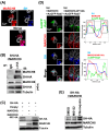
Lysosomal degradation of RSV-SH ubiquitinated by MARCH8. (A) HEp-2 cells were transfected with MARCH8-HA expression plasmid. After 24 h, the cells were then infected with wt-RSV at MOI of 1 for 72 h. Squares indicate co-localization areas. (B) For the co-immunoprecipitation assay, 293T cells expressing SH-HA and MARCH8 were incubated with anti-HA antibody or control IgG followed by the subsequent addition of Protein A/G agarose. (C) 293T cells were co-transfected with RSV-SH-HA and wt-MARCH8, and MARCH8-W114A mutant expression plasmids. At 48 hpt, the cells were subjected to western blotting using anti-HA or MARCH8 antibodies. (D) HEp-2 cells were co-transfected with expression plasmids for a combination of wt-SH plus MARCH8, and wt-SH plus MARCH8-W114A mutant along with late endosome/lysosome marker AcGFP-Rab7. Nuclei were stained with DAPI. Insets represent an enlargement of the areas indicated by a small square. Fluorescence profiles are shown with fluorescence intensity (y-axis) and distance (x-axis) according to the direction of the arrows within the enlarged squares. (E) 293T cells were co-transfected with SH and MARCH8 expression plasmids. At 24 hpt, the cells were untreated (DMSO control) or treated with MG132 or chloroquine for 16 h before harvest.
Figure 3
Figure 3
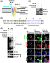
MARCH8 ubiquitinates RSV-SH on Lysine 13. (A) The complex structure of pentamer RSV-SH (yellow) and partial MARCH8 (cyan) is predicted using Alphafold 3 software. Transmembrane domains of MARCH8 and SH predicted by SOSUI system were highlighted in blue and orange colors, respectively. Tryptophan at amino acid position 114 (W114) in MARCH8 is the E2 enzyme binding site, and RSV-K13, which MARCH8 ubiquitinates, was predicted to be present together on the cytoplasmic side. (B) Amino acid alignment of human RSV-SH subgroup A and B and bovine RSV-SH. K13 residues in the cytoplasmic tail of SH and the transmembrane domain predicted by SOSUI are shown in red and purple, respectively. (C) 293T cells were co-transfected with wt-SH and SH-K13R mutant together with MARCH8 expression plasmids. (D) For ubiquitination of SH by MARCH8, 293T cells were co-transfected with wt-SH and wt-MARCH8, SH-K13R mutant and wt-MARCH8, and wt-SH and MARCH8-W114A mutant along with FLAG-tagged ubiquitin (FLAG-Ubi). Ubiquitination of SH was detected by western blotting using anti-FLAG antibody. * heavy chain, ** light chain. (E) HEp-2 cells were co-transfected with expression plasmids of wt-SH and a combination of wt-SH plus MARCH8, and wt-SH plus MARCH8-W114A mutant along with AcGFP-Rab7. Insets represent an enlargement of the areas indicated by a small square and arrowheads show the co-localization area of SH and Rab7.
Figure 4
Figure 4
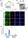
MARCH8-mediated ubiquitination of RSV-SH promotes apoptosis and inhibits virus persistence. HEp-2 cells were transfected with the MARCH8 expression plasmid or an empty plasmid. The cells were infected with the wt-RSV or RSV-K13R mutant virus. At various time points (indicated), aliquots of the cell culture medium were collected, and the viral titer in the medium was measured as TCID50. Each day after infection, viral titers in transfected cells were compared with viral titers in samples transfected with empty vectors and infected with wt-RSV. Asterisks indicate statistically significant differences using the Student’s t-test (* p < 0.05, ** p < 0.01) (A). HEp-2 cells were transfected and infected with wt-RSV or RSV-K13R viruses, as described in Figure 4A (B). HEp-2 cells were transfected and infected with wt-RSV or RSV-K13R mutant viruses. The cells were subjected to a TUNEL assay to detect cell death by apoptosis (C). The apoptosis rate was calculated as the ratio of apoptotic cells to the total number of cells using the Analyze Particles plug-in of the ImageJ software. Asterisks indicate statistically significant differences using the Student’s t-test (* p < 0.01) (D).
Figure 5
Figure 5
MARCH8 knockdown inhibits virus-induced apoptosis and enhances viral replication. MARCH8 mRNA expression levels in control (sh-control) and MARCH8-knockdown (KD) (sh-MARCH8#1 and sh-MARCH8#2) A549 cell lines were examined by conventional RT-PCR using primers specific for MARCH8 (A). Control and MARCH8-KD A549 cells were infected with wt-RSVand the cells were collected and subjected to western blotting with the appropriate antibodies to detect each protein (B). Control and MARCH8-KD A549 cells were infected with wt-RSV. At various time points (indicated), aliquots of the cell culture medium were collected, and the viral titer was measured as TCID50 (C). The virus-infected cells were fixed and stained with Hoechst stain, and the number of viable cells from more than 10 images was counted (D). The TUNEL assay was performed to quantify cell death by apoptosis. Representative images in TUNEL assays of control and MARCH8-KD cells (E). Viral titers and the number of viable MARCH8-KD cells were compared with those in control cells. Asterisks indicate statistically significant differences using the Student’s t-test (*: p < 0.05, **: p < 0.01).

Similar articles

Cited by

References

    1. Li Y., Wang X., Blau D.M., Caballero M.T., Feikin D.R., Gill C.J., A Madhi S., Omer S.B., Simões E.A.F., Campbell H., et al. Global, regional, and national disease burden estimates of acute lower respiratory infections due to respiratory syncytial virus in children younger than 5 years in 2019: A systematic analysis. Lancet. 2022;399:2047–2064. doi: 10.1016/S0140-6736(22)00478-0. - DOI - PMC - PubMed
    1. Kenmoe S., Nair H. The disease burden of respiratory syncytial virus in older adults. Curr. Opin. Infect. Dis. 2024;37:129–136. doi: 10.1097/QCO.0000000000001000. - DOI - PMC - PubMed
    1. Peng R., Chen C., Chen Q., Zhang Y., Huang R., Zhang Y., Li J. Global progress in clinical research on human respiratory syncytial virus vaccines. Front. Microbiol. 2024;15:1457703. doi: 10.3389/fmicb.2024.1457703. - DOI - PMC - PubMed
    1. Blanken M.O., Rovers M.M., Molenaar J.M., Winkler-Seinstra P.L., Meijer A., Kimpen J.L., Bont L. Respiratory Syncytial Virus and Recurrent Wheeze in Healthy Preterm Infants. N. Engl. J. Med. 2013;368:1791–1799. doi: 10.1056/NEJMoa1211917. - DOI - PubMed
    1. Hammitt L.L., Dagan R., Yuan Y., Cots M.B., Bosheva M., Madhi S.A., Muller W.J., Zar H.J., Brooks D., Grenham A., et al. Nirsevimab for Prevention of RSV in Healthy Late-Preterm and Term Infants. N. Engl. J. Med. 2022;386:837–846. doi: 10.1056/NEJMoa2110275. - DOI - PubMed

Publication types

MeSH terms

LinkOut - more resources