

PD1-Targeted Transgene Delivery to Treg Cells
- PMID: 39772246
- PMCID: PMC11680301
- DOI: 10.3390/v16121940
PD1-Targeted Transgene Delivery to Treg Cells
Abstract
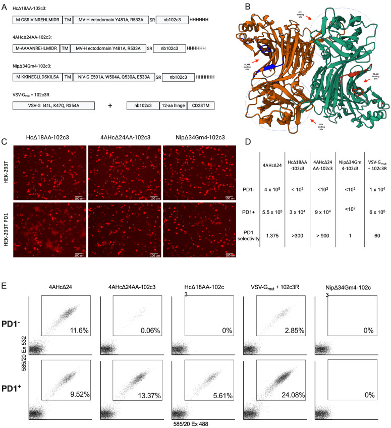
Achieving the precise targeting of lentiviral vectors (LVs) to specific cell populations is crucial for effective gene therapy, particularly in cancer treatment where the modulation of the tumor microenvironment can enhance anti-tumor immunity. Programmed cell death protein 1 (PD-1) is overexpressed on activated tumor-infiltrating T lymphocytes, including regulatory T cells that suppress immune responses via FOXP3 expression. We developed PD1-targeted LVs by incorporating the anti-PD1 nanobody nb102c3 into receptor-blinded measles virus H and VSV-Gmut glycoproteins. We assessed the retargeting potential of nb102c3 and evaluated transduction efficiency in activated T lymphocytes. FOXP3 expression was suppressed using shRNA delivered by these LVs. Our results demonstrate that PD1-targeted LVs exerted pronounced tropism towards PD1+ cells, enabling the selective transduction of activated T lymphocytes while sparing naive T cells. The suppression of FOXP3 in Tregs reduced their suppressive activity. PD1-targeted glycoprotein H provided greater specificity, whereas the VSV-Gmut, together with the anti-PD1 pseudoreceptor, achieved higher viral titers but was less selective. Our study demonstrates that PD1-targeted LVs may offer a novel strategy to modulate immune responses within the tumor microenvironment with the potential for developing new therapeutic strategies aimed at enhancing anti-tumor immunity.
Keywords: FOXP3; PD1; Treg; lentivector; nanobody; retargeting.
Conflict of interest statement
The authors declare no conflicts of interest.
Figures


Similar articles
-
Antigen-presenting cell-targeted lentiviral vectors do not support the development of productive T-cell effector responses: implications for in vivo targeted vaccine delivery.Gene Ther. 2017 Jun;24(6):370-375. doi: 10.1038/gt.2017.30. Epub 2017 May 25. Gene Ther. 2017. PMID: 28540936
-
Remodeling of the tumor microenvironment via disrupting Blimp1+ effector Treg activity augments response to anti-PD-1 blockade.Mol Cancer. 2021 Nov 20;20(1):150. doi: 10.1186/s12943-021-01450-3. Mol Cancer. 2021. PMID: 34798898 Free PMC article.
-
Anti-PD-1 amplifies costimulation in melanoma-infiltrating Th1-like Foxp3+ regulatory T cells to alleviate local immunosuppression.J Immunother Cancer. 2025 Jan 6;13(1):e009435. doi: 10.1136/jitc-2024-009435. J Immunother Cancer. 2025. PMID: 39762077 Free PMC article.
-
Gene Therapy With Regulatory T Cells: A Beneficial Alliance.Front Immunol. 2018 Mar 19;9:554. doi: 10.3389/fimmu.2018.00554. eCollection 2018. Front Immunol. 2018. PMID: 29616042 Free PMC article. Review.
-
Lentiviral vectors for immune cells targeting.Immunopharmacol Immunotoxicol. 2010 Jun;32(2):208-18. doi: 10.3109/08923970903420582. Immunopharmacol Immunotoxicol. 2010. PMID: 20085508 Free PMC article. Review.
References
Publication types
MeSH terms
Substances
Grants and funding
LinkOut - more resources
Full Text Sources
Research Materials
