. 2024 Dec 23;16(12):1964.
doi: 10.3390/v16121964.

A Subunit Vaccine Harboring the Fusion Capsid Proteins of Porcine Circovirus Types 2, 3, and 4 Induces Protective Immune Responses in a Mouse Model

Affiliations

A Subunit Vaccine Harboring the Fusion Capsid Proteins of Porcine Circovirus Types 2, 3, and 4 Induces Protective Immune Responses in a Mouse Model

Qikai Wang et al. Viruses. .

Abstract

Coinfections with porcine circovirus types 2, 3, and 4 (PCV2, PCV3, and PCV4) are increasingly being detected in the swine industry. However, there is no commercially available vaccine which prevents coinfection with PCV2, PCV3, and PCV4. The development of a vaccine expressing capsid (Cap) fusion proteins of multiple PCVs represents a promising approach for broadly preventing infection with PCVs. In this study, we developed a PCV subunit vaccine candidate (Cap 2-3-4) by predicting, screening, and fusing antigenic epitopes of Cap proteins of PCV2, PCV3, and PCV4. Immunoprotection assays showed that the prokaryotic expression of Cap 2-3-4 could effectively induce high levels of PCV2, PCV3, and PCV4 Cap-specific antibodies and successfully neutralize both PCV2 and PCV3. Furthermore, Cap 2-3-4 demonstrated a potent ability to activate cellular immunity and thus prevent lung damage in mice. This study provides a new option for the development of broad vaccines against PCVs.

Keywords: antigenic epitope; capsid; fusion protein; immunogenicity; porcine circovirus; subunit vaccine.


Conflict of interest statement

The authors declare no conflicts of interest.

Figures

Figure 1
Figure 1
Plasmid construction process and protein purification. (A,B) Schematic diagram of vector construction. T7 and AcMNPV represent the promoters in the vectors. SCap 2-3-4 represents a combination of shorter predicted B-cell epitopes from Cap2, Cap3, and Cap4. LCap 2-3-4 is a combination of longer B-cell epitopes. ECap 2-3-4 is a combination of Cap2, Cap3, and Cap4 without NLS. (C) SDS–PAGE results for purified proteins. M: protein marker; 1: pSCap 2-3-4; 2: pLCap 2-3-4; 3: pECap 2-3-4; 4: rSCap 2-3-4; 5: rLCap2-3-4. The positions of purified protein were indicated by the location marked with the red arrow.
Figure 2
Figure 2
Immunogenicity evaluation of recombinant protein vaccines. (A) The mouse immunization and challenge experimental process. (BD) The levels of specific antibodies against Cap2, Cap3, and Cap4 in the serum were measured at the indicated times using ELISAs. (E) The harvested serum samples at 28 d and 35 d post-inoculation were incubated with PCV2 and PCV3, respectively. PCV2- and PCV3-neutralizing antibody levels in PK-15 cells were analyzed after stimulation with the treated serum for 72 h. ND indicates no detection. * p < 0.05, ** p < 0.01 versus rSCap 2-3-4 immunized group at the same time point (n = 5); # p < 0.05, ## p < 0.01 versus pSCap 2-3-4 immunized group at the same time point (n = 5); ns: no significant difference.
Figure 3
Figure 3
Detection of splenic lymphocyte proliferation and cytokine levels. (A) The isolated splenic lymphocytes were further stimulated with PCV2 and PCV3 for 48 h following ConA; this served as a positive control while the medium served as a negative control. A CCK-8 assay was employed to detect lymphocyte proliferation. (B,C) The levels of IL-4 and IFN-γ in lymphocyte supernatants were measured from different groups after challenging with PCV2 and PCV3. * p < 0.05, ** p < 0.01 versus rSCap 2-3-4 immunized group at the same time point (n = 5); # p < 0.05, ## p < 0.01 versus pSCap 2-3-4 immunized group at the same time point (n = 5); ns: no significant difference.
Figure 4
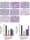
Figure 4
Detection of pathological changes in mouse lung tissue and virus copy numbers. (A) These lungs were processed and embedded in paraffin and sectioned for histological staining. Lung sections were stained with HE and examined microscopically to observe pathological changes, with a 100 μm scale bar. (B) The copies of PCV2 and PCV3 in lungs from each group were quantified using qPCR. ND indicates no detection. && p < 0.01 versus PBS immunized group at the same time point (n = 5); ns: no significant difference.

Similar articles

References

    1. Finsterbusch T., Mankertz A. Porcine circoviruses—Small but powerful. Virus Res. 2009;143:177–183. doi: 10.1016/j.virusres.2009.02.009. - DOI - PubMed
    1. Ma Z., Liu M., Liu Z., Meng F., Wang H., Cao L., Li Y., Jiao Q., Han Z., Liu S. Epidemiological investigation of porcine circovirus type 2 and its coinfection rate in Shandong province in China from 2015 to 2018. BMC Vet. Res. 2021;17:17. doi: 10.1186/s12917-020-02718-4. - DOI - PMC - PubMed
    1. Ouyang T., Zhang X., Liu X., Ren L. Co-Infection of Swine with Porcine Circovirus Type 2 and Other Swine Viruses. Viruses. 2019;11:185. doi: 10.3390/v11020185. - DOI - PMC - PubMed
    1. Chen N., Huang Y., Ye M., Li S., Xiao Y., Cui B., Zhu J. Co-infection status of classical swine fever virus (CSFV), porcine reproductive and respiratory syndrome virus (PRRSV) and porcine circoviruses (PCV2 and PCV3) in eight regions of China from 2016 to 2018. Infect. Genet. Evol. 2019;68:127–135. doi: 10.1016/j.meegid.2018.12.011. - DOI - PubMed
    1. Tan C.Y., Lin C.N., Ooi P.T. What do we know about porcine circovirus 3 (PCV3) diagnosis so far?: A review. Transbound. Emerg. Dis. 2021;68:2915–2935. doi: 10.1111/tbed.14185. - DOI - PubMed

Publication types

MeSH terms

LinkOut - more resources