

G protein regulation by RGS proteins in the pathophysiology of dilated cardiomyopathy
- PMID: 39772618
- PMCID: PMC12103878
- DOI: 10.1152/ajpheart.00653.2024
G protein regulation by RGS proteins in the pathophysiology of dilated cardiomyopathy
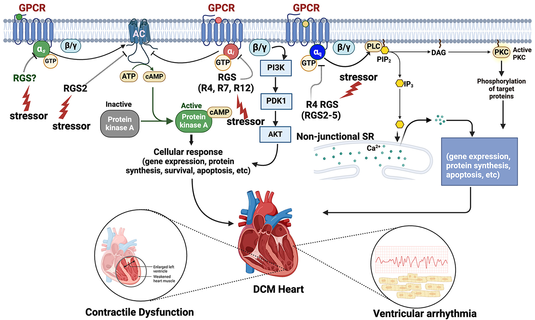
Abstract
Regulators of G protein signaling (RGS) proteins fine-tune signaling via heterotrimeric G proteins to maintain physiologic homeostasis in various organ systems of the human body including the brain, kidney, heart, and vasculature. Impaired regulation of G protein signaling by RGS proteins is implicated in the pathogenesis of several human diseases including various forms of cardiomyopathy such as hypertrophic cardiomyopathy and dilated cardiomyopathy (DCM). Both genetic and nongenetic changes that impinge on G protein signaling in cardiomyocytes are implicated in the etiology of DCM, and there is accumulating evidence that such genetic and nongenetic changes affecting G protein signaling in cell types other than cardiomyocytes could serve as a DCM trigger in humans. This review discusses and highlights mammalian RGS proteins and their roles in cardiac physiology and disease, with a specific focus on the current understanding of the etiology of DCM and the pathogenic roles of RGS proteins that are prominently expressed in the cardiovascular system. Growing evidence suggests that defects in G protein regulation by RGS proteins in the cardiovascular system likely contribute to cardiomyocyte structural damage and decreased contractile function that hallmark DCM. Further studies that enhance the understanding of the dynamics of G protein regulation by RGS proteins in several cell types in the myocardium and the vasculature are critical to gaining more insight into the etiology of DCM and heart failure, and to the identification of novel therapeutic targets.
Keywords: G protein signaling; RGS proteins; dilated cardiomyopathy; pathological mechanisms; vascular dysfunction.
Conflict of interest statement
DISCLOSURES
No conflicts of interest, financial or otherwise, are declared by the authors.
Figures

References
Publication types
MeSH terms
Substances
Grants and funding
- R01 HL174004/HL/NHLBI NIH HHS/United States
- R56DK132859-01A1/HHS | NIH | NIDDK | Division of Diabetes, Endocrinology, and Metabolic Diseases (DEM)
- GM143493/HHS | NIH | National Institute of General Medical Sciences (NIGMS)
- R01 GM143493/GM/NIGMS NIH HHS/United States
- R01 HL139754/HL/NHLBI NIH HHS/United States
LinkOut - more resources
Full Text Sources