. 2025 Feb 25;99(2):e0130724.
doi: 10.1128/jvi.01307-24. Epub 2025 Jan 7.

Structural insights into hybridoma-derived neutralizing monoclonal antibodies against Omicron BA.5 and XBB.1.16 variants of SARS-CoV-2

Affiliations

Structural insights into hybridoma-derived neutralizing monoclonal antibodies against Omicron BA.5 and XBB.1.16 variants of SARS-CoV-2

Hengrui Hu et al. J Virol. .

Abstract

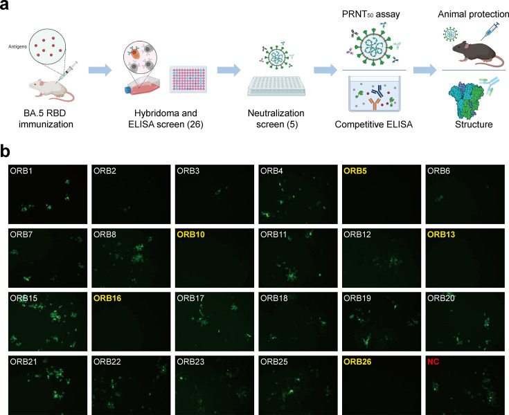
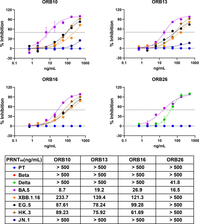
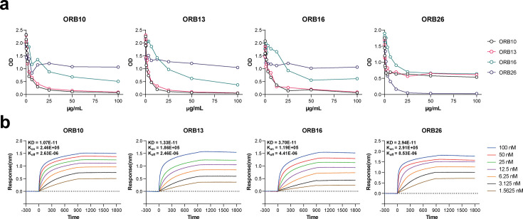
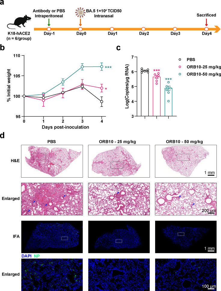
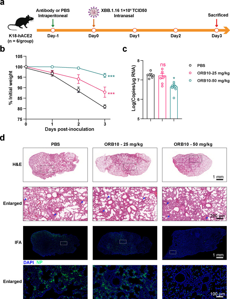
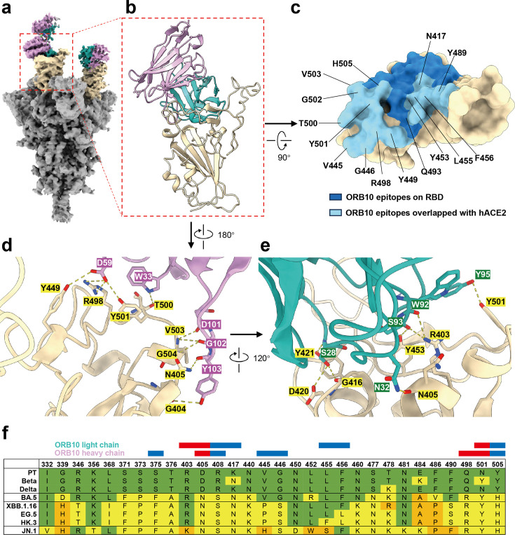
The emergence of novel variants of severe acute respiratory syndrome coronavirus-2 (SARS-CoV-2) continues to pose an ongoing challenge for global public health services, highlighting the urgent need for effective therapeutic interventions. Neutralizing monoclonal antibodies (mAbs) are a major therapeutic strategy for the treatment of COVID-19 and other viral diseases. In this study, we employed hybridoma technology to generate mAbs that target the BA.5 receptor-binding domain (RBD) of the SARS-CoV-2 spike protein. Through a comprehensive screening process, we identified four mAbs capable of effectively neutralizing BA.5, XBB.1.16, and related variant infections in vitro, among which ORB10 was found to neutralize BA.5 variants with a plaque reduction neutralization test (PRNT50) of 8.7 ng/mL. Additionally, competitive binding assays, sequencing of heavy and light chain variable regions, and binding kinetics characterization provided insights into the epitopes and binding affinities of the identified mAbs. Moreover, in vivo experiments in the K18-hACE2 mouse model demonstrated the protective efficacy of ORB10 against both BA.5 and XBB.1.16 variants. Finally, cryo-electron microscopy structural analysis of the ORB10-RBD complex identified key residues involved in the antibody-antigen interactions, providing insights into the molecular mechanisms of neutralization and immune escape of SARS-CoV-2 Omicron variants from mAbs.

Importance: The ongoing evolution of SARS-CoV-2 has led to the emergence of variants capable of evading immune responses elicited by natural infection and vaccination, especially the highly transmissible and immune-evasive Omicron variants. This study generated and characterized a panel of monoclonal antibodies (mAbs) specifically targeting the RBD of the Omicron BA.5 variant, of which the ORB10 showed efficacy against Omicron BA.5 and XBB.1.16 variants both in vitro and in vivo. Cryo-EM structural analysis further elucidated the binding epitope interactions and neutralization mechanism between ORB10 and the BA.5 RBD protein. This study enhances our understanding of antibody-mediated neutralization of SARS-CoV-2 and provides valuable insights into the development of effective therapeutic strategies to combat ongoing SARS-CoV-2 variant infections.

Keywords: K18-hACE2 mouse model; Omicron variants; SARS-CoV-2; cryo-EM structure; neutralizing antibody.


Conflict of interest statement

The authors declare no conflict of interest.

Figures

Fig 1
Fig 1
Flow charts of the study and primary screening of mAbs. (a) Flowchart illustrating the process of screening and characterizing mAbs. ELISA was used to screen cloned hybridoma cells, identifying a total of 35 mAbs. Five mAbs with high neutralization activity were further identified using microneutralization assays. The selected mAbs were subsequently subjected to a plaque reduction neutralization test (PRNT) assay, analysis of the protective effects in mice, and cryo-EM structure analysis. (b) IFA results of the primary neutralization activity screening. The mAbs that completely neutralize the BA.5 variant at a concentration of 500 ng/mL are denoted in yellow in the upper left corner; “NC” represents the negative control of the vehicle. Mouse mAbs against NP were used as the primary antibody in the IFA.
Fig 2
Fig 2
PRNT assay of ORB10, ORB13, ORB16, and ORB26 mAbs against different SARS-CoV-2 strains. Neutralizing activities of the four mAbs were detected using the PRNT assay. These mAbs were serially diluted and evaluated for neutralizing activity by counting plaque numbers. PRNT50 values are listed in the table below. Data are presented as the mean ± SEM of two independent experiments. PT represents the SARS-CoV-2 prototype strain.
Fig 3
Fig 3
Competitive ELISA and binding kinetics of ORB10, ORB13, ORB16, and ORB26. (a) Competition between the four neutralizing mAbs. These mAbs were labeled with biotin and serially diluted, then label-free mAbs were added to compete with the labeled mAbs. (b) Binding kinetics of the four neutralizing mAbs to BA.5 RBD proteins were evaluated using BLI assays; additionally, Kon and Koff values of the mAbs were measured for each mAb.
Fig 4
Fig 4
Analysis of the protective effects of ORB10 against the BA.5 variant in vivo. (a) Flow chart showing the analysis of the protective effects of ORB10 against BA.5 in mice. K18-hACE2 mice were intranasally infected with BA.5 at 1 × 10⁴ TCID50. Body weights of mice were monitored daily; mice were euthanized at 4 d.p.i. (b) Changes in the body weights of mice after infection with BA.5. Body weight is normalized to day 0 and presented as the mean ± SEM. (c) Viral copies in the lungs of mice. The same lung tissue was collected and tested for viral copy number using qRT-PCR; data are presented as the mean ± SEM. (d) Pathological analysis of the lungs of mice infected with BA.5. H&E staining and IFA were used to evaluate the pathological changes in murine lungs. Rabbit sera against SARS-CoV-2 NP protein was used as the primary antibody in the IFA. Scale bars are provided in the respective microscopic images. Dotted boxes represent the enlarged area. Blue arrows denote inflammatory cell infiltration.
Fig 5
Fig 5
Analysis of the protective effects of ORB10 against the XBB.1.16 variant in vivo. (a) Flow chart showing the analysis of the protective effects of ORB10 against XBB.1.16 in mice. K18-hACE2 mice were intranasally infected with XBB.1.16 at 1 × 10⁴ TCID50. Body weights of mice were monitored daily; mice were euthanized at 3 d p.i. (b) Changes in the body weights of mice after infection with XBB.1.16. Body weight is normalized to day 0 and presented as the mean ± SEM. (c) Viral copies in the lungs of mice. The same lung tissue was collected and tested for viral copy number using qRT-PCR; data are presented as the mean ± SEM. (d) Pathological analysis of the lungs of mice infected with XBB.1.16. H&E staining and IFA were used to evaluate the pathological changes in murine lungs. Rabbit sera against NP was used as the primary antibody in the IFA. Scale bars are provided in the respective microscopic images. Dotted boxes denote the enlarged area. Blue arrows represent inflammatory cell infiltration.
Fig 6
Fig 6
Binding stoichiometry between BA.5 S trimers and ORB10 Fab. (a) Side view of the BA.5 S trimer complexed with two ORB10 Fabs. The variable heavy and light chains of ORB10 Fab are colored purple and green, respectively; the two “up” RBDs of the BA.5 S trimer are colored in cream. (b) Magnified view of the RBD in complex with two ORB10 Fabs (PDB:8ZPP). (c) Surface illustration of the interface between the RBD and ORB10. Residues colored light and dark blue are those recognized by ORB10. Among these, residues overlapping the binding sites for ACE2 are labeled and colored light blue. (d and e) Magnified view of hydrogen bonding (yellow dashed lines) between the RBD of the BA.5 S trimer and the variable heavy chain (d) or light chain (e) of ORB10 Fab. (f) Sequence alignment of RBD mutated sites of different SARS-CoV-2 variants. The red and blue boxes above the sequence alignment represent the RBD residues involved in hydrogen bonds and interaction surface between ORB10 and BA.5 RBD, respectively.

Similar articles

References

    1. Supasa P, Zhou D, Dejnirattisai W, Liu C, Mentzer AJ, Ginn HM, Zhao Y, Duyvesteyn HME, Nutalai R, Tuekprakhon A, et al. . 2021. Reduced neutralization of SARS-CoV-2 B.1.1.7 variant by convalescent and vaccine sera. Cell 184:2201–2211. doi:10.1016/j.cell.2021.02.033 - DOI - PMC - PubMed
    1. Zhou D, Dejnirattisai W, Supasa P, Liu C, Mentzer AJ, Ginn HM, Zhao Y, Duyvesteyn HME, Tuekprakhon A, Nutalai R, et al. . 2021. Evidence of escape of SARS-CoV-2 variant B.1.351 from natural and vaccine-induced sera. Cell 184:2348–2361. doi:10.1016/j.cell.2021.02.037 - DOI - PMC - PubMed
    1. Dejnirattisai W, Zhou D, Supasa P, Liu C, Mentzer AJ, Ginn HM, Zhao Y, Duyvesteyn HME, Tuekprakhon A, Nutalai R, et al. . 2021. Antibody evasion by the P.1 strain of SARS-CoV-2. Cell 184:2939–2954. doi:10.1016/j.cell.2021.03.055 - DOI - PMC - PubMed
    1. Liu C, Ginn HM, Dejnirattisai W, Supasa P, Wang B, Tuekprakhon A, Nutalai R, Zhou D, Mentzer AJ, Zhao Y, et al. . 2021. Reduced neutralization of SARS-CoV-2 B.1.617 by vaccine and convalescent serum. Cell 184:4220–4236. doi:10.1016/j.cell.2021.06.020 - DOI - PMC - PubMed
    1. Roemer C, Sheward DJ, Hisner R, Gueli F, Sakaguchi H, Frohberg N, Schoenmakers J, Sato K, O’Toole Á, Rambaut A, Pybus OG, Ruis C, Murrell B, Peacock TP. 2023. SARS-CoV-2 evolution in the Omicron era. Nat Microbiol 8:1952–1959. doi:10.1038/s41564-023-01504-w - DOI - PubMed

Substances

Supplementary concepts

LinkOut - more resources