. 2025 Jan 10;11(2):eadq8336.
doi: 10.1126/sciadv.adq8336. Epub 2025 Jan 8.

Plasticity of human resilience mechanisms

Affiliations

Plasticity of human resilience mechanisms

Giovanni Leone et al. Sci Adv. .

Abstract

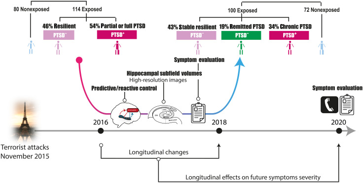
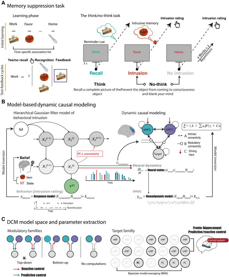
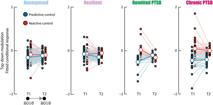
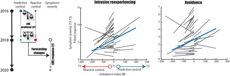
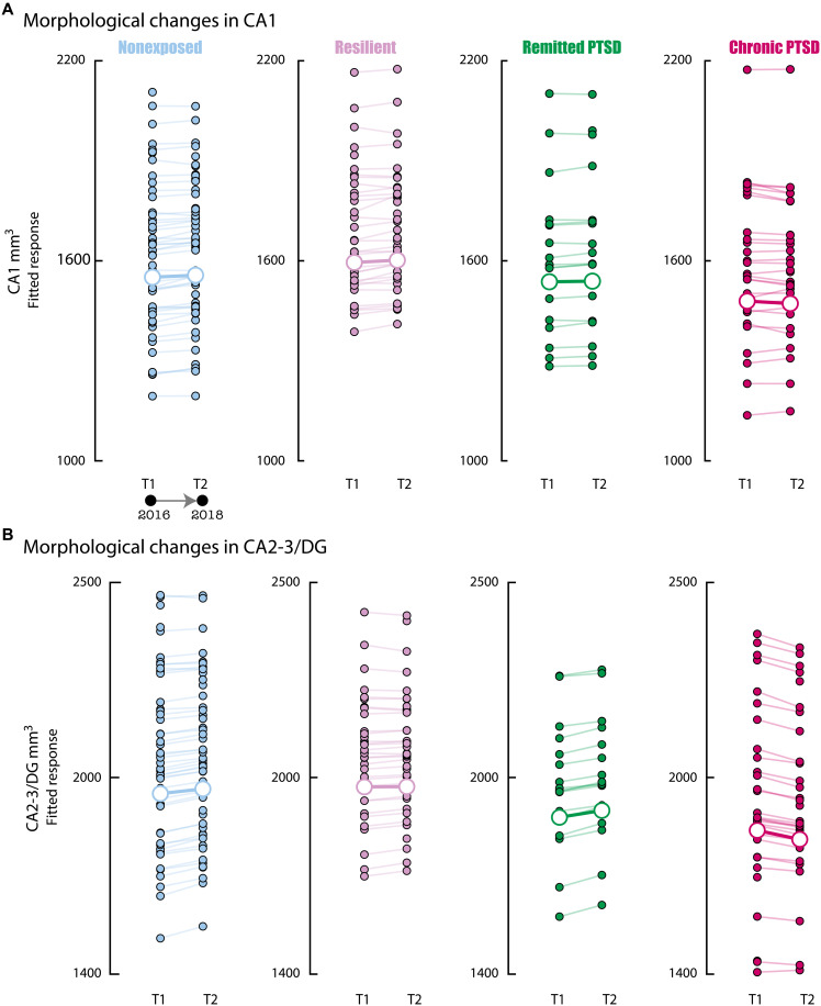
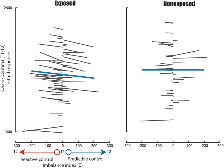
The hippocampus's vulnerability to trauma-induced stress can lead to pathophysiological disturbances that precipitate the development of posttraumatic stress disorder (PTSD). The mechanisms of resilience that foster remission and mitigate the adverse effects of stress remain unknown. We analyzed the evolution of hippocampal morphology between 2016/2017 and 2018/2019, as well as the memory control mechanisms crucial for trauma resilience. Participants were individuals exposed to the 2015 Paris terrorist attacks (N = 100), including chronic (N = 34) and remitted (N = 19) PTSD, and nonexposed (N = 72). We found that normalization of inhibitory control processes, which regulate the resurgence of intrusive memories in the hippocampus, not only predicted PTSD remission but also preceded a reduction in traumatic memories. Improvement in control mechanisms was associated with the interruption of stress-induced atrophy in a hippocampal region that includes the dentate gyrus. Human resilience to trauma is characterized by the plasticity of memory control circuits, which interacts with hippocampal neuroplasticity.


Figures

Fig. 1.
Fig. 1.. Longitudinal design.
Indexes of memory control and hippocampal integrity were collected at two time points after the 2015 Paris terrorist attack: 8 to 18 months (time 1) and 30 to 42 months (time 2). Participants with a similar degree of exposure were diagnosed in both time points as non-PTSD (PTSD), chronic PTSD (PTSD+), or remitted PTSD (i.e., recovered from an initial PTSD at time 1). The intensity of PTSD symptoms was also measured in a third time point collected 5 years after the attacks. We analyzed longitudinal changes between time 1 and time 2 in the markers of interest: (i) the top-down regulation of hippocampal activity during intrusion control originating from the right DLPFC and (ii) the volumes of hippocampal subfields, including the CA1 and a mixture region composed of the close by DG, CA2, and CA3 subfields. We also investigated how those changes preceded the future evolution of symptoms using a third time point.
Fig. 2.
Fig. 2.. Memory suppression task and model-based dynamic causal modeling (DCM).
(A) Participants learned pairs of words and pictures of an object in a series of test-feedback cycles until they reached 90% of correct response. A new study list was proposed at each time point. Lists were counterbalanced between time points and conditions. Then, participants performed the TNT task in the MRI. For think items, participants recalled a detailed image of the associated object. For No-think items, participants had to prevent the picture from entering awareness and to remove it from consciousness if it came to mind. After each trial, participants reported the extent to which the associated image entered their awareness (intrusion). (B) Binary intrusion ratings were fed into a HGF (116) including two levels in which the dynamic updating of beliefs is weighted by uncertainty. The parameter ω regulates the speed of beliefs adjustment. Beliefs are assumed to arise from the precision-weighted combination of the memories about previous trials and specific word-object pairs. Beliefs and positive PEs were then used as parametric modulators of the top-down coupling between control and memory systems (B matrix). (C) DCM space expressing different hypotheses about computational influence on the modulation of the coupling between control regions [anterior and posterior middle frontal gyrus (aMFG and pMFG, respectively)] and memory target regions, including the rostral hippocampus (rHIP), caudal hippocampus (cHIP), and precuneus (PCu). Null models were also estimated but are not shown here. The computational model and the winning DCM family describing a top-down influence of computational indexes were identified at time 1 in (44). Here, we implemented Bayesian model averaging (BMA) in the family of models that won at time 1 and focused on fronto-hippocampal coupling previously associated with a control effect in PTSD.
Fig. 3.
Fig. 3.. Remission from PTSD is associated with the plastic restoration of the balance between predictive and reactive memory control.
Small blue and red circles represent the modulation of the top-down coupling from the MFG to the hippocampus during predictive control and reactive control, respectively, at time 1 and time 2, for each individual (as predicted by the LME, i.e., fitted conditional response). Larger dots and bold lines represent the fixed effects of control and time at the group level. Remitted PTSD was characterized by a gain balance between predictive control and reactive control at time 2, while no significant evolution was observed in the other groups.
Fig. 4.
Fig. 4.. Control plasticity forecasts traumatic memory evolution.
The left panel shows the LME predictors and response for the forecasting analysis. On middle and right panels, the x axes show the evolution of the control index reflecting the balance between predictive control and reactive control at time 2 relative to time 1, expressed in degrees (see Materials and Methods). Across time points, the balance index can either evolve toward a gain in reactive control (left part of the plots, negative shift from T1) or toward a gain in predictive control (right part of the plots, positive shift from T1). We display the difference in imbalance between T1 and T2 for visualization purposes. On the y axes, symptoms’ severity, predicted by the LME model (i.e., conditional response). Gray lines represent individual data points for each chronic PTSD participant, while the blue line represents the fixed effect at the population level. Improvement of reactive control forecasted the future reductions in intrusive reexperiencing (left) or avoidance (right) symptoms severity, while increase in predictive control had the opposite effect.
Fig. 5.
Fig. 5.. Chronic PTSD is associated with atrophy of CA2-3/DG.
Evolution of hippocampal subfields’ volumes of CA1 (A) and CA2-3/DG (B) with time in nonexposed, resilient, remitted, and chronic PTSD groups (the y axes reflect the fitted conditional response of the LME model). Small dots and pale lines indicate individual data points. Larger dots and bold lines reflect the fixed effect of time at the group level. No morphological changes were observed in CA1 in any of the four groups. Chronic PTSD was associated with the atrophic reduction of CA2-3/DG volumes at time 2 compared to those at time 1, while no significant variations were observed in the other three groups.
Fig. 6.
Fig. 6.. Relationship between control plasticity and morphological changes in the hippocampus.
On the x axes, the evolution of the control index, reflecting the balance between predictive control and reactive control at time 2 relative to that at time 1, expressed in degrees (see Materials and Methods). Across time points, the balance index can either evolve toward a gain in reactive control (left part of the plots) or toward a gain in predictive control (right part of the plots). On the y axes, CA2-3/DG volumes, predicted by the LME model (i.e., conditional response). Gray lines represent individual data points for each exposed (left) or nonexposed (right) participant, while the blue line represents the fixed effect at the population level. Improvement of reactive control in exposed individuals predicted parallel CA2-3/DG volumetric plastic changes, while no significant relationship was observed in nonexposed individuals.

Similar articles

Cited by

References

    1. Kalisch R., Baker D. G., Basten U., Boks M. P., Bonanno G. A., Brummelman E., Chmitorz A., Fernàndez G., Fiebach C. J., Galatzer-Levy I., Geuze E., Groppa S., Helmreich I., Hendler T., Hermans E. J., Jovanovic T., Kubiak T., Lieb K., Lutz B., Müller M. B., Murray R. J., Nievergelt C. M., Reif A., Roelofs K., Rutten B. P. F., Sander D., Schick A., Tüscher O., Diest I., van Harmelen A.-L., Veer I. M., Vermetten E., Vinkers C. H., Wager T. D., Walter H., Wessa M., Wibral M., Kleim B., The resilience framework as a strategy to combat stress-related disorders. Nat. Hum. Behav. 1, 784–790 (2017). - PubMed
    1. Pascual-Leone A., Bartres-Faz D., Human brain resilience: A call to action. Ann. Neurol. 90, 336–349 (2021). - PMC - PubMed
    1. Russo S. J., Murrough J. W., Han M.-H., Charney D. S., Nestler E. J., Neurobiology of resilience. Nat. Neurosci. 15, 1475–1484 (2012). - PMC - PubMed
    1. van Rooij S. J. H., Santos J. L., Hinojosa C. A., Ely T. D., Harnett N. G., Murty V. P., Lebois L. A. M., Jovanovic T., House S. L., Bruce S. E., Beaudoin F. L., An X., Neylan T. C., Clifford G. D., Linnstaedt S. D., Germine L. T., Bollen K. A., Rauch S. L., Haran J. P., Storrow A. B., Lewandowski C., Musey P. I., Hendry P. L., Sheikh S., Jones C. W., Punches B. E., Swor R. A., Pascual J. L., Seamon M. J., Harris E., Pearson C., Peak D. A., Merchant R. C., Domeier R. M., Rathlev N. K., O’Neil B. J., Sanchez L. D., Joormann J., Pizzagalli D. A., Sheridan J. F., Harte S. E., Kessler R. C., Koenen K. C., McLean S. A., Ressler K. J., Stevens J. S., Defining the r factor for post-trauma resilience and its neural predictors. Nat. Ment. Health 2, 680–693 (2024).
    1. Yehuda R., LeDoux J., Response variation following trauma: A translational neuroscience approach to understanding PTSD. Neuron 56, 19–32 (2007). - PubMed