. 2025 Jan 14;122(2):e2419712122.
doi: 10.1073/pnas.2419712122. Epub 2025 Jan 7.

The single-stranded DNA-binding factor SUB1/PC4 alleviates replication stress at telomeres and is a vulnerability of ALT cancer cells

Affiliations

The single-stranded DNA-binding factor SUB1/PC4 alleviates replication stress at telomeres and is a vulnerability of ALT cancer cells

Jean-Christophe Dubois et al. Proc Natl Acad Sci U S A. .

Abstract

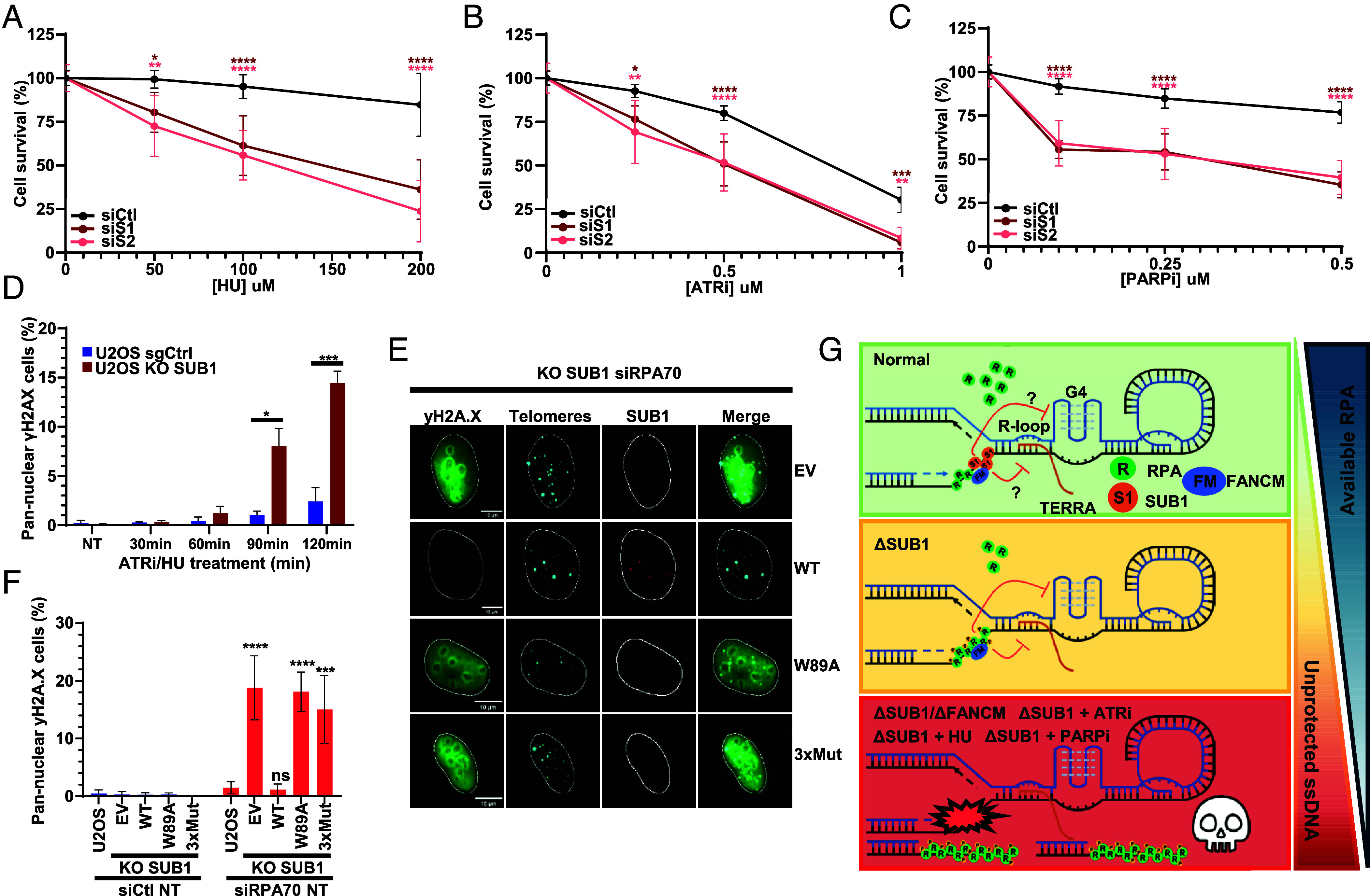
To achieve replicative immortality, cancer cells must activate telomere maintenance mechanisms. In 10 to 15% of cancers, this is enabled by recombination-based alternative lengthening of telomeres pathways (ALT). ALT cells display several hallmarks including heterogeneous telomere length, extrachromosomal telomeric repeats, and ALT-associated PML bodies. ALT cells also have high telomeric replication stress (RS) enhanced by fork-stalling structures (R-loops and G4s) and altered chromatin states. In ALT cells, telomeric RS promotes telomere elongation but above a certain threshold becomes detrimental to cell survival. Manipulating RS at telomeres has thus been proposed as a therapeutic strategy against ALT cancers. Through analysis of genome-wide CRISPR fitness screens, we identified ALT-specific vulnerabilities and describe here our characterization of the roles of SUB1, a ssDNA-binding protein, in telomere stability. SUB1 depletion increases RS at ALT telomeres, profoundly impairing ALT cell growth without impacting telomerase-positive cells. During RS, SUB1 is recruited to stalled forks and ALT telomeres via its ssDNA-binding domain. This recruitment is potentiated by RPA depletion, suggesting that these factors may compete for ssDNA. The viability of ALT cells and their resilience toward RS also requires ssDNA binding by SUB1. SUB1 depletion accelerates cell death induced by FANCM depletion, triggering unsustainable levels of telomeric damage in ALT cells. Finally, combining SUB1 depletion with RS-inducing drugs rapidly induces replication catastrophe in ALT cells. Altogether, our work identifies SUB1 as an ALT susceptibility with roles in the mitigation of RS at ALT telomeres and suggests advanced therapeutic strategies for a host of still poorly managed cancers.

Keywords: DNA damage; DNA replication; alternative lenghtening of telomeres; replication stress; telomeres.


Conflict of interest statement

Competing interests statement:The authors declare no competing interest.

Figures

Fig. 1.
Fig. 1.
Identification of ALT-specific Vulnerabilities. (A) Volcano plot of the differential genetic dependencies in experimentally validated ALT cell lines. Mean subtracted CERES scores obtained from the Cancer Dependency Map (21Q1) for a panel of validated ALT cell lines (n = 7) were compared with the rest of the available cell lines (n = 801). Differential dependencies are plotted against the false discovery rate (FDR)-corrected Q value, calculated from multiple unpaired t tests. The dashed horizontal line represents an FDR of 1%. Red dots represent components of the FANCM complex. (B) Box and whiskers plots of preferentially essential genes of ALT cells. Horizontal lines represent the median and whiskers show 1 to 99 percentile cell lines. Significance was determined by Mann–Whitney tests. ****P < 0.0001, ***P < 0.001, and **P < 0.01. (C and D) Codependency analysis of SUB1/PC4 (Depmap 21Q1) reveals association with the Fanconi Anemia DNA repair pathway and nucleic acid metabolism. (E) Interactome of top 100 SUB1/PC4 codependent genes created using STRINGdb [medium confidence setting (0.4)] and Cytoscape for network formatting.
Fig. 2.
Fig. 2.
SUB1 localizes to damaged telomeres. (A) Rod representation and sequence of the different domains of SUB1 and position of ssDNA-binding domain mutations. (BE) U2OS and HeLa LT cells transfected with indicated siRNAs were exposed to 10 mM ATRi (AZD6738) for 6 h and processed for immunofluorescence. Telomeric SUB1 foci were automatically counted using CellProfiler. (B) Representative U2OS cells are shown. White arrows show colocalization events. (C) Graphical representation of the number of telomeric SUB1 foci per nuclei. Each dot represents one nucleus and the red line is the mean number of foci/nucleus for each condition. The graph represents the merged data from two biological replicates. Statistical significance was established by Ordinary one-way ANOVA and Dunnett’s multiple comparison tests. (D and E) Histogram representing the percentage of cells with 0 to 3, 4 to 20, 21 to 40, or >40 telomeric SUB1 foci in U2OS or HeLa cells. The graph represents the merged data from two biological replicates. (F) U2OS cells synchronized by double thymidine block were exposed to ATRi for 3 h prior to TEL-FISH and DAPI staining. Quantitative image-based cytometry was performed to assess cell cycle distribution of cells containing ≥ 4 telomeric SUB1 foci. (G) Scatterplot representing the number of SUB1 telomeric foci in KO SUB1 cells (KO3) complemented with WT or mutant HA-SUB1. The SUB1 KO3 cell line is described in more detail in Fig. 3 and SI Appendix, Fig. S3. The graph represents the merged data from two biological replicates. Statistical significance was established by Kruskal–Wallis and Dunn’s multiple comparison tests (*P < 0.05, **P < 0.01, ***P < 0.001, and ****P < 0.0001).
Fig. 3.
Fig. 3.
SUB1 alleviates RS at ALT telomeres. (AC) A panel of ALT and TEL+ cell lines were transfected with the indicated siRNAs against SUB1 or FANCM and processed for RPA32pS33 and BLM IF along with telomeric FISH. (A) Representative IF-Tel-FISH in U2OS (ALT) and HeLa LT (TEL+). Quantification of RPA32pS33+ (B) or BLM+ (C) telomeres in ALT and TEL+ cell lines transfected with the indicated siRNAs was performed automatically using Cell Profiler. Graphs represent the merged data from three biological replicates. Statistical significance was established by one-way ANOVA and Holm–Šidák’s multiple comparison tests (DF) sgCtl and KO SUB1 U2OS cells were processed for RPA32pS33 and BLM IF along with telomeric FISH. (D) Representative IF-FISH results for sgCtl and KO SUB1 cells. Histograms representing the percentage of cells with 0, 1 to 3, 4 to 6, 7 to 9, or ≥10 (E) telomeric RPA32 pS33 foci and (F) telomeric BLM foci. (G and H) sgCtl and KO SUB1 cells were transfected with the indicated siRNAs and 48 h later processed for Tel-FISH and IF. Graphs represent the merged data from three biological replicates. Statistical significance was established by Kruskal–Wallis and Dunn’s multiple comparison tests (*P < 0.05, **P < 0.01, ***P < 0.001, and ****P < 0.0001).
Fig. 4.
Fig. 4.
SUB1 is a vulnerability of ALT cells, and its Codepletion with FANCM is Highly Toxic. (A) U2OS, CAL72, Saos-2, HeLa LT, or RPE1 cells were transfected with the indicated siRNAs. 500 cells per condition were seeded and colonies were counted after 10 to 14 d. The graph represents the merged data from two biological replicates each consisting of two technical replicates. (B) U2OS cells stably transduced with the indicated siRNA-resistant HA-SUB1 constructs were transfected with siRNAs and colony formation assays were performed 10 to 14 d posttransfection. The graph represents the merged data from two biological replicates each consisting of two technical replicates. (C) U2OS, CAL72, Saos-2, HeLa LT, or RPE1 were transfected with the indicated siRNAs and processed for immunofluorescence and telomere FISH. Telomeric RPA32pS33 foci were automatically counted using CellProfiler. The data presented for siCtl, siS1, and siFM1 are the same as in Fig. 3B as SUB1/FANCM codepletion experiments were performed simultaneously. Experiments were performed in biological duplicates and a representative replicate is shown here. Statistical significance was established by one-way ANOVA and Holm–Šidák’s multiple comparison tests (D) Representative images of telomeric foci in U2OS and HeLa cells. (E and F) G-rich ssDNA TRF analysis of ALT (U2OS) and telomerase+ (HeLa) siRNA-transfected cells. Genomic DNA was extracted 48 h after siRNA transfection, digested with a restriction enzyme mix, and underwent native in-gel hybridization with radiolabeled [CCCTAA]5 oligonucleotides targeting the G-rich overhang. A denaturing Southern blot with a radiolabeled telomeric probe (Telo800) was used for normalization. ssDNA and dsDNA controls of TRF enzyme-digested from a plasmid indicate that the probe hybridized only to ssDNA. Experiments were performed in biological replicates (G and H) CCA analysis of genomic DNA from the indicated sgCtl or SUB1 KO U2OS or HeLa LT cell lines transfected with control or FANCM-targeting siRNAs. Reactions were performed with or without Phi29 polymerase (Φ29±). CCA products were dot-blotted and hybridized with a radiolabeled [CCCTAA]5 oligonucleotide probe. C-circle values were quantified relative to siCtl. Biological replicates were performed. Statistical significance was established by one-way ANOVA and Holm–Šidák’s multiple comparison tests (P-values: *P < 0.05, **P < 0.01, ***P < 0.001, and ****P < 0.0001).
Fig. 5.
Fig. 5.
The ss DNA–binding protein SUB1 associates with stalled and collapsed DNA replication forks. (AC) U2OS WT or HA-SUB1 cells were treated with 2 mM HU or HU combined with 1 uM ATRi for the indicated times. After fixation, IF against SUB1/HA and RPA32 was performed. RPA32- and SUB1-foci were automatically counted using CellProfiler. (A) Representative images of WT U2OS cells stained for endogenous SUB1 and RPA32 IF. Nuclei were outlined with the “Outliner” ImageJ plugin. (B) Histogram representing the fraction of HU- and HU/ATRi-treated cells containing SUB1 foci. Cells with >30 RPA and SUB1 foci were considered positive. (C) Graphical representation of the number of foci contained in each nucleus. Graphs represent the merged data from two biological replicates (D) SUB1 mutants were stably transduced into U2OS cells that were then treated with HU and ATRi. Representative immunofluorescence images are shown. (E) Graphs represent the merged data from two biological replicates (F) Schematic representation of in situ analysis of protein interactions at DNA replication forks (SIRF). (G) Cells were exposed for 30 min to EdU (100 μM) to label ongoing replication forks. Cells were then treated or not with HU or a combination of HU and ATRi to induce fork stalling and collapse respectively. Cells were processed for SIRF as outlined and EdU-SUB1 proximity foci were automatically acquired using CellProfiler. Statistical significance was established by one-way ANOVA and Šidák’s multiple comparison tests. Experiments were performed in biological triplicates and a representative replicate is shown here (***P < 0.001 and ****P < 0.0001).
Fig. 6.
Fig. 6.
SUB1 Protects Against RS and Replication Catastrophe in ALT cells. (AC) U2OS cells were transfected with the indicated siRNAs and incubated with HU, ATRi, or PARPi at the indicated concentrations for 4 d. Cells were then released into fresh media and allowed to form colonies over 10 d prior to fixation and viability assessment. Graphs represent cell viability compared to nontreated cells. Statistical significance was established by ordinary two-way ANOVA and Tukey’s multiple comparison tests. (A) Graphs represent the merged data from four biological replicates each consisting of two technical replicates. (B and C) Graphs represent the merged data from three biological replicates each consisting of two technical replicates. (D) U2OS sgCtl and KO SUB1 cells were exposed to ATRi and HU for the indicated times and processed for γ-H2A.X immunofluorescence. γ-H2A.X nuclear intensity was automatically counted using Cell Profiler. Cells with nuclear γ-H2A.X ≥250 were considered pan-nuclear. Experiments were performed in biological duplicates and a representative replicate is shown here. (E) Representative images and (F) quantification of pan-nuclear γ-H2A.X cells. WT and KO SUB1 cells complemented with the indicated HA-SUB1 constructs were transfected with the indicated siRNAs and 48 h late processed for γ-H2A.X and SUB1 IF along with telomere FISH. The SUB1 IF and telomere fish staining from these cells are also shown in SI Appendix, Fig. S2D to support the recruitment of WT SUB1 to telomeres in stressed cells. The graph represents the merged data from three biological replicates. Statistical significance was established by ordinary two-way ANOVA and Tukey’s multiple comparison tests. (G) A model for the roles of SUB1 in the resilience of ALT cells toward RS. In ALT cells, SUB1 binds ssDNA at stressed telomeres and ongoing replication forks and collaborates with RPA, FANCM, and other factors to alleviate RS at ALT telomeres and elsewhere in the genome. Due to its roles at the interface between transcription and replication, its affinity for G4-forming DNA, and its potential role in limiting TERRA levels, SUB1 might help mitigate R-loop and G4 formation at ALT telomeres. SUB1 depletion enhances RS at telomeres, making ALT cells more dependent on RPA and prone to undergo replication catastrophe. Codepleting SUB1 and FANCM or combining SUB1 depletion with RS-inducing drugs is highly toxic to ALT cells. (*P < 0.05, **P < 0.01, ***P < 0.001, and ****P < 0.0001).

Similar articles

References

    1. Maciejowski J., De Lange T., Telomeres in cancer: Tumour suppression and genome instability. Nat. Rev. Mol. Cell Biol. 18, 175–186 (2017). - PMC - PubMed
    1. Gilson E., Géli V., How telomeres are replicated. Nat. Rev. Mol. Cell Biol. 8, 825–38 (2007). - PubMed
    1. Lalonde M., Chartrand P., TERRA, a multifaceted regulator of telomerase activity at telomeres. J. Mol. Biol. 432, 4232–4243 (2020). - PubMed
    1. Dilley R. L., Greenberg R. A., Alternative telomere maintenance and cancer. Trends Cancer 1, 145–156 (2015). - PMC - PubMed
    1. Sfeir A., et al. , Mammalian telomeres resemble fragile sites and require TRF1 for efficient replication. Cell 138, 90–103 (2009). - PMC - PubMed

LinkOut - more resources