. 2025 Jan 14;122(2):e2419196122.
doi: 10.1073/pnas.2419196122. Epub 2025 Jan 7.

The cGAS-STING, p38 MAPK, and p53 pathways link genome instability to accelerated cellular senescence in ATM-deficient murine lung fibroblasts

Affiliations

The cGAS-STING, p38 MAPK, and p53 pathways link genome instability to accelerated cellular senescence in ATM-deficient murine lung fibroblasts

Majd Haj et al. Proc Natl Acad Sci U S A. .

Abstract

Ataxia-telangiectasia (A-T) is a pleiotropic genome instability syndrome resulting from the loss of the homeostatic protein kinase ATM. The complex phenotype of A-T includes progressive cerebellar degeneration, immunodeficiency, gonadal atrophy, interstitial lung disease, cancer predisposition, endocrine abnormalities, chromosomal instability, radiosensitivity, and segmental premature aging. Cultured skin fibroblasts from A-T patients exhibit premature senescence, highlighting the association between genome instability, cellular senescence, and aging. We found that lung fibroblasts derived from ATM-deficient mice provide a versatile experimental system to explore the mechanisms driving the premature senescence of primary fibroblasts lacking ATM. Atm-/- fibroblasts failed to proliferate under ambient oxygen conditions (21%). Although they initially proliferated under physiological oxygen levels (3%), they rapidly entered senescence. In contrast, wild-type (WT) lung fibroblasts did not senesce under 3% oxygen and eventually underwent immortalization and neoplastic transformation. However, rapid senescence could be induced in WT cells either by Atm gene ablation or persistent chemical inhibition of ATM kinase activity, with senescence induced by ATM inhibition being reversible upon inhibitor removal. Moreover, the concomitant loss of ATM and p53 led to senescence evasion, vigorous growth, rampant genome instability, and subsequent immortalization and transformation. Our findings reveal that the rapid senescence of Atm-/- lung fibroblasts is driven by the collaborative action of the cGAS-STING, p38 MAPK, and p53 pathways in response to persistent DNA damage, ultimately leading to the induction of interferon-α1 and downstream interferon-stimulated genes. We propose that accelerated cellular senescence may exacerbate specific A-T symptoms, particularly contributing to the progressive, life-threatening interstitial lung disease often observed in A-T patients during adulthood.

Keywords: ATM; ataxia–telangiectasia; cGAS-STING; p53; senescence.


Conflict of interest statement

Competing interests statement:The authors declare no competing interest.

Figures
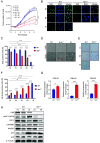
Fig. 1.
Fig. 1.
Premature senescence in murine Atm−/− lung fibroblasts at 3% O2 concentration. (A) Growth curves of WT and Atm−/− cells across passage levels P2 to P8. (B) Representative EdU staining images of WT and Atm−/− cells at P2 and P8 (EdU: green; nuclei: blue). (C) Quantification of EdU-positive cells for WT and Atm−/− cells at various passage levels (P2 to P8). IR denotes WT cells exposed to 10 Gy of ionizing radiation and analyzed 10 d postexposure. (D) Bright-field microscopy images of cells at P8, with red arrows indicating cells exhibiting senescent morphology. (E) SA-β-Gal activity staining in cells at P2 and P8. (F) Quantitative analysis of SA-β-Gal-positive cells at different passage levels. (G) RT-qPCR analysis of cell cycle arrest mediators’ genes at P8. (H) Western blotting analysis of senescence-associated markers in WT and Atm−/− cells at P2 and P8, along with irradiated WT controls. Data in panels C, F, and G are represented as mean + SEM from at least three independent experiments. Statistical significance determined using a two-tailed t test is denoted as follows: *P < 0.05, ***P < 0.001, and ns = not significant.
Fig. 2.
Fig. 2.
p53 loss leads to early senescence escape in ATM-deficient murine lung fibroblasts. (A) Growth curves of cells with different Atm/Trp53 genotypes at different passage levels. (B) Quantification of EdU in cells with different Atm/Trp53 genotypes at different passage levels (P2 to P15). (C) Quantification of SA-β-Gal in cells with the different Trp53/Atm genotypes at different passage levels (P2 to P15). (D) WB analysis of cell cycle arrest and senescence markers in cells with different Atm/Trp53 genotypes at P2, P8, and P15. (E) PCR analysis of Trp53 (Upper) and Atm (Lower) alleles in cells with the different Atm/Trp53 genotypes at different passage levels. Note that by P15, the cells with the initial genotype, Atm−/−;Trp53+/−, were already dKO. Mean and SEM of at least three independent experiments are shown. P-values were calculated using the two-tailed t test (*P < 0.05, **P < 0.01, ***P < 0.001, ns = not significant).
Fig. 3.
Fig. 3.
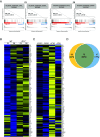
Atm−/− lung fibroblasts exhibit high, sustained expression of ISGs. (A) GSEA plots for hallmark interferon gene sets when global gene expression was compared between Atm−/− and WT cells at P2 (Upper) and P8 (Lower). (B) A representative heat map of IFN response genes in Atm−/− and WT cells at P2 and P8. Blue represents downregulation, and yellow–upregulation. (C) Representative heat map of IFN-response genes in WT cells following treatment with ATMi (+ATMi) or DMSO (+DMSO) for 15 d, and 20 d after ATMi (−ATMi) or DMSO removal (−DMSO). Blue represents downregulation and yellow–upregulation. (D) Venn diagram of up-regulated ISGs in both datasets (yellow; up-regulated genes in Atm−/− cells, blue; up-regulated genes in WT cells during ATMi treatment). The overlap represents ISGs that were up-regulated in both setups. The upregulation in Atm−/− cells at P8 is compared to WT cells at P8, and in WT cells the comparison is between cells treated with ATMi and those treated with DMSO.
Fig. 4.
Fig. 4.
The p38 MAPK is a major regulator of the premature senescence of Atm−/− lung fibroblasts. (A) CPD curves of WT and Atm−/− cells cultured in the presence with cGASi, STINGi, and p38i. (B) Quantification of the percentage of SA-β-Gal-positive cells from figure (A) at P.8 (day 32). (C) Quantification of average γH2AX nuclear foci per cell from (A) at P8. (D) Quantification of MN in cells from experiment (A) at P8. A cell was considered micronuclei-positive if it contained at least one micronucleus. (E) WB analysis of various IFN-related proteins, senescence markers, and cell cycle arrest mediators in cellular extracts from cells in experiment (A), at P8. Mean and SEM of at least three independent experiments are shown. P-values were calculated using the unpaired t test (*P < 0.05, ***P < 0. 01, ***P < 0.001, ns = not significant).
Fig. 5.
Fig. 5.
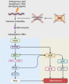
Model depicting cellular pathways involved in premature senescence of ATM-deficient lung fibroblasts. Persistent genome instability, coupled with ongoing DNA damage, leads to MN formation and the release of nuclear DNA into the cytoplasm. Central to this process is the signaling cascade that begins with the activation of cGAS and STING, ultimately leading to the activation of IFN genes and downstream ISGs. The model also considers potential additional nuances, such as STING activation by other triggers and cross talk with the p38 MAPK and p53 pathways. Collectively, this network halts cell cycle progression and activates various features of senescence, most notably the SASP. Figure created with BioRender.

Similar articles

Cited by

  • The cold immunological landscape of ATM-deficient cancers.
    Sinha S, Ng V, Novaj A, Zhu Y, Yazaki S, Pei X, Derakhshan F, Pareja F, Setton J, Naulin F, Beltrán-Visiedo M, Shin E, Longhini ALF, Gardner R, Ma J, Ma K, Roulston A, Morris S, Koehler M, Powell S, Rosen E, Galluzzi L, Reis-Filho J, Khan A, Riaz N. Sinha S, et al. J Immunother Cancer. 2025 May 11;13(5):e010548. doi: 10.1136/jitc-2024-010548. J Immunother Cancer. 2025. PMID: 40350205 Free PMC article.
  • ZC3H4 safeguards genome integrity by preventing transcription-replication conflicts at noncoding RNA loci.
    Frey Y, Goehring L, Haj M, Rona G, Fijen C, Pagano M, Huang TT, Rothenberg E, Ziv Y, Shiloh Y. Frey Y, et al. Sci Adv. 2025 Jun 20;11(25):eadt8346. doi: 10.1126/sciadv.adt8346. Epub 2025 Jun 18. Sci Adv. 2025. PMID: 40531993 Free PMC article.
  • Profile of Yosef Shiloh.
    Viegas J. Viegas J. Proc Natl Acad Sci U S A. 2025 Jan 21;122(3):e2426242122. doi: 10.1073/pnas.2426242122. Epub 2025 Jan 14. Proc Natl Acad Sci U S A. 2025. PMID: 39808656 Free PMC article. No abstract available.
  • The cGAS-STING pathway in atherosclerosis.
    Wang SY, Chen YS, Jin BY, Bilal A. Wang SY, et al. Front Cardiovasc Med. 2025 Apr 25;12:1550930. doi: 10.3389/fcvm.2025.1550930. eCollection 2025. Front Cardiovasc Med. 2025. PMID: 40351606 Free PMC article. Review.
  • The STING Signaling: A Novel Target for Central Nervous System Diseases.
    Song M, Ren J, Zhu Z, Yi Z, Wang C, Liang L, Tian J, Mao G, Mao G, Chen M. Song M, et al. Cell Mol Neurobiol. 2025 Apr 7;45(1):33. doi: 10.1007/s10571-025-01550-4. Cell Mol Neurobiol. 2025. PMID: 40195137 Free PMC article. Review.

References

    1. Taylor A. M. R., et al. , Chromosome instability syndromes. Nat. Rev. Dis. Primers 5, 64 (2019). - PMC - PubMed
    1. Chatterjee N., Walker G. C., Mechanisms of DNA damage, repair, and mutagenesis. Environ. Mol. Mutagen 58, 235–263 (2017), 10.1002/em.22087. - DOI - PMC - PubMed
    1. Thada V., Greenberg R. A., Unpaved roads: How the DNA damage response navigates endogenous genotoxins. DNA Repair (Amst) 118, 103383 (2022). - PMC - PubMed
    1. de Almeida L. C., Calil F. A., Machado-Neto J. A., Costa-Lotufo L. V., DNA damaging agents and DNA repair: From carcinogenesis to cancer therapy. Cancer Genet. 252–253, 6–24 (2021). - PubMed
    1. Rothblum-Oviatt C., et al. , Ataxia telangiectasia: A review. Orphanet. J. Rare Dis. 11, 159 (2016). - PMC - PubMed

MeSH terms