. 2025 Apr;39(9):1064-1083.
doi: 10.1177/08853282241312040. Epub 2025 Jan 8.

Impact of composition and surfactant-templating on mesoporous bioactive glasses structural evolution, bioactivity, and drug delivery property

Affiliations

Impact of composition and surfactant-templating on mesoporous bioactive glasses structural evolution, bioactivity, and drug delivery property

Dana Almasri et al. J Biomater Appl. 2025 Apr.

Abstract

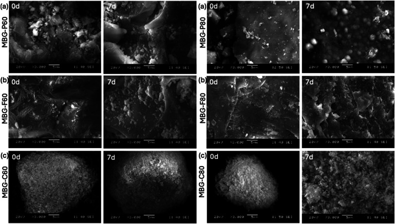
This study explores mesoporous bioactive glasses (MBGs) that show promise as advanced therapeutic delivery platforms owing to their tailorable porous properties enabling enhanced drug loading capacity and biomimetic chemistry for localized, sustained release. This work systematically investigates the complex relationship between MBG composition and surfactant templating on structural evolution, in vitro bioactive response, resultant drug loading efficiency and release. A total of 12 samples of sol-gel-derived MBG were synthesized using cationic and non-ionic structure-directing agents (cetyltrimethylammonium bromide, Pluronic F127 and P123) while modulating the SiO2/CaO content, generating MBG with surface areas of 60-695 m2/g. Electron microscopy and nitrogen desorption studies verified the successful synthesis of the 12 ordered MBG formulations. Assessment of hydroxyapatite conversion kinetics via FTIR spectroscopy and SEM demonstrated accelerated deposition for 70-80% SiO2 formulations, independent of the surfactant used. However, the templating agent had an impact on drug loading as observed in this study where MBG synthesized by the templating agent Pluronic P123 had higher drug loading compared to the other surfactants. To determine the drug release mechanisms, the in vitro kinetic profiles were fitted to various mathematical models including ze-ro. Most compositions exhibited release properties closest to zero-order, indicating a concentration-independent drug elution rate. These results in this study explain the relationship between tailored hierarchical architecture and intrinsic ion release rates to enable advanced functionality.

Keywords: Mesoporous; and bioactivity; bioactive glass; chemical synthesis; drug loading; drug release; kinetic modelling; sol-gel; surfactants.


Conflict of interest statement

Declaration of conflicting interestsThe author(s) declared no potential conflicts of interest with respect to the research, authorship, and/or publication of this article.

Figures

Figure 1.
Figure 1.
X-ray patterns of (a) MBG-F including all the different compositions as indicated in the figure, (b) MBG-C and all its different compositions, and (c) MBG-P also including all its compositions. The Figure shows that sol-gel synthesized bioactive glass samples have an amorphous structure indicated by the broad dispersive peaks. Each figure shows the highlighted region where the peaks start and end around 10° to 30° on average for MBG-F85, MBG-C85 and MBG-P85.
Figure 2.
Figure 2.
FTIR spectra of MBG-P, MBG-F and MBG-C before and after immersion in SBF for 7 days. The figure shows the development of HCA on the surface of MBG with time. MBG-P shows the most discernable peaks while MBG-C shows the lowest intensity peaks observed.
Figure 3.
Figure 3.
XRD of MBG-F60, MBG-P60, MBG-C60, MBG-F80, MBG-P80 and MBG-C80 before and after immersion in SBF for 7 days. The XRD shows peaks that are representative of the development of HCA on the surface which indicates bioactivity. The peaks that represent the planes of HCA are represented by an *.
Figure 4.
Figure 4.
SEM images of MBG that show the glass particle after immersion in SBF for 7 days (a) shows MBG-P60 at day 0 and day 7, (b) shows MBG-F60 at day 0 and day 7, (c) shows MBG-C60 at day 0 and day 7. While (d) shows MBG-P80 at day 0 and day 7, (e) shows MBG-F80 at day 0 and day 7, (f) shows MBG-C80 at day 0 and day 7.
Figure 5.
Figure 5.
Si, Ca, and P ion release after immersion of MBGs in SBF for different periods. (a) The release of silicon ions from the different compositions is compared to each other to show any clear trends that indicate biodegradation of the MBG. (b) Release of calcium ions, which is indicative of bioactivity and the formation of HA, and (c) release of phosphate, which, similar to calcium, shows the bioactivity of the synthesized bioglass with an error average < 5%.
Figure 6.
Figure 6.
Nitrogen adsorption-desorption isotherm showing the quantity of gas adsorbed (cm3/g) as a function of pressure (p/po). (a) is the N2 adsorption isotherm for MBG-P. (b) is the N2 adsorption isotherm for MBG-F. (c) is the N2 adsorption isotherm for MBG-C.
Figure 7.
Figure 7.
Drug loading (%) of vancomycin (orange) and ciprofloxacin (green) on MBG-P80, MBG-C80 and MBG-F80. Ciprofloxacin had a higher loading rate for all compositions.
Figure 8.
Figure 8.
Cumulative vancomycin release from (a) MBG-P85, -F85, and -C85, (b) MBG-P80, -F80, and -C80, (c) MBG-P70, -F70, and -C70, and (d) MBG-P60, -F60, and -C60 with error average of 6%.
Figure 9.
Figure 9.
Cumulative ciprofloxacin release from MBG-P80 (black), MBG-C80 (red) and MBG-F80 (blue) with error average of 5%.
Figure 10.
Figure 10.
Fitting of vancomycin release data from MBG-P85 to various mathematical models. The coefficient of determination (R2) was determined for each model to determine the goodness of fit. Vancomycin release from all MBG compositions was fitted to (a) zero order, (b) first order, (c) Higuchi, and (d) Korsmeyer-Peppas models similar to this example fitting.
Figure 11.
Figure 11.
Images of antibacterial testing for vancomycin-loaded MBG against gram-positive E. coli and gram-negative S. aureus. The images show the zone of inhibition as observed after 24 hours of incubation at 37°C (all measurements ±0.2 cm).

Similar articles

Cited by

References

    1. Hench LL. Bioactive glasses and glass-ceramics. Mater Sci Forum 1998; 293: 37–64. DOI: 10.4028/www.scientific.net/msf.293.37. - DOI
    1. Wu C, Chang J, Xiao Y. Mesoporous bioactive glasses for drug delivery and bone tissue regeneration. Advanced Bioactive Inorganic Materials for Bone Regeneration and Drug Delivery 2013; 1: 1–24. DOI: 10.1201/b13926-2. - DOI
    1. Itälä A, Ylänen HO, Yrjans J, et al. Characterization of microrough bioactive glass surface: surface reactions and osteoblast responses in vitro. J Biomed Mater Res 2002; 62(3): 404–411. DOI: 10.1002/jbm.10273. - DOI - PubMed
    1. Itälä A, Koort J, Ylänen HO, et al. Biologic significance of surface microroughing in bone incorporation of porous bioactive glass implants. J Biomed Mater Res 2003; 67(2): 496–503. DOI: 10.1002/jbm.a.10501. - DOI - PubMed
    1. Aguilar-Rabiela AE, Leal-Egaña A, Nawaz Q, et al. Integration of mesoporous bioactive glass nanoparticles and curcumin into PHBV microspheres as biocompatible composite for drug delivery applications. Molecules 2021; 26(11): 3177. DOI: 10.3390/molecules26113177. - DOI - PMC - PubMed