. 2025 Jan 31;99(1):e0185924.
doi: 10.1128/jvi.01859-24. Epub 2025 Jan 8.

Platelet factor 4-derived C15 peptide broadly inhibits enteroviruses by disrupting viral attachment

Affiliations

Platelet factor 4-derived C15 peptide broadly inhibits enteroviruses by disrupting viral attachment

Shuai Lv et al. J Virol. .

Abstract

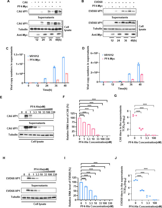
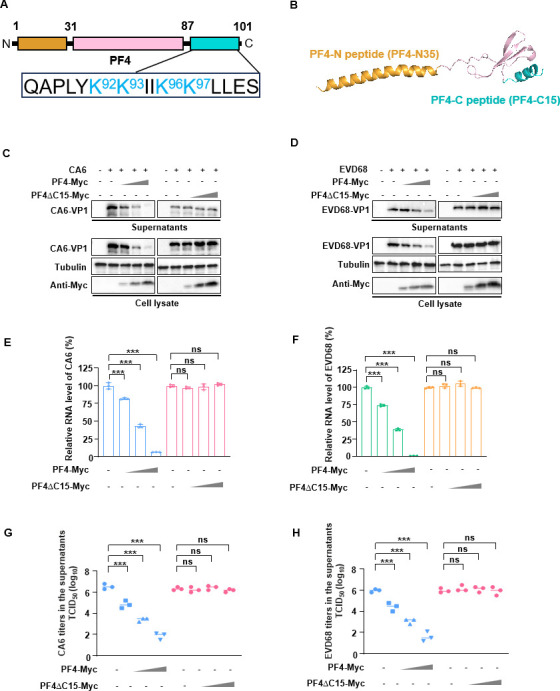
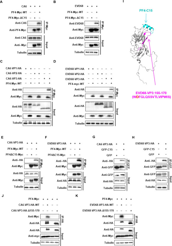
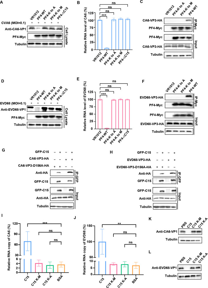
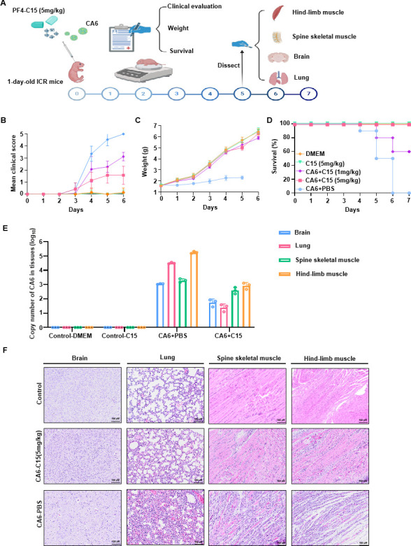
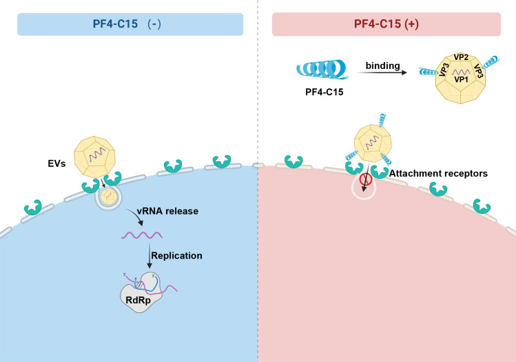
Platelet factor 4 (PF4) has been shown to regulate several viral infections. Our previous study demonstrated that PF4 inhibits the entry of enterovirus A 71 (EV71) and coxsackievirus A16 (CA16), which cause hand, foot, and mouth disease (HFMD). In this study, we report that PF4 also inhibits the circulating HFMD pathogen coxsackievirus A6 (CA6) and the re-emerging enterovirus D68 (EVD68). A 15-amino acid peptide, C15, at the C-terminus of PF4 confers anti-viral activity against multiple enteroviruses (EVs) besides CA6 and EVD68, including EV71 and CA16. Mechanistic studies revealed that wild-type C15 with a net-positive charge (+3), but not its mutants C15M and C15A (both -1), specifically binds to the VP3 capsid protein of CA6 and EVD68, thereby disrupting their attachment to the host cell surface. In addition, VP3 of EVs contains a conserved domain (residues 155-170) crucial for binding to C15. An aspartic acid residue at position 156 imparts a net-negative charge to this domain, which, when substituted with a neutrally charged amino acid, reduces the binding affinity of VP3 for C15. Additionally, C15 protects neonatal mice from lethal challenge upon a CA6 infection. These results suggest that C15 is a promising broad-spectrum anti-viral candidate against multiple EVs.

Importance: EVs, which pose a significant public health threat, can be classified into 15 species, with EV-A, -B, -C, and -D infecting humans and causing a wide range of diseases, from mild illnesses, such as HFMD, to more severe conditions, such as acute flaccid paralysis. The emergence of new and alternative strains highlights the urgent need for broad-spectrum anti-viral agents. In this study, we identified that the C15 of PF4 exhibits potent anti-viral activity against multiple EVs by binding to their surface and blocking their entry into host cells. Furthermore, C15 provides significant protection in vivo. These findings highlight the potential of C15 as a broad-spectrum anti-viral candidate. Our study opens a new avenue for developing treatments to combat the diverse and evolving threats posed by EVs.

Keywords: PF4; broad-spectrum; enterovirus; inhibitor; peptide C15.


Conflict of interest statement

The authors declare no conflict of interest.

Figures

Fig 1
Fig 1
PF4 inhibits the replication of CA6 and EVD68. (A and B) HEK-293T cells transfected with cDNA-PF4-Myc or the VR1012 expression vector serving as a control. After 12 h, the cells were infected with CA6 (MOI = 0.5) or EVD68 (MOI = 1). At the indicated time points, viral loads in both the cells and the supernatant (concentrated 100-fold) were assessed using Western blot analysis with an anti-VP1 antibody, while tubulin was used as a loading control. (C and D) Viral loads in the supernatant were evaluated by measuring VP1 gene mRNA levels using reverse transcription quantitative PCR (RT-qPCR). (E and H) PF4-His proteins expressed in Escherichia coli were added to the cell supernatant at the indicated concentration, and the cells were simultaneously infected with CA6 or EVD68. Cell lysates and supernatants (concentrated 100-fold) were collected at 48 h. Western blot was performed to detect the viral VP1 protein in the cells and supernatant, with tubulin serving as a loading control. (F and I) Intracellular mRNA levels of VP1 were determined by RT-qPCR, using GAPDH as a control. (G and J) 50% tissue culture infective dose (TCID50) assays were performed using supernatants collected from infected rhabdomyosarcoma cells at 48 h, containing various concentrations of PF4-His. All data are derived from three independent experiments and are presented as mean ± SD. Statistical significance was determined using one-way analysis of variance. ***P < 0.001.
Fig 2
Fig 2
PF4 lacking 15 amino acids at the C-terminus loses its anti-viral activity. (A) Schematic representation of the PF4 gene. (B) Prediction of the three-dimensional (3D) structure of the PF4 protein using AlphaFold2. (C and D) HEK-293T cells were transfected with varying concentrations of cDNA-PF4-Myc, with the VR1012 expression vector as a control. Twelve hours later, the cells were infected with CA6 or EVD68. Western blot analysis was performed to detect the expression levels of various proteins using the corresponding antibodies. (E and F) RT-qPCR was used to measure the mRNA levels of VP1 of CA6 or EVD68 in cells transfected with different concentrations of cDNA-PF4-Myc. GAPDH was used as a normalization control. (G and H) TCID50 assays were performed using supernatants collected from infected rhabdomyosarcoma cells at 48 h, containing various concentrations of PF4-Myc. All data are derived from three independent experiments and are presented as mean ± SD. Statistical significance was determined using one-way analysis of variance. **P < 0.001. ns, no significance.
Fig 3
Fig 3
C15 inhibits the replication of multiple EVs. (A) Cell viability of rhabdomyosarcoma (RD) cells after 48 h treatment with different concentrations of C15 was assessed using the Cell Counting Kit-8 assay (n = 3). (B, E, H, and K) RD cells were infected with indicated enterovirus (at various MOIs) and treated with different concentrations of C15 for 48 h. Western blot analysis was conducted to detect the VP1 protein levels in cell lysates and supernatants (concentrated 100-fold), with tubulin used as a loading control. (C, F, I, and L) Intracellular mRNA levels of the VP1 gene from different enterovirus strains were quantified by RT-qPCR, with GAPDH as a normalization control. All data are from three independent experiments and are presented as mean ± SD. The EC50 values were determined using GraphPad Prism version 9 with a variable slope (four parameters). (D, G, J, and M) TCID50 assays were performed using supernatants collected from infected RD cells at 48 h, containing various concentrations of C15. All data are from three independent experiments and are presented as mean ± SD. Statistical significance was assessed using one-way analysis of variance. ***P < 0.001. (N, O, and P) Intracellular mRNA levels of different enterovirus strains were quantified by RT-qPCR, with GAPDH as a normalization control. All data are from three independent experiments and are presented as mean ± SD. The EC50 values were determined using GraphPad Prism version 9 with a variable slope (four parameters).
Fig 4
Fig 4
C15 inhibits viral entry into cells by affecting virus binding to cells. (A and B) A time-of-addition assay with C15 was performed. RD cells were treated with C15 at different stages: pretreatment, incubation treatment, or post-entry of CA6 (MOI = 0.05) and EVD68 (MOI = 0.1). The inhibitory effect was assessed by measuring intracellular vRNA concentrations at 12 h post-infection (n = 3). (C and D) CA6 and EVD68 were incubated with C15 in an Eppendorf (EP) tube for 4 h at 4°C. The mixture was then concentrated 1:100 and concentrated using a 100 kDa protein concentrator tube, with three exchanges to remove excess C15. The final virus solution was re-suspended in cell culture medium and used to infect RD cells. After 48 h, cells were collected and analyzed by RT-qPCR. (E) RD cells in six-well plates were infected with CA6 (MOI = 0.5) and EVD68 viruses containing C15 (100 and 1,000 nM) at 4°C for 1 h. The cells were then washed three times with cold phosphate-buffered saline, lysed, and vRNA levels were measured by RT-qPCR. (F) EVD68 virus bound to the surface of HeLa cells was detected by immunofluorescence. Multicellular (flat image) and single cell (stereoscopic 3D image) are shown. Bars of flat image are 100 μm, and bars for 3D image are as follows: length, 58 µm; width, 58 µm; height, 12 µm. The histogram shows the fluorescence ratio. All data are from three independent experiments and are presented as mean ± SD. Statistical significance was assessed using one-way analysis of variance. ***P < 0.001. ns, no significance.
Fig 5
Fig 5
PF4 interacts with the VP3 proteins of CA6 and EVD68. (A and B) HEK293T cells were transfected with cDNA-PF4-Myc or PF4△C15-Myc. The cell lysates were incubated with protein G containing Myc antibody, then CA6 and EVD68 suspensions were added for 4 h, followed by co-IP analysis. VR1012 expression vector served as a control. (C and D) Co-IP analysis of the interaction between CA6/EVD68-VP1/2/3-HA and PF4-Myc in HEK293T cells transfected with the indicated plasmids. VR1012 expression vector was used as a control. (E and F) Co-IP analysis of the interaction between CA6/EVD68-VP3-HA and PF4-Myc or PF4ΔC15-Myc in HEK293T cells transfected with the indicated plasmids. VR1012 empty vector served as a control. (G and H)Co-IP analysis of the interaction between CA6/EVD68-VP3-HA and GFP-C15 in HEK293T cells transfected with the indicated plasmids. p-EGFP expression vector was used as a control. (I)Docking result showing the interaction between C15 and EVD68-VP3. Blue, C15; gray, EVD68-VP3; purple, EVD68-VP3 interaction region (155–170). (J and K) Co-IP analysis of the interaction between CA6/EVD68-VP3-HA (wild type or Δ155–170) and PF4-Myc in HEK293T cells transfected with the indicated plasmids. VR1012 expression vector was used as a control.
Fig 6
Fig 6
The interaction between C15 and the VP3 proteins of CA6 and EVD68 via a direct ionic interaction. (A and D) HEK-293T cells were transfected with cDNA-PF4-Myc or its mutants, with the VR1012 empty vector serving as a control. After 12 h, the cells were infected with CA6 (MOI = 0.5) and EVD68 (MOI = 1). Viral productions were assessed using Western blot analysis with an anti-VP1 antibody at 48 h, while tubulin was used as a loading control. (B and E) Viral mRNA levels in the cells were evaluated by quantifying VP1 mRNA levels using RT-qPCR. (C and F) Co-IP analysis of the interaction between CA6/EVD68-VP3-HA and PF4-Myc or its mutants in HEK293T cells transfected with the indicated plasmids. VR1012 expression carriers were used as a control. (G and H) Co-IP analysis was conducted to investigate the interaction between CA6/EVD68-VP3-HA or their D156A mutants and PF4-Myc in HEK293T cells transfected with the indicated plasmids. The VR1012 expression vector served as a control. (I and J) The binding ability of peptide C15 and its mutants to CA6 and EVD68 was assessed by ELISA and RT-qPCR analysis (n = 3). The relative RNA copy number of the virus binding to peptides was normalized to the binding of the virus to C15 and its mutants. (K and L) At a concentration of 100 nM, peptide C15 effectively inhibited the replication of CA6 and EVD68, whereas its mutant did not exhibit such inhibitory effects. Three independent experiments are presented as mean ± SD. Statistical significance was assessed using one-way analysis of variance. **P < 0.01, ***P < 0.001. ns, no significance.
Fig 7
Fig 7
PF4 protects neonatal mice from CA6 lethal challenge. (A) The CA6 virus (107 TCID50/mL) with varying concentrations of C15 (1 and 5 mg/kg) was intracranially injected into 1 day-old ICR mice (10 µL/mouse). Various indicators were observed over 7 days, and samples were collected for analysis. (B) Clinical scores and survival rates were monitored for 6 days post-infection. Clinical disease severity was categorized as follows: 0, healthy; 1, lethargy and inactivity; 2, wasting; 3, limb tremor and weakness; 4, hind-limb paralysis; and 5, moribund or dead. (C) Changes in body weight of the mice over a 7 day period were recorded. (D) Survival rates of the mice over 7 days were evaluated. (E) Copy numbers of CA6 in brain, lung, spinal skeletal muscle, and hind-limb muscle tissues of infected mice were assessed by RT-qPCR on day 4. (F) Representative images of hematoxylin and eosin-stained tissues from mice subjected to various treatments were captured on day 4. Magnification: ×200. Scale bars: 100 µm.
Fig 8
Fig 8
Inhibition mechanism of C15 on enteroviruses. C15 with positive charge binds to the VP3 proteins of EVs, which possess the conserved domain 155–170 with negative charge via a direct ionic interaction, thereby blocking the attachment and entry of EVs into cells.

Similar articles

References

    1. Baggen J, Thibaut HJ, Strating J, van Kuppeveld FJM. 2018. The life cycle of non-polio enteroviruses and how to target it. Nat Rev Microbiol 16:368–381. doi:10.1038/s41579-018-0005-4 - DOI - PubMed
    1. Brown DM, Zhang Y, Scheuermann RH. 2020. Epidemiology and sequence-based evolutionary analysis of circulating non-polio enteroviruses. Microorganisms 8:1856. doi:10.3390/microorganisms8121856 - DOI - PMC - PubMed
    1. Xing W, Liao Q, Viboud C, Zhang J, Sun J, Wu JT, Chang Z, Liu F, Fang VJ, Zheng Y, Cowling BJ, Varma JK, Farrar JJ, Leung GM, Yu H. 2014. Hand, foot, and mouth disease in China, 2008-12: an epidemiological study. Lancet Infect Dis 14:308–318. doi:10.1016/S1473-3099(13)70342-6 - DOI - PMC - PubMed
    1. Chen Y, Chen S, Shen Y, Li Z, Li X, Zhang Y, Zhang X, Wang F, Jin Y. 2024. Molecular evolutionary dynamics of coxsackievirus A6 causing hand, foot, and mouth disease from 2021 to 2023 in China: genomic epidemiology study. JMIR Public Health Surveill 10:e59604. doi:10.2196/59604 - DOI - PMC - PubMed
    1. Brown DM, Hixon AM, Oldfield LM, Zhang Y, Novotny M, Wang W, Das SR, Shabman RS, Tyler KL, Scheuermann RH. 2018. Contemporary circulating enterovirus D68 strains have acquired the capacity for viral entry and replication in human neuronal cells. mBio 9:e01954-18. doi:10.1128/mBio.01954-18 - DOI - PMC - PubMed

Publication types

MeSH terms

LinkOut - more resources