

Analysis and characterization of interhypothalamic adhesions in adults: No longer only a pediatric finding
- PMID: 39773032
- PMCID: PMC11713944
- DOI: 10.1177/19714009251313513
Analysis and characterization of interhypothalamic adhesions in adults: No longer only a pediatric finding
Abstract
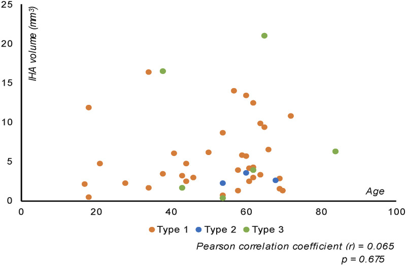
Background and purpose: Inter-hypothalamic adhesions (IHAs) are parenchymal tissue bridges traversing the third ventricle, previously reported only in the pediatric population. We aim to understand the prevalence of IHA in the adult population, assess their size and location, and ultimately investigate whether IHA volumes correlate with age.
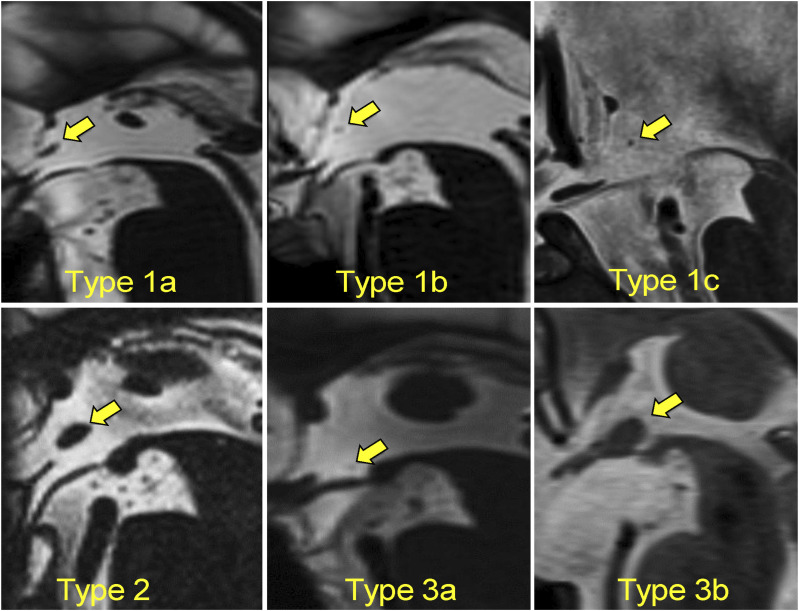
Materials and methods: Patients who underwent routine high-resolution 3D T2WI MRI studies of the temporal bone/internal auditory canal at an otolaryngology clinic between 2008 and 2014 were consecutively selected. The presence of IHAs was confirmed when a parenchymal structure could be traced across the third ventricle, connecting bilateral hypothalamus, and was visible in at least two planes. They were classified based on their location within the third ventricle, considering their connection with hypothalamic nuclei. Patient clinical information were collected from electronic charts. The prevalence and volumes of IHAs were calculated. Associations between age and IHA volume, as well as between IHA type, age, volume, and gender, were analyzed.
Results: 779 patients, with a mean age of 54.7 years were included. Among them, 44 IHAs were identified within 41 patients, resulting in a prevalence of 5.26% in our cohort. Type 1 IHA was the most frequently encountered type, comprising 70.5% of all cases. No significant correlation was observed between IHA volumes and age. Additionally, no associations were found between IHA type and age, volume, or gender.
Conclusion: IHAs are not exclusive to the pediatric population; they are also present in adults, with a prevalence of 5.26% in patients undergoing temporal bone/internal auditory canal MRI.
Keywords: Inter-hypothalamic adhesion; hypothalamic adhesion; magnetic resonance imaging; prevalence; volumetric assessment.
Conflict of interest statement
Declaration of conflicting interestsThe author(s) declared no potential conflicts of interest with respect to the research, authorship, and/or publication of this article.
Figures


Similar articles
-
Interhypothalamic adhesions: prevalence, structure, and location-based classification map in pediatric patients undergoing MRI.Neuroradiology. 2025 Jan;67(1):277-285. doi: 10.1007/s00234-024-03505-w. Epub 2024 Nov 15. Neuroradiology. 2025. PMID: 39542912
-
Interhypothalamic adhesions in endoscopic third ventriculostomy.Childs Nerv Syst. 2019 Sep;35(9):1565-1570. doi: 10.1007/s00381-019-04231-y. Epub 2019 Jun 6. Childs Nerv Syst. 2019. PMID: 31172270
-
Interhypothalamic Adhesion as Cause of Aborted Third Ventriculostomy: Neuroradiologic and Neuroendoscopic Considerations in Pediatric Case.World Neurosurg. 2019 Apr;124:214-218. doi: 10.1016/j.wneu.2019.01.018. Epub 2019 Jan 21. World Neurosurg. 2019. PMID: 30677576
-
Structural magnetic resonance imaging for the early diagnosis of dementia due to Alzheimer's disease in people with mild cognitive impairment.Cochrane Database Syst Rev. 2020 Mar 2;3(3):CD009628. doi: 10.1002/14651858.CD009628.pub2. Cochrane Database Syst Rev. 2020. PMID: 32119112 Free PMC article.
-
Diagnostic approaches for inherited hemolytic anemia in the genetic era.Blood Res. 2017 Jun;52(2):84-94. doi: 10.5045/br.2017.52.2.84. Epub 2017 Jun 22. Blood Res. 2017. PMID: 28698843 Free PMC article. Review.
References
LinkOut - more resources
Full Text Sources
