

Efficacy of two radiographic algorithms for detection of peri-implant bone defects on cone-beam computed tomography scans
- PMID: 39773233
- PMCID: PMC11708108
- DOI: 10.1186/s12903-024-05397-x
Efficacy of two radiographic algorithms for detection of peri-implant bone defects on cone-beam computed tomography scans
Abstract
Background: Early detection of peri-implant bone defects can improve long-term durability of dental implants. By the advances in cone-beam computed tomography (CBCT) scanners and introduction of new algorithms, it is important to find the most efficient protocol for detection of bone defects. This study aimed to assess the efficacy of metal artifact reduction (MAR) and advanced noise reduction (ANR) algorithms for detection of peri-implant bone defects.
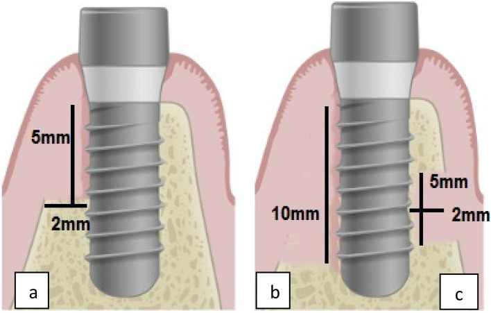
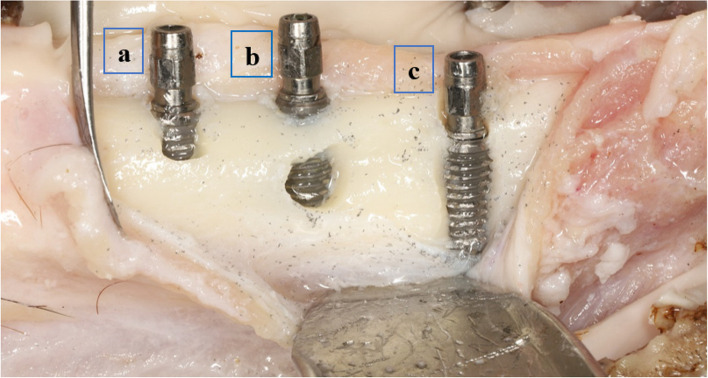
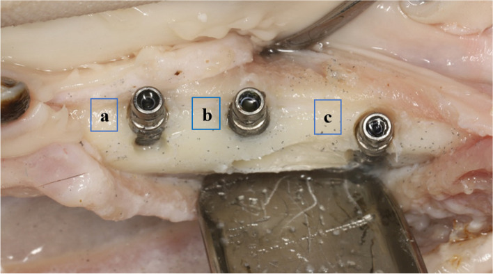
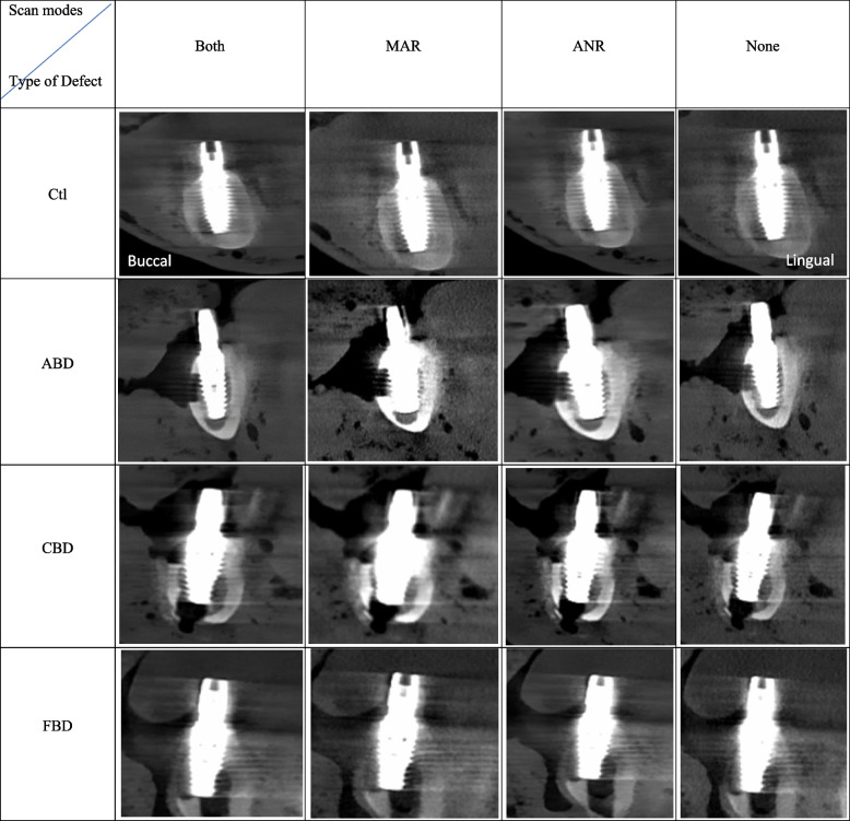
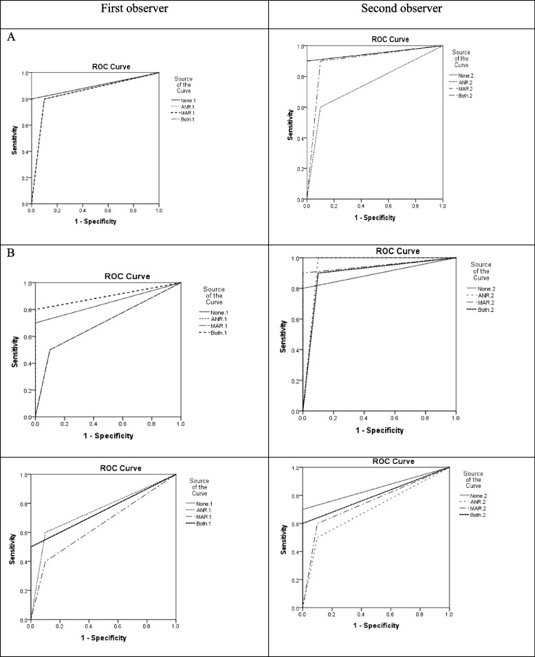
Materials and methods: In this in vitro study, 40 titanium implants were placed in 7 sheep mandibles. Crestal, apical, and Full defects (n = 10 from each type) were created around the implants, and 10 implants were also placed as controls. CBCT scans were obtained in four modes: with MAR, with ANR, with both MAR and ANR, and without any filter. Totally, 28 scans were obtained and evaluated by a radiologist and a maxillofacial surgeon. The observers recorded their observations in a checklist, and data were analyzed by SPSS version 21 using the kappa coefficient of agreement, sensitivity and specificity values, area under the receiver operating characteristic (ROC) curve (AUC), intraclass correlation coefficient, t-test and paired t-test (P < 0.05).
Results: The inter-observer agreement was high for detection of all defects in all modes except with ANR. No significant difference was found in AUC and diagnostic accuracy of different scan modes (P > 0.05). The most common diagnostic error was related to misdiagnosis of control group with full defect with ANR filter, such that the existing bone was not detected. Defect depth was averagely over-estimated while defect length was under-estimated. Correct diagnosis of defects had the highest frequency when both filters were on.
Conclusion: The diagnostic accuracy and sensitivity for detection of different defect types were not significantly different in different scan modes but activation of ANR filter significantly decreased the specificity and positive predictive value compared with no use of filter.
Keywords: Advanced noise reduction; Bone defect; Cone-Beam Computed Tomography; Dental implants; Metal artifact reduction.
© 2025. The Author(s).
Conflict of interest statement
Declarations. Ethics approval and consent to participate: This in vitro experimental study was carried out after obtaining ethical approval from the ethics committee of Hamadan University of Medical Sciences (IR.UMSHA.REC.1401 − 671(. Consent for publication: Not applicable. Competing interests: The authors declare no competing interests.
Figures
References
-
- Dave M, Davies J, Wilson R, Palmer R. A comparison of cone beam computed tomography and conventional periapical radiography at detecting peri-implant bone defects. Clin Oral Implants Res. 2013;24(6):671–8. - PubMed
-
- Razavi T, Palmer RM, Davies J, Wilson R, Palmer PJ. Accuracy of measuring the cortical bone thickness adjacent to dental implants using cone beam computed tomography. Clin Oral Implants Res. 2010;21(7):718–25. - PubMed
MeSH terms
Substances
LinkOut - more resources
Full Text Sources
