. 2025 Jan 7;44(1):7.
doi: 10.1186/s13046-024-03238-x.

Antibody-based delivery of interleukin-2 modulates the immunosuppressive tumor microenvironment and achieves cure in pancreatic ductal adenocarcinoma syngeneic mice

Affiliations

Antibody-based delivery of interleukin-2 modulates the immunosuppressive tumor microenvironment and achieves cure in pancreatic ductal adenocarcinoma syngeneic mice

Carmine Carbone et al. J Exp Clin Cancer Res. .

Abstract

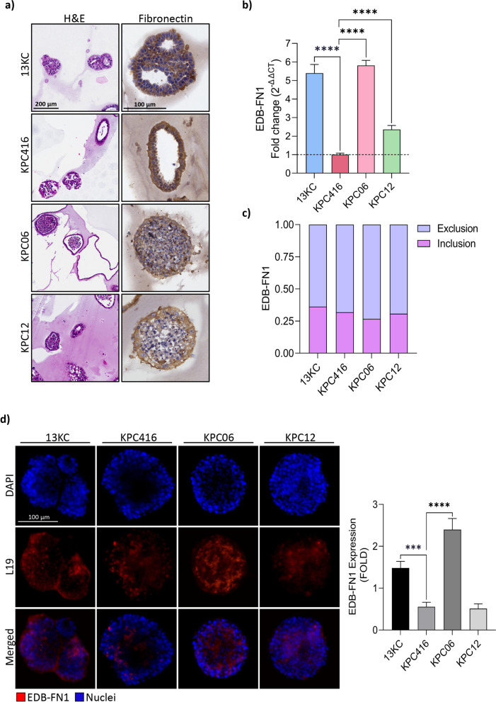
Background: Pancreatic ductal adenocarcinoma (PDAC) is one of the most aggressive and deadly type of cancer, with an extremely low five-year overall survival rate. To date, current treatment options primarily involve various chemotherapies, which often prove ineffective and are associated with substantial toxicity. Furthermore, immunotherapies utilizing checkpoint inhibitors have shown limited efficacy in this context, highlighting an urgent need for novel therapeutic strategies. This study investigates the preclinical efficacy of an innovative targeted therapy based on antibody-cytokine fusion proteins, specifically interleukin-2 (IL-2), a pivotal driver of cell-mediated immunity, fused to L19 antibody, which selectively binds to extra domain B of fibronectin (EDB-FN1) expressed in the tumor microenvironment.

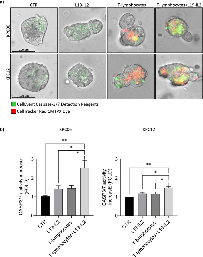
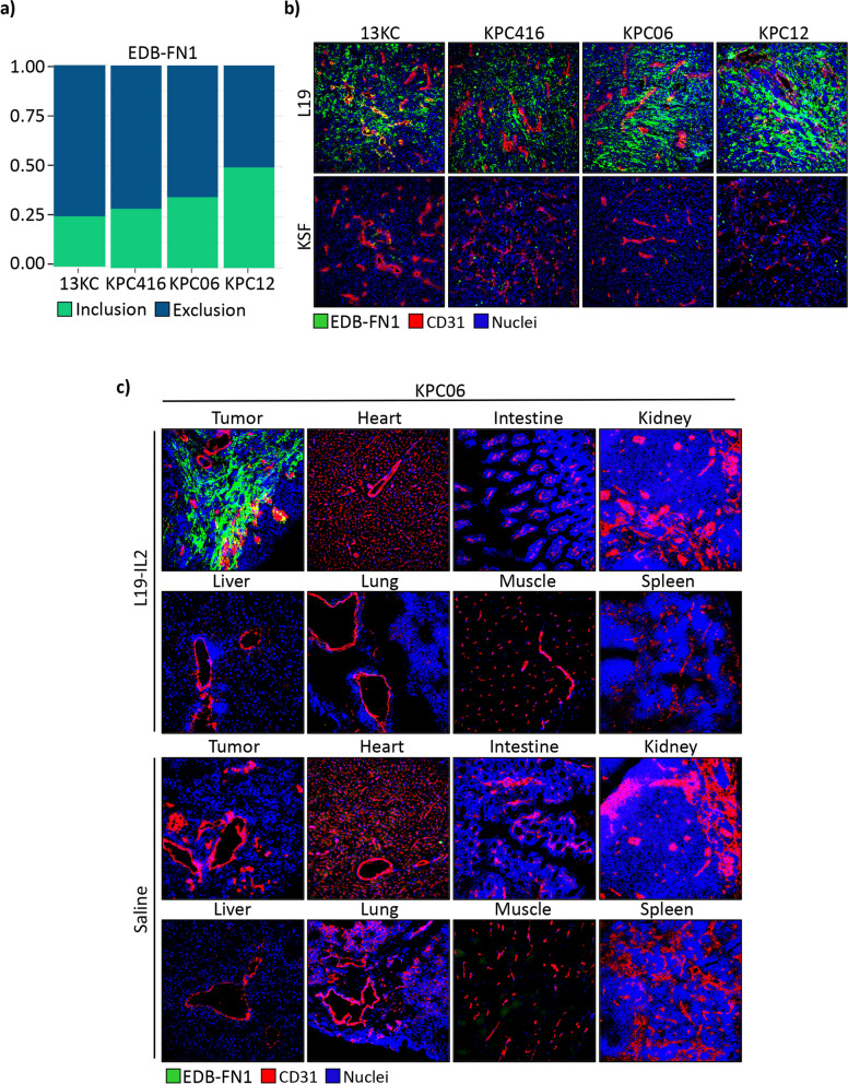
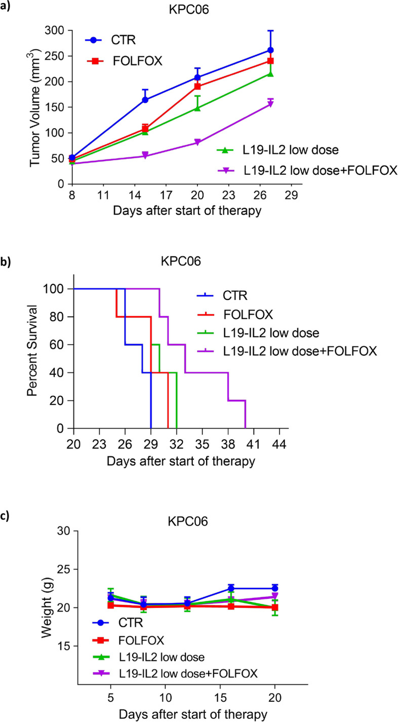
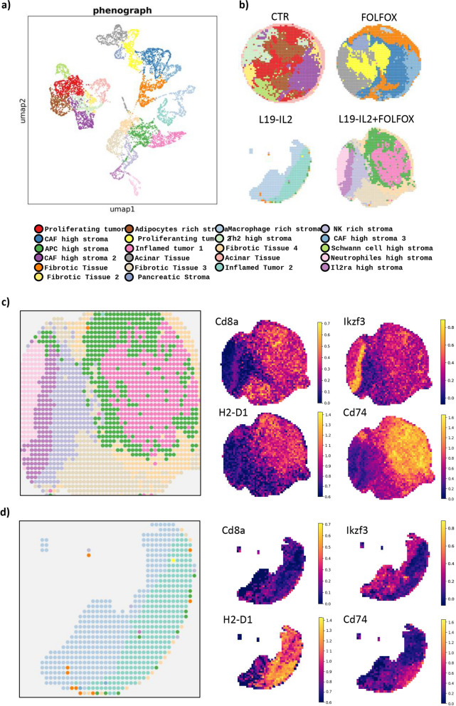
Methods: We tested the effectiveness of different immunocytokines through in vivo characterization in syngeneic C57BL/6J orthotopic mouse models of PDAC. Based on these results, we decided to focus on L19-IL2. To assess the efficacy of this immunocytokine we developed an ex-vivo immune-spheroid interaction platform derived from murine 3D pancreatic cultures, and telomerase reverse transcriptase (TERT) specific T-lymphocytes. Moreover, we evaluated the anti-cancer effect of L19-IL2 in combination with standard therapy in vivo experiments in PDAC mouse models. Tumor samples collected after the treatments were characterized for tumor infiltrating immune cell components by bulk RNA sequencing (RNA-seq) and spatial transcriptomics (Stereo-seq) analysis.

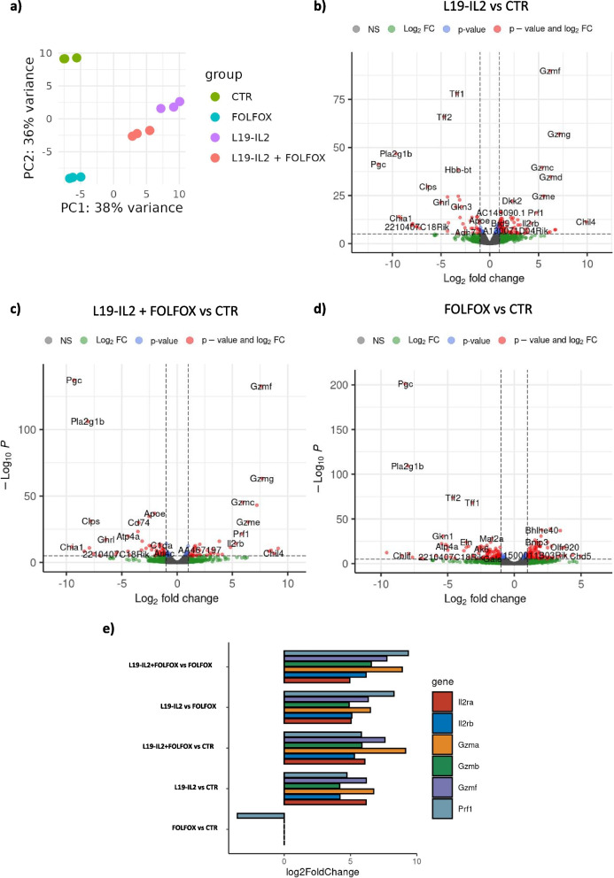
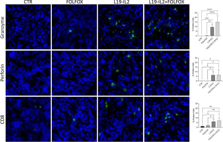
Results: The tumor-targeted L19-IL2 fusion protein demonstrated potent, dose-dependent anti-tumor activity in mice with pancreatic tumors resistant to standard chemotherapy. Spatial Transcriptomics (ST) and RNA-seq analyses indicated that L19-IL2 treatment induced a significant influx of immune cells into the tumor microenvironment, with these cells expressing activation markers like granzymes, perforins, and the IL-2 receptors.

Conclusions: Our results demonstrated that L19-IL2 enhances immune infiltration and cytotoxicity, remodeling the "cold" tumor microenvironment (TME) in PDAC. This innovative antibody-cytokine fusion protein improves therapeutic outcomes, paving the way for novel targeted treatment strategies in PDAC.

Keywords: Chemotherapy; Immunocytokines; Orthotopic syngeneic mouse models; Pancreatic cancer.


Conflict of interest statement

Declarations. Ethics approval and consent to participate: Animal experiments were approved by animal ethics committee of Catholic University of the Sacred Heart of Rome and by Ministry of Health with approval number n°593/2019-PR. This study was approved by the ethics committee of AOUI Verona Hospital (Territorial Ethics Committee for the South-West Veneto Area) with approval number Prot. n°53732. The patients provided written consent. This study was carried out in accordance with Declaration of Helsinki. Consent for publication: All patients provided consent for publication. Competing interests: Dario Neri is a co-founder and shareholder of Philogen ( www.philogen.com ), a Swiss-Italian Biotech company that operates in the field of ligand-based pharmacodelivery. Roberto De Luca, and Emanuele Puca are employees of Philochem AG, a daughter company of Philogen acting as discovery unit of the group.

Figures

Fig.1
Fig.1
Characterization of the expression of EDB-FN1 in mouse pancreatic cancer models. a Histochemical analysis of Fibronectin expression in different 3D models of PDAC. 10X images of Hematoxylin/Eosin, 40X images of Fibronectin. Images shown are representative of 1 out of more than 10 fields acquired. b Real-time analysis of EDB-FN1 in spheroid models of PDAC. c Analysis of inclusion splice junction (pink bars) and exclusion splice junction (light blue bars) reads of EDB-FN1. d IF analysis on spheroids confirmed L19 was able to recognize EDB-FN1. Protein analyzed (in red) and nuclei (in blue) were reported. Images shown are representative of 1 out of more than 10 fields acquired. Bar plot showing the fold increase in EDB-FN1 fluorescence calculated as the ratio between the mean of CTCF quantified in each group and the mean of CTR. P-value < 0.05 was indicated in figures with one asterisk (*), P-value < 0.01 with two asterisks (**), P-value < 0.001 with three asterisks (***) and P-value < 0.0001 with four asterisks (****)
Fig. 2
Fig. 2
Evaluation of the L19-IL2 effect on ex-vivo interaction platforms. a Immunity-spheroid interaction platforms with TERT specific T-lymphocytes and KPC06 and KPC12 treated with L19-IL2. The induction of apoptosis was evaluated using the CellEvent Caspase-3/7 Detection Reagent (green), while T-lymphocytes were stained with the vital staining CellTracker Red CMPTX Dye (Red). The platforms were monitored daily, and fluorescence images were acquired using the EVOS FL Auto 2 Cell Imaging System over a 48 h period. Images shown are representative of 1 out of more than 10 fields acquired. b Bar plot showing the fold increase in Caspase 3/7 activity in comparison to CTR. The fold increase is calculated as the ratio between the mean of CTCF quantified in each group and the mean of CTR. P-value < 0.05 was indicated in figures with one asterisk (*), P-value < 0.01 with two asterisks (**), P-value < 0.001 with three asterisks (***) and P-value < 0.0001 with four asterisks (****)
Fig. 3
Fig. 3
The L19-targeted antibody specifically hits EDB-FN1 of mouse cancer tissues. a Graphic representation of inclusion splice junction (green bars) and exclusion splice junction (blue bars) reads of EDB-FN1 from RNA-seq raw data in PDAC model tissues. b L19-targeted antibody in IgG1 format specifically target EDB-FN1 (Green) in vivo mice tumor tissues, while KSF antibody (specific for hen egg lysozyme, an irrelevant antigen) was used as negative CTR. c IF-based biodistribution analysis in orthotopic KPC06 mice. EDB-FN1 shown in green, saline was used as negative CTR
Fig.4
Fig.4
L19-IL2 treatment effects in combination with FOLFOX in KPC06 model. Plot showing tumor growth curves of KPC06 tumor-bearing mice randomly assigned to receive vehicle, as CTR, standard therapy (FOLFOX i.p., once a week for 2 weeks), and L19-IL2 (30 µg/mouse i.v., once a week for 2 weeks) alone or in combination with FOLFOX; b Kaplan–Meier survival analysis of KPC06 mice, grouped according to each experimental condition. c Variation of body weight in the different treatment groups
Fig. 5
Fig. 5
Differential expression analysis on KPC06 treated mice show immune response activation. a Plot showing Principal Component Analysis on RNA-seq data. b Volcano plot showing the genes differentially expressed (log2 Fold Change ≦ −1.5 ≧ 1.5, FDR < 0.05) in the comparison between L19-IL2 treated mice and control (CTR). c Volcano plot showing the genes differentially expressed (log2 Fold Change ≦ −1.5 ≧ 1.5, FDR < 0.05) between mice treated with a combination of L19-IL2 and FOLFOX and CTR. d Volcano plot showing the genes differentially expressed (log2 Fold Change ≦ −1.5 ≧ 1.5, FDR < 0.05) between FOLFOX treated mice and CTR. e Bar plot showing log2 Fold Change value for T-lymphocyte activation genes resulting from DEA between the different treatment groups
Fig. 6
Fig. 6
Stereo-Seq OMNI spatial transcriptomics analysis. a,b Spatial clustering of Stereo-seq OMNI data on the four cores analyzed. c Spatially resolved clusters of L19-IL2 + FOLFOX sample and heatmap showing an enhanced inflammation in the tumor core with a major representation of Cd8a+ Activated T-lymphocytes, Cd74+ H2-D1+ APCs and Ikzf3+ NK. d Spatially resolved clusters of L19-IL2 sample and heatmap showing an inflamed tumor core
Fig. 7
Fig. 7
Immunofluorescence analysis on KPC06 models. IF analysis for CD8.+ TILs, GRZB and PRF1 in KPC06 tumor tissues. Protein analyzed (in green) and nuclei (in blue) are reported. Images shown are representative of 1 out of more than 10 fields acquired. Bar plots show percentage of positive cells grouped by treatments. P-value < 0.05 was indicated in figures with one asterisk (*), P-value < 0.01 with two asterisks (**), P-value < 0.001 with three asterisks (***) and P-value < 0.0001 with four asterisks (****)

Similar articles

Cited by

References

    1. Halbrook CJ, Lyssiotis CA, di Magliano MP, Maitra A. Pancreatic cancer: advances and challenges. Cell. 2023;186(8):1729–54. - PMC - PubMed
    1. Jiang Y, Sohal DPS. Pancreatic adenocarcinoma management. JCO Oncol Pract. 2023;19(1):19–32. - PubMed
    1. Kleeff J, Korc M, Apte M, La Vecchia C, Johnson CD, Biankin AV, et al. Pancreatic cancer. Nat Rev Dis Primers. 2016;2(1):16022. - PubMed
    1. Conroy T, Desseigne F, Ychou M, Bouché O, Guimbaud R, Bécouarn Y, et al. FOLFIRINOX versus gemcitabine for metastatic pancreatic cancer. N Engl J Med. 2011;364(19):1817–25. - PubMed
    1. Von Hoff DD, Ervin T, Arena FP, Chiorean EG, Infante J, Moore M, et al. Increased survival in pancreatic cancer with nab-Paclitaxel plus Gemcitabine. N Engl J Med. 2013;369(18):1691–703. - PMC - PubMed

MeSH terms

LinkOut - more resources