. 2025 Jan 17;14(2):e240714.
doi: 10.1530/EC-24-0714. Print 2025 Feb 1.

Metformin reverts aortic calcifications and elastin loss induced by an experimental metabolic syndrome

Metformin reverts aortic calcifications and elastin loss induced by an experimental metabolic syndrome

Lucas Streckwall et al. Endocr Connect. .

Abstract

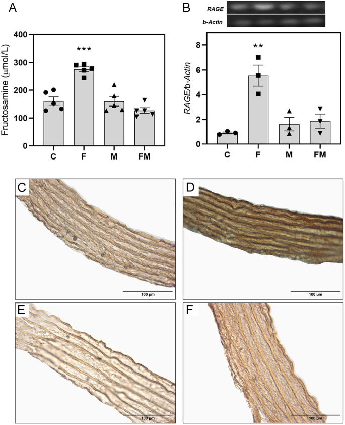
Metabolic syndrome (MetS) is associated with osteogenic transdifferentiation of vascular smooth muscle cells (VSMCs) and accumulation of arterial calcifications (ACs). Metformin (MET) inhibits this transdifferentiation in vitro. Here, we evaluate the in vivo efficacy of oral MET to reduce AC in a model of MetS. Twenty young male Wistar rats were divided into two groups: one received water and the other received water plus 20% fructose to induce MetS. After 14 days, and for another 4 weeks, MET (100 mg/kg per day) was added to half of each group's drinking source, thus C (water), F (fructose), M (MET) and FM (fructose + MET). Serum and adipose tissue were collected. Aortas were dissected for histomorphometric and immunohistochemical analysis, ex vivo calcification studies and isolation of VSMCs to measure their alkaline phosphatase activity (ALP), collagen production, extracellular mineralization, gene expression of RUNX2 and receptor for advanced glycation end-products (AGEs) (RAGE), and elastic fiber production. F group showed parameters compatible with MetS. Aortic tunica media from F showed decreased elastic-to-muscular layer ratio, increased collagen content and increased levels of the AGEs structure carboxymethyl-lysine. Aortic arches from F presented a tendency for higher ex vivo calcification. VSMCs from F showed increased ALP, collagen secretion, mineralization and expression of RUNX2 and RAGE, and decreased elastic fiber production. All these effects were reverted by MET cotreatment (FM group). In vitro, AGEs-modified bovine serum albumin upregulated RAGE expression of control VSMCs, and this was prevented by MET in an AMP kinase-dependent manner. Thus, experimental MetS induces RAGE upregulation and osteogenic transdifferentiation of aortic VSMCs curbed by oral treatment with MET.

Keywords: advanced glycation end-products; fructose; metabolic syndrome; metformin; osteogenic transdifferentiation; receptor for advanced glycation end-products; vascular smooth muscle cells.


Conflict of interest statement

The authors declare that there is no conflict of interest that could be perceived as prejudicing the impartiality of the work reported. Dr Antonio McCarthy is a Senior Editor of Endocrine Connections. Dr McCarthy was not involved in the review or editorial process for this paper, in which he is listed as an author.

Figures

Figure 1
Figure 1
Histomorphometric analysis of aortic sections. (A) The black dotted line shows the distance between external and internal elastic membranes, and the black and white lines represent the width of individual muscular and elastic layers, respectively (H&E), obj. ×100. (B) Comparison of average tunica media thickness. (C) Comparison of average elastic-to-muscular layer ratio. (D) Representative micrography of aortic section stained with Sirius Red, obj. ×40. (E) The red area represents the area covered by collagen, obj. ×40. (F) Quantitative analysis of area covered by collagen. C: control group; F: rats receiving 20% fructose solution; M: rats receiving 100 mg/kg per day metformin; FM: rats receiving fructose plus metformin. Differences: *P < 0.05 vs C, M and FM. Results are expressed as mean ± SEM. n = 3 per experimental group.
Figure 2
Figure 2
Effects of fructose and/or metformin treatments on different markers of VSMC osteogenic potential: (A) alkaline phosphatase activity, (B) type 1 collagen production, (C) mineralization of extracellular matrix, (D) relative gene expression of RUNX2 by RT-PCR and (E) quantification of elastic fibers. C: control group; F: rats receiving 20% fructose solution; M: rats receiving 100 mg/kg per day metformin; FM: rats receiving fructose plus metformin. Differences: *P < 0.05 vs C, M and FM; **P < 0.01 vs C, M and FM. Results are expressed as mean ± SEM. n = 3–5 per experimental group.
Figure 3
Figure 3
Effect of fructose and/or metformin treatment on (A) serum fructosamine levels, (B) VSMC relative gene expression of RAGE by RT-PCR. Carboxymethyl-lysine extracellular accumulation in the aortic wall (by immunohistochemistry, obj. ×40) of (C) control rats (C group), (D) rats receiving 20% fructose solution (F group), (E) rats receiving 100 mg/kg per day metformin (M group) and (F) rats receiving fructose plus metformin (FM group). Immunohistochemical staining of CML was performed in aortic sections of all animals, and representative photographs are shown in panels (C–F). Differences: **P < 0.01 vs C, M and FM; ***P < 0.001 vs C, M and FM. Quantitative results are expressed as mean ± SEM. n = 3–5 per experimental group.
Figure 4
Figure 4
In vitro effects of metformin and/or the AMPK inhibitor compound C on RAGE relative gene expression of control VSMCs (from C group). B: 100 μg/mL non-glycated bovine serum albumin (BSA); BM: 100 μg/mL BSA plus 500 μM metformin; BC: 100 μg/mL BSA + 0.5 μM compound C; BMC: 100 μg/mL BSA +500 μM metformin + 0.5 μM compound C; A: 100 μg/mL AGEs–BSA; AM: 100 μg/mL AGEs–BSA + 500 μM metformin; AC: 100 μg/mL AGEs–BSA +0.5 μM compound C; AMC: 100 μg/mL AGEs–BSA + 500 μM metformin + 0.5 μM compound C. Differences: ***P < 0.001. Results are expressed as mean ± SEM. n = 4 per experimental group.

Similar articles

References

    1. Martini N, Streckwall L & McCarthy AD. Osteoporosis and vascular calcifications. Endocr Connections 2023. 12 e230305. (10.1530/ec-23-0305) - DOI - PMC - PubMed
    1. Vidavsky N, Kunitake JAMR & Estroff LA. Multiple pathways for pathological calcification in the human body. Adv Healthc Mater 2020. 10 2001271. (10.1002/adhm.202001271) - DOI - PMC - PubMed
    1. Juutilainen A, Lehto S, Suhonen M, et al. . Thoracoabdominal calcifications predict cardiovascular disease mortality in type 2 diabetic and nondiabetic subjects: 18-year follow-up study. Diabetes Care 2009. 33 583–585. (10.2337/dc09-1813) - DOI - PMC - PubMed
    1. Demer LL & Tintut Y. Inflammatory, metabolic, and genetic mechanisms of vascular calcification. Arterioscler Thromb Vasc Biol 2014. 34 715–723. (10.1161/atvbaha.113.302070) - DOI - PMC - PubMed
    1. Tyson J, Bundy K, Roach C, et al. . Mechanisms of the osteogenic switch of smooth muscle cells in vascular calcification: WNT signaling, BMPs, mechanotransduction, and EndMT. Bioengineering 2020. 7 88. (10.3390/bioengineering7030088) - DOI - PMC - PubMed

LinkOut - more resources