

Ultrasound super-resolution imaging for non-invasive assessment of microvessel in prostate lesion
- PMID: 39773358
- PMCID: PMC11706184
- DOI: 10.1186/s40644-024-00819-z
Ultrasound super-resolution imaging for non-invasive assessment of microvessel in prostate lesion
Abstract
Background: Prostate cancer (PCa) is the leading cause of cancer-related morbidity and mortality in men worldwide. An early and accurate diagnosis is crucial for effective treatment and prognosis. Traditional invasive procedures such as image-guided prostate biopsy often cause discomfort and complications, deterring some patients from undergoing these necessary tests. This study aimed to explore the feasibility and clinical value of using ultrasound super-resolution imaging (US SRI) for non-invasively assessing the microvessel characteristics of prostate lesion.
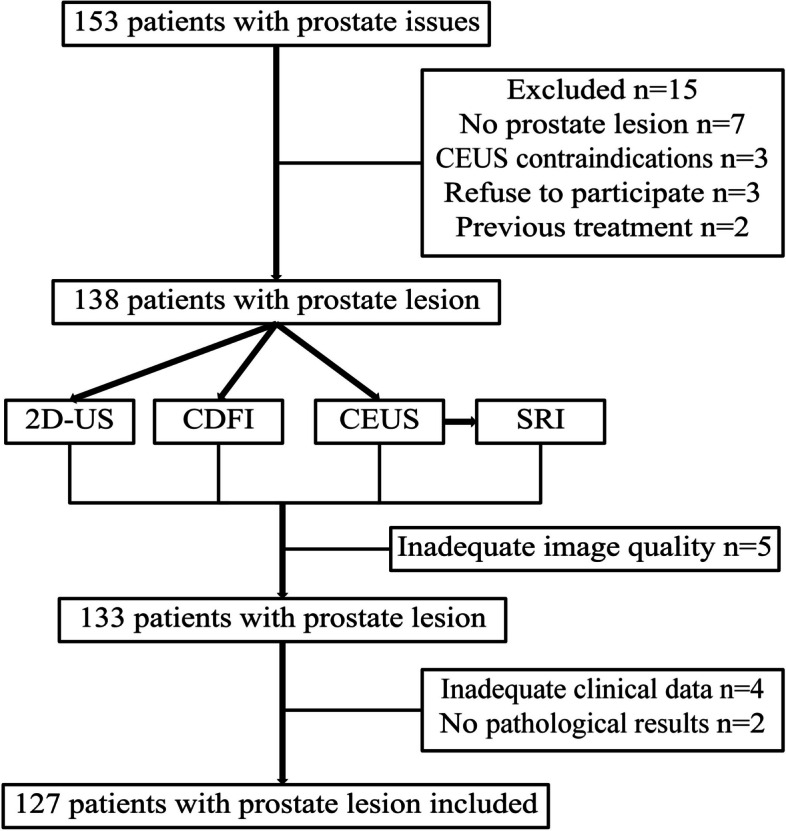
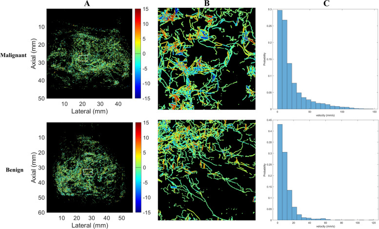
Methods: This study included 127 patients with prostate lesion who presented at Renmin Hospital of Wuhan University between November 2023 and June 2024 were included in this study. All the patients underwent transrectal US (TRUS), contrast-enhanced US (CEUS), and US SRI. CEUS parameters of time-intensity curve (TIC): arrival time (AT), rising time (RT), time to peak (TTP), peak intensity (PKI), falling time (FT), mean transit time (MTT), ascending slope (AS), descending slope (DS), D/A slope ratio (SR), and area under the TIC (AUC). US SRI parameters: microvessel density (MVD), microvessel diameter (D), microvessel velocity (V), microvessel tortuosity (T), and fractal number (FN), were analyzed and compared between prostate benign and malignant lesion.
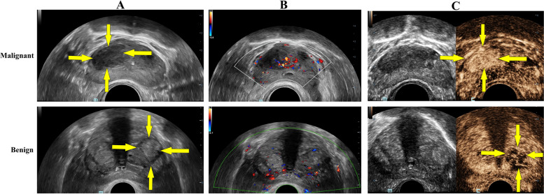
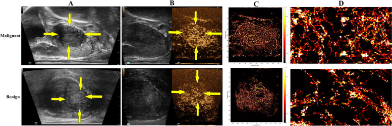
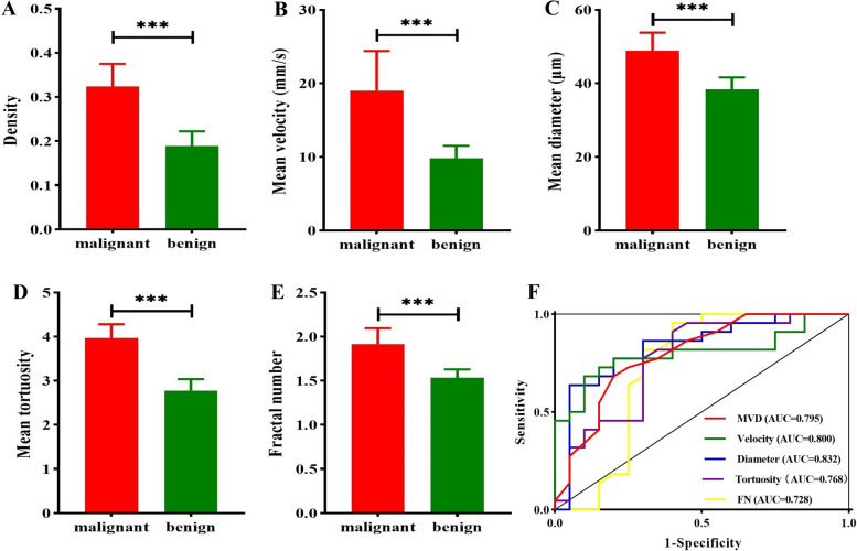
Results: The tumor markers of prostate in the malignant group were all higher than those in the benign group, and the differences were statistically significant (P < 0.001). The TIC parameters of CEUS revealed that the PKI, AS, DS, and AUC were significantly higher in the malignant group than in the benign group (P < 0.001), whereas the RT, TTP and FT in the malignant group were significantly lower (P < 0.001). Malignant lesion exhibited significantly higher MVD, larger D, faster V, greater T, and more complex FN than benign lesion (P < 0.001).
Conclusions: US SRI is a promising non-invasive imaging modality that can provide detailed microvessel characteristics of prostate lesion, offering an advancement in the differential diagnosis for prostate lesion. And, US SRI may be a valuable tool in clinical practice with its ability to display and quantify microvessel with high precision.
Keywords: Microvessel; Non-invasive diagnosis; Prostate lesion; Ultasound super-resolution imaging.
© 2025. The Author(s).
Conflict of interest statement
Declarations. Ethics approval and consent to participate: The studies involving human participants were reviewed and approved by the Institutional Review Board of Renmin Hospital of Wuhan University (Approval Number: WDRY2024-K109). The patients/participants provided their written informed consent to participate in this study. Consent for publication: Not applicable. Competing interests: The authors declare no competing interests.
Figures
References
-
- Zheng RS, Chen R, Han BF, Wang SM, Li L, Sun KX, Zeng HM, Wei WQ, He J. Cancer incidence and mortality in China, 2022. Zhong Hua Zhong Liu Za Zhi. 2024;46:221–31. - PubMed
MeSH terms
Substances
LinkOut - more resources
Full Text Sources
Medical
Research Materials
Miscellaneous
