

Long-term observation of the in vivo safety of a new design of phakic refractive lens
- PMID: 39773390
- PMCID: PMC11705939
- DOI: 10.1186/s12886-024-03803-0
Long-term observation of the in vivo safety of a new design of phakic refractive lens
Abstract
Background: To evaluate the biosafety, reduction in anterior capsule opacification, and fluctuation in intraocular pressure (IOP) of a new phakic refractive lens (PRL) with a sinusoidal drainage groove design.
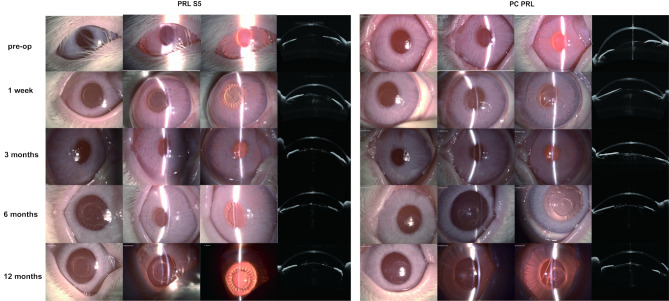
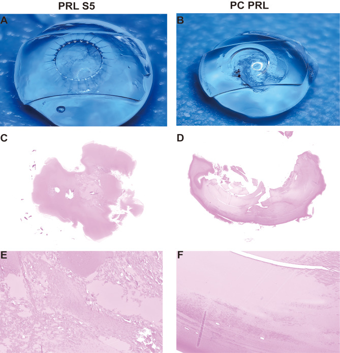
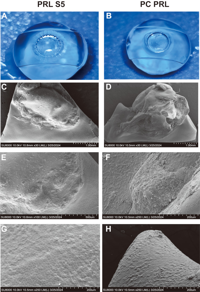
Methods: This self-controlled experiment was performed on eight eyes of four rabbits. Each rabbit was implanted with a sinusoidal PRL (PRL-S5) in the right eye and a conventional posterior chamber PRL (PC-PRL) in the left eye. Slit-lamp examinations, optical coherence tomography, and IOP evaluation were performed before surgery and at 1 day, 1 week, 3 months, 6 months, and 1 year postoperatively in each eye. Gross examination, histopathology, and electron microscopy of the capsule and PRL were performed 1 year postoperatively.
Results: On slit-lamp examination, the inflammatory reactions recovered one week after surgery. The PC-PRL group developed anterior subcapsular cataracts at 3 months postoperatively and diffuse and dense opacification of the cortex at 1 year. PRL-S5 showed mild local opacification in the optical zone 6 months postoperatively, which did not progress significantly. At 1 year, PC-PRLs had greater opacification (27.37-72.17%) than PRL S5 (6.63-66.96%). Three months after surgery, one eye in the PC-PRL group had scleral staphyloma, one eye had corneal edema, and one eye experienced nasal hepatic prolapse into the anterior chamber. One eye in the PRL-S5 group had papillary membranes but recovered 6 months postoperatively. Histopathological examination revealed liquefaction and necrosis of the opacified area in the center of the subcapsular membrane in both groups. Numerous granular bodies and fibrous precipitates were observed in epithelial cells in the opaque area. Electron microscopy showed that epithelial cells proliferated on the surface of all anterior capsule membranes, with no significant differences between the two groups. The capsular PC-PRL group showed anterior cortical proliferation and fibrosis. An IOP elevation was noted on the first postoperative day (18.8 to 37.9 mmHg). However, both the PC-PRL and PRL-S5 groups exhibited relatively stable IOP levels 1 week, 3 months, 6 months, and 1 year postoperatively.
Conclusions: The new PRL exhibited robust long-term biocompatibility. The sinusoidal groove design facilitated the maintenance of IOP stability without necessitating iridectomy and effectively mitigated the onset and progression of cataracts.
Keywords: Cataract; Complications; Intraocular pressure; Phakic Intraocular Lens; Phakic Refractive Lens; Refractive surgery.
© 2024. The Author(s).
Conflict of interest statement
Declarations. Ethics approval and consent to participate: The animals were purchased from Shanghai Jiagan Biotechnology Co. and we have obtained their informed consent. All animal experiments were approved by the Animal Ethics Committee of the Eye and ENT Hospital of Fudan University, Shanghai, China (No. 2022114), and all experimental protocols, including care, transportation, and experiments of the animals, complied with the guidelines of the Animal Care and Use Committee of Fudan University. Consent for publication: Not applicable. Competing interests: The authors declare no competing interests.
Figures


References
-
- Shi X, Gao Z, Leng L, Guo Z. Temporal and spatial characterization of myopia in China. Front Public Health [Internet]. 2022 Aug 16 [cited 2024 Mar 16];10. https://www.frontiersin.org/journals/public-health/articles/10.3389/fpubh.2022.896926/full - PMC - PubMed
MeSH terms
Grants and funding
- 82171095/National Natural Science Foundation of China
- 81770955/National Natural Science Foundation of China
- 23XD1400500/Shanghai Science and Technology Development Foundation
- 20410710100/Shanghai Science and Technology Development Foundation
- SHDC12024148/Shanghai Shenkang Hospital Development Center
LinkOut - more resources
Full Text Sources
