. 2025 Jan 7;13(1):e009316.
doi: 10.1136/jitc-2024-009316.

Targeted inhibition of Aurora kinase A promotes immune checkpoint inhibition efficacy in human papillomavirus-driven cancers

Affiliations

Targeted inhibition of Aurora kinase A promotes immune checkpoint inhibition efficacy in human papillomavirus-driven cancers

Soma Ghosh et al. J Immunother Cancer. .

Abstract

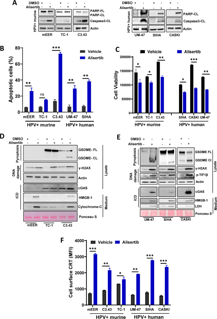
Background: Human papillomavirus (HPV)-driven cancers include head and neck squamous cell carcinoma and cervical cancer and represent approximately 5% of all cancer cases worldwide. Standard-of-care chemotherapy, radiotherapy, and immune checkpoint inhibitors (ICIs) are associated with adverse effects and limited responses in patients with HPV-driven cancers. The integration of targeted therapies with ICIs may improve outcomes. In a previous study, we demonstrated that Aurora kinase A (AURKA, Aurora A) inhibitors lead to apoptosis of human HPV-positive cancer cells in vitro and in vivo. Here, we explored the potential of Aurora A inhibition to enhance response to ICIs in immune-competent preclinical models of HPV-driven cancers.

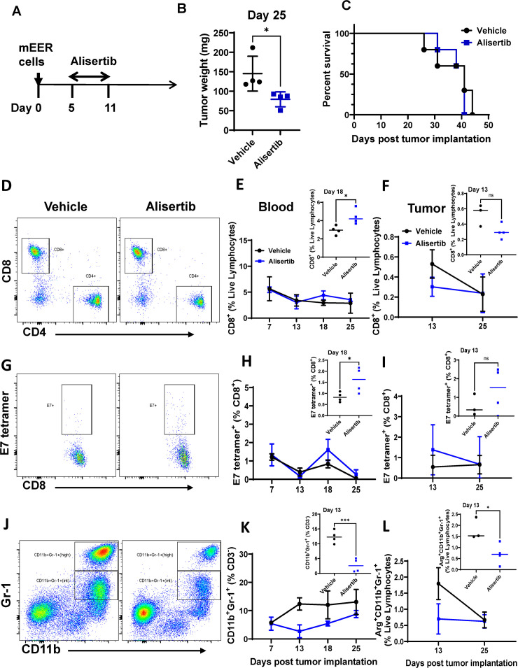
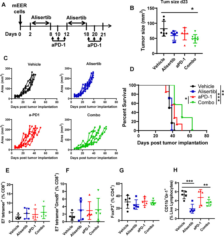
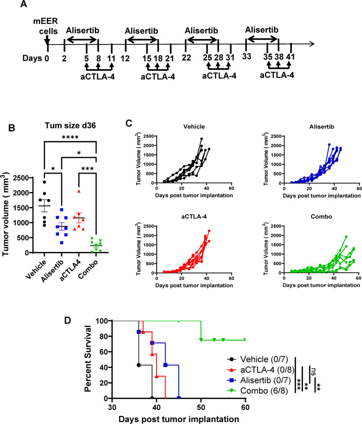
Methods: We assessed the induction of apoptosis, DNA damage, and immunogenic cell death (ICD) in response to treatment with the Aurora A inhibitor alisertib in vitro and antitumor efficacy of alisertib as a monotherapy and in combination with ICIs that inhibit programmed cell death protein-1 (PD-1) or cytotoxic T-lymphocyte associated protein 4 (CTLA-4) in murine HPV-positive immune-competent tumor models. In each treatment group, we determined the tumor growth kinetics and long-term survival and assessed the tumor immune microenvironment using polychromatic flow cytometry.

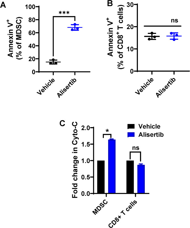
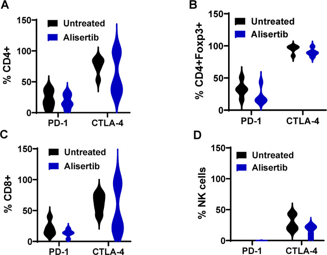
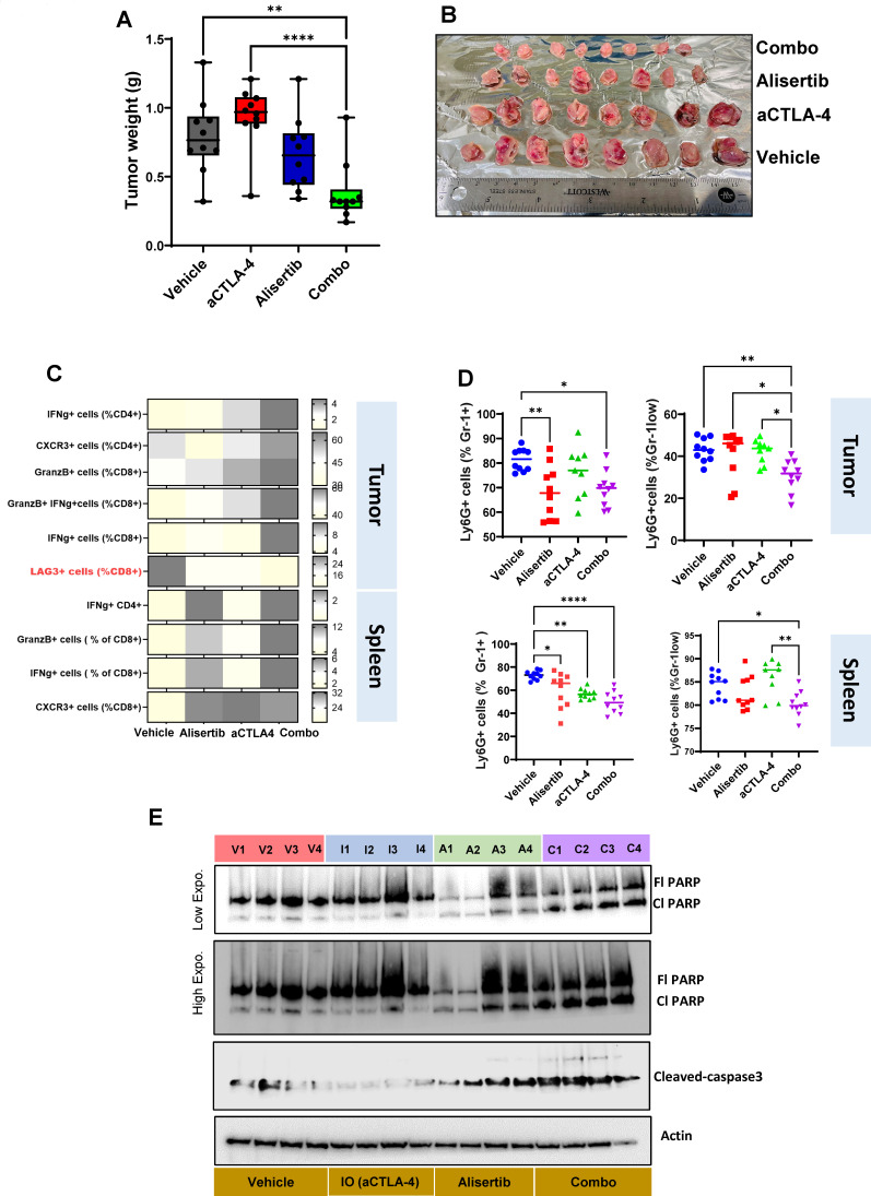
Results: Aurora A inhibition induced apoptosis, DNA damage, and ICD in vitro in multiple human and murine HPV-positive cancer cell lines. Importantly, Aurora A inhibition induced selective apoptotic depletion of myeloid-derived suppressor cells (MDSCs). In vivo experiments demonstrated that the combination of alisertib with ICIs, specifically anti-CTLA4, resulted in improved survival outcomes by altering the tumor immune microenvironment. This combination enhanced CD8 T-cell infiltration and decreased the frequencies of MDSCs, whereas neither alisertib nor ICIs (anti-PD-1/anti-CTLA-4) alone showed such effects.

Conclusion: Our study establishes the potential of Aurora A inhibition to sensitize HPV-positive tumors to ICIs, specifically anti-CTLA-4 treatment. This combination strategy resulted in enhanced antitumor efficacy, driven by systemic and intratumoral increases in CD8 T-cell responses and reduced immunosuppressive cell populations, specifically MDSCs. These findings offer insights into the synergistic effects of Aurora A inhibition and ICIs and argue for further investigation and optimization of this combination approach in HPV-driven cancers.

Keywords: Head and Neck Cancer; Immune Checkpoint Inhibitor; Immunotherapy; Myeloid-derived suppressor cell - MDSC; Viral-specific T cells.


Conflict of interest statement

Competing interests: FMJ and JKS have received research funding from Takeda Pharmaceuticals. All other authors declare no potential conflicts of interest.

Figures

Figure 1
Figure 1. Aurora kinase A inhibition induces cytotoxicity, apoptosis, DNA damage, and immunogenic cell death in human papillomavirus (HPV)-positive murine and human cancer cell lines in vitro. (AC) Murine and human cancer cells were treated with 300 nmol/L alisertib for 48 hours before being subjected to lysis and immunoblotting with the indicated antibodies (A), annexin-FITC staining to measure apoptosis (B), or a CellTiter-Glo assay to measure cell viability (C). (DE) Murine (D) and human (E) cells were treated with 300 nmol/L alisertib for 48 hours before immunoblotting with antibodies that indicate the presence of pyroptosis, DNA damage, and immunogenic cell death (ICD). To determine the amount of released HMGB-1 (D, E), cytochrome C (D), and lactate dehydrogenase (LDH) (E), equal volumes of conditioned medium were subjected to immunoblotting analysis; Ponceau S staining was used as a loading control. (F) Calreticulin (CRT) expression on the cell surface was analyzed by flow cytometry on murine and human cell lines treated with 300 nmol/L alisertib for 48 hours. The significance of differences was determined using an unpaired, two-tailed Student’s t-test. *p≤0.05, **p≤0.001, ***p≤0.0001. CL, cleaved; DMSO, dimethyl sulfoxide; FL, full; GDSME, gasdermin E; MFI, mean fluorescence intensity; UM-47, UMSCC47.
Figure 2
Figure 2. Aurora kinase A inhibition has differential effects on T cells and MDSCs in vivo in HPV-positive tumor-bearing mice. (AC) Groups of C57BL/6 mice were implanted with mEER cells (1×10⁶) subcutaneously on the flank and treated with alisertib (10 mg/kg) daily for 6 days starting at day 5 after tumor cell implantation (A) and monitored for tumor growth (weight) (B) and survival (C). (DL) Changes in total CD8 T cells (D–F), HPV E7 antigen-specific CD8 T cells (G–I), and MDSCs (CD11b+ Gr-1+) (J–L) were monitored by flow cytometry at the indicated time points in the blood (day 18 for E7 and CD8 and day 13 for MDSCs) (E, H, K) and tumor (day 13 for MDSCs and day 25 for E7+ and CD8 cells) (F, I, L). Representative flow profiles for CD8 T cells (D), HPV E7 antigen-specific CD8 T cells (G), and MDSCs (J) in the blood from vehicle-treated and alisertib-treated mice are shown. The significance of differences was determined using an unpaired, two-tailed Student’s t-test. ns, p≤0.05*, ***p<0.009. HPV, human papillomavirus; MDSCs, myeloid-derived suppressor cells.
Figure 3
Figure 3. Aurora kinase A inhibition ex vivo induces apoptosis in HPV-positive tumor-induced MDSCs but not CD8 T cells. (AB) Splenocytes isolated from mEER tumor-bearing mice were treated ex vivo with vehicle or alisertib (300 nM) for 6 hours, and apoptotic cells (annexin V+) within the MDSC (A) and CD8 T cell (B) fractions were identified by flow cytometry. (C) MDSCs and CD8 T cells (C) from the mEER tumors were purified and treated ex vivo with vehicle or with alisertib (300 nmol/L) for 6 hours. Protein extracts were analyzed for apoptosis and survival pathway proteins using an antibody array. The significance of differences was determined using an unpaired, two-tailed Student’s t-test. ns (not significant), *p≤0.01, **p≤0.004, ***p≤0.0001. Cyto-C, cytochrome C; HPV, human papillomavirus; MDSCs, myeloid-derived suppressor cells.
Figure 4
Figure 4. Aurora kinase A inhibition in combination with PD-1 blockade extends the survival of HPV-positive tumor-bearing mice. (AD) Groups of C57BL/6 mice were implanted with mEER cells (1×10⁶) subcutaneously on the flank and were treated sequentially with 10 mg/kg of alisertib and anti-PD-1 antibody (aPD-1) at 250 µg per dose individually or in combination (combo) as shown (A) and monitored for tumor growth (area) (B, C) and survival (D). Significant differences between the survival curves were determined using the log-rank Mantel-Cox test *p<0.05, **p<0.005, ***p<0.0005. (EH) Tumor-infiltrating leukocytes were separated from tumors on day 23 after the tumor initiation when all the treatments were completed. Frequencies of total CD8 T cells (E), HPV E7 antigen-specific granzyme B-expressing (GrnzB+) CD8 T cells (F), regulatory T cells (FoxP3+) (G), and myeloid-derived suppressor cell (H) were measured using one-way analysis of variance. **p<0.005, ***p<0.0005. HPV, human papillomavirus; PD-1, programmed cell death protein-1.
Figure 5
Figure 5. Aurora kinase A inhibition leads to persistently higher frequencies of CTLA-4-positive lymphocyte subsets than PD-1-positive lymphocyte subsets in mice with human papillomavirus-positive tumors. Single-cell suspensions of mEER tumor cells from vehicle-treated and alisertib-treated mice were analyzed by flow cytometry for the expression of PD-1 and CTLA-4 in CD4+ (A), regulatory T (B), CD8 (C), and natural killer (NK) (D) cells. CTLA-4, cytotoxic T-lymphocyte associated protein 4; PD-1, programmed cell death protein-1.
Figure 6
Figure 6. Aurora kinase A inhibition in combination with anti-CTLA-4 treatment extends the survival of human papillomavirus-positive tumor-bearing mice. Groups of C57BL/6 mice were implanted with mEER cells (1×10⁶) subcutaneously on the flank and were treated sequentially with 10 mg/kg each of alisertib and anti-CTLA-4 antibody (aCTLA-4) individually or in combination (combo) (A) and monitored for tumor growth (B, C) and survival (D). Significant differences between the survival curves were determined using the log-rank (Mantel-Cox) test. ns, p≥0.05, *p<0.05, **p<0.005, ***p<0.0006, ****p<0.0001. CTLA-4, cytotoxic T-lymphocyte associated protein 4.
Figure 7
Figure 7. Aurora kinase A inhibition in combination with CTLA-4 blockade promotes antitumor immunity and cancer cell apoptosis. (AB) Groups of C57BL/6 mice with mEER tumors were treated sequentially with alisertib (10 mg/kg) and anti-CTLA-4 antibody (aCTLA-4; 100 µg per dose) individually or in combination (combo). All mice were euthanized after two treatment cycles on day 23, and tumors were weighed (A, B). (C) Tumor-infiltrating lymphocytes and circulating lymphocytes from the tumor and spleen were isolated and analyzed by polychromatic flow cytometry to assess the frequencies of different immune cell populations. Changes in their expression are represented as a heatmap. The negative immune regulator LAG3 is highlighted in red. (D) Frequencies of circulating myeloid-derived suppressor cell levels for the tumor and spleen. The significance of differences in immune cell subsets among the treatment groups were assessed using one-way analysis of variance. *p<0.05, **p<0.005, ***p<0.0001, ****p<0.00005. (E) Immunoblotting was performed on tumor tissues from four mice per treatment group at day 23. The expression of cleaved caspase-3 (cl caspase-3), full-length PARP (Fl PARP), and cleaved PARP (cl PARP) were analyzed to assess apoptosis. The treatment groups are as follows: V – vehicle, I – anti-CTLA-4, A – alisertib, C – combination. CTLA-4, cytotoxic T-lymphocyte associated protein 4, IO-Immunotherapy; PARP, poly(ADP-ribose) polymerase .

Similar articles

Cited by

References

    1. Mahal BA, Catalano PJ, Haddad RI, et al. Incidence and Demographic Burden of HPV-Associated Oropharyngeal Head and Neck Cancers in the United States. Cancer Epidemiol Biomarkers Prev. 2019;28:1660–7. doi: 10.1158/1055-9965.EPI-19-0038. - DOI - PubMed
    1. Aggarwal P, Zaveri JS, Goepfert RP, et al. Swallowing-related outcomes associated with late lower cranial neuropathy in long-term oropharyngeal cancer survivors: cross-sectional survey analysis. Head Neck. 2019;41:3880–94. doi: 10.1002/hed.25923. - DOI - PMC - PubMed
    1. Ghosh S, Shah PA, Johnson FM. Novel Systemic Treatment Modalities Including Immunotherapy and Molecular Targeted Therapy for Recurrent and Metastatic Head and Neck Squamous Cell Carcinoma. Int J Mol Sci. 2022;23:7889. doi: 10.3390/ijms23147889. - DOI - PMC - PubMed
    1. Burtness B, Harrington KJ, Greil R, et al. Pembrolizumab alone or with chemotherapy versus cetuximab with chemotherapy for recurrent or metastatic squamous cell carcinoma of the head and neck (KEYNOTE-048): a randomised, open-label, phase 3 study. Lancet. 2019;394:1915–28. doi: 10.1016/S0140-6736(19)32591-7. - DOI - PubMed
    1. Monk BJ, Enomoto T, Kast WM, et al. Integration of immunotherapy into treatment of cervical cancer: Recent data and ongoing trials. Cancer Treat Rev. 2022;106:102385. doi: 10.1016/j.ctrv.2022.102385. - DOI - PMC - PubMed

MeSH terms

LinkOut - more resources