. 2025 Jan 7;13(1):e010618.
doi: 10.1136/jitc-2024-010618.

Differential impact of TIM-3 ligands on NK cell function

Affiliations

Differential impact of TIM-3 ligands on NK cell function

Juncheng Wang et al. J Immunother Cancer. .

Abstract

Background: The transmembrane protein T-cell immunoglobulin and mucin-domain containing molecule 3 (TIM-3) is an immune checkpoint receptor that is expressed by a variety of leukocyte subsets, particularly in the tumor microenvironment. An effective TIM-3-targeting therapy should account for multiple biological factors, including the disease setting, the specific cell types involved and their varying sensitivities to the four putative TIM-3 ligands (galectin-9, phosphatidylserine, high mobility group protein B1 and carcinoembryonic antigen cell adhesion molecule 1), each of which engages a unique binding site on the receptor's variable immunoglobulin domain. The primary objectives of this study were to assess the prevalence and function of TIM-3+ natural killer (NK) cells in patients with head and neck squamous cell carcinoma (HNSCC), determine whether the four TIM-3 ligands differentially affect TIM-3+ NK cell functions, identify the most immunosuppressive ligand, and evaluate whether targeting ligand-mediated TIM-3 signaling enhances NK cell effector functions.

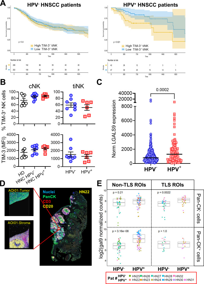
Methods: Single-cell RNA sequencing and flow cytometry were used to study the prevalence, phenotypes and function of TIM-3+ NK cells in HNSCC patient tumors and blood. In vitro killing, proliferation and cytokine production assays were implemented to evaluate whether the four TIM-3 ligands differentially modulate TIM-3+ NK cell functions, and whether disruption of TIM-3/ligand interaction can enhance NK cell-mediated antitumor effector mechanisms. Finally, The Cancer Genome Atlas survival analysis and digital spatial profiling were employed to study the potential impact of etiology-associated differences on patients with HNSCC outcomes.

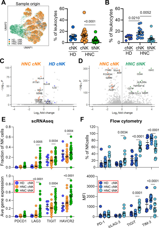
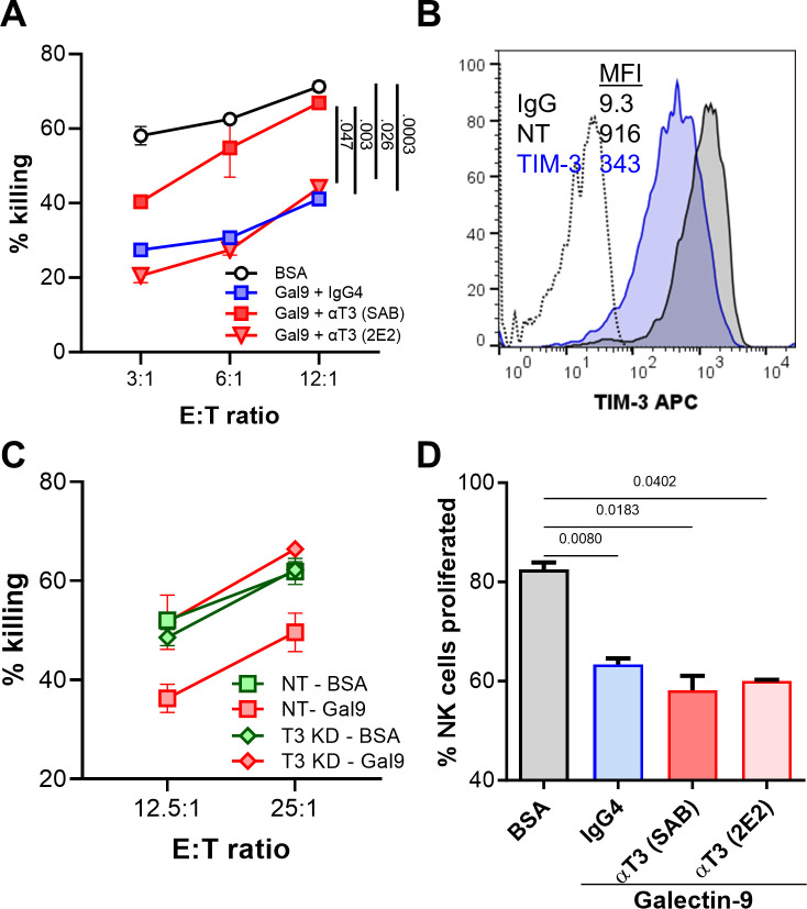
Results: We demonstrate that TIM-3 is highly prevalent on circulating and tumor-infiltrating NK cells. It co-expresses with CD44 and marks NK cells with heightened effector potential. Among the four putative TIM-3 ligands, galectin-9 most consistently suppresses NK cell-mediated cytotoxicity and proliferation through TIM-3 and CD44 signaling, respectively, but promotes IFN-γ release in a TIM-3-dependent manner. Among patients with HNSCC, an elevated intratumoral TIM-3+ NK cell gene signature associates with worse outcomes, specifically in those with human papillomavirus (HPV)+ disease, potentially attributable to higher galectin-9 levels in HPV+ versus HPV- patients.

Conclusions: Our findings underscore the complex functional impact of TIM-3 ligand signaling, which is consistent with recent clinical trials suggesting that targeting TIM-3 alone is suboptimal as an immunotherapeutic approach for treating malignancies.

Keywords: Head and Neck Cancer; Immune Checkpoint Inhibitor; Innate; Natural killer - NK; Tumor Microenvironment.


Conflict of interest statement

Competing interests: RLF is co-founder and stockholder of Novasenta, and declares competing interests with Aduro Biotech (consulting), AstraZeneca/MedImmune (clinical trial, research funding), Bristol Myers Squibb (advisory board, clinical trial, research funding), EMD Serono (advisory board), MacroGenics (advisory board), Merck (advisory board, clinical trial), Novasenta (consulting, stock, research funding), Numab Therapeutics AG (advisory board), Pfizer (advisory board), Sanofi (consultant), Tesaro (research funding) and Zymeworks (consultant). TCB serves on the scientific advisory board of Walking Fish Therapeutics, Bespoke Therapeutics, Mestag Therapeutics, Kalivir Therapeutics, and Galvanize Therapeutics. TCB is a consultant for Attivare Therapeutics and Tabby Therapeutics. LV declares US Patent Number: 10,543,264, a methodology licensed to INmune Bio where DN-TNF can be used to prevent or treat malignancies. RB declares PCT/US15/612657 (Cancer Immunotherapy), PCT/US18/36052 (Microbiome Biomarkers for Anti-PD-1/PD-L1 Responsiveness: Diagnostic, Prognostic and Therapeutic Uses Thereof), PCT/US63/055227 (Methods and Compositions for Treating Autoimmune and Allergic Disorders).

Figures

Figure 1
Figure 1. TIM-3 is prevalent in NK cells in PBL (cNK) and TIL (tiNK). (A) NK cells from blood and tumors were bioinformatically extracted from our previously published scRNAseq data sets of healthy donors (HD) and patients with HNSCC (HNC), and visualized as a UMAP plot. NK cell frequencies within PBL and TIL are also shown (% of leukocytes). (B) cNK and tiNK frequencies identified by scRNAseq were validated by flow cytometry. Transcriptomic differences were highlighted by volcano plots between (C) HNC and HD cNK, and (D) HNC cNK and tiNK, showing DEGs. The inhibitory ICR landscape of cNK and tiNK cells was assessed by (E) scRNAseq and (F) flow cytometry, measuring the fraction of cells expressing each marker or average gene/protein expression levels. Each circle in dot-plot summaries represents a single donor. P values based on Mann-Whitney U (HD vs HNC cNK) and Wilcoxon signed-rank (HNC cNK vs tiNK) tests. cNK, circulating natural killer cells; DEGs, differentially-expressed genes; HNSCC, head and neck squamous cell carcinoma; ICR, immune checkpoint receptor; MFI, mean fluorescence intensity; NK, natural killer; PBL, peripheral blood lymphocyte; scRNAseq, single-cell RNA sequencing; TIL, tumor-infiltrating lymphocyte; TIM-3, T-cell immunoglobulin and mucin-domain containing molecule 3; tiNK, tumor-infiltrating NK cells; UMAP, Uniform Manifold Approximation and Projection.
Figure 2
Figure 2. CD56dimCD16dim/− NK cells, predominant in HNSCC tumors, express the lowest TIM-3 levels among subsets. NK cell subsets were profiled by CD56 and CD16 expression using flow cytometry (online supplemental figure 2). (A) Representative CD56/CD16 staining in HD cNK, HNSCC cNK and HNSCC tiNK is shown. (B) The distribution of the three major NK cell subsets in cNK and tiNK is summarized. (C) TIM-3 staining examples for patient-matched cNK and tiNK subsets (pt.14; online supplemental table 1) are shown. TIM-3 expression, measured by (D) the frequency of TIM-3+ cells and (E) the mean fluorescence intensity (MFI), is shown for the three major NK subsets in cNK and tiNK, as assessed by flow cytometry. Each circle in dot-plot summaries represents a single donor. P values based on Mann-Whitney U (HD vs HNC cNK) and Wilcoxon signed-rank (HNC cNK vs tiNK) tests. cNK, circulating natural killer cells; HD, healthy donors; HNC, patients with HNSCC; HNSCC, head and neck squamous cell carcinoma; NK, natural killer; PBL, peripheral blood lymphocyte; TIL, tumor-infiltrating lymphocyte; TIM-3, T-cell immunoglobulin and mucin-domain containing molecule 3; tiNK, tumor-infiltrating NK cells.
Figure 3
Figure 3. TIM-3 marks NK cells with higher effector potential in blood and tumors. Flow cytometry compared TIM-3+ and TIM-3 NK cells from HD cNK, HNSCC cNK and HNSCC tiNK. Median antigen frequency and scaled expression values for 7 HD cNK, as well as 14 HNC patient-matched cNK and tiNK are visualized using dot plots (explored in greater detail in online supplemental figures 4-6). Circle size represents median frequency, and color indicates scaled median MFI values. Summaries for (A) CD56brightCD16, (B) CD56dimCD16+ and (C) CD56dimCD16dim/− NK cells are shown. Graphical summaries of TIM-3 and TIM-3+ (D) cNK and (E) tiNK phenotypes were created with BioRender.com. cNK, circulating natural killer cells; HD, healthy donors; HNC, patients with HNSCC; HNSCC, head and neck squamous cell carcinoma; MFI, mean fluorescence intensity; NK, natural killer; TIM-3, T-cell immunoglobulin and mucin-domain containing molecule 3; tiNK, tumor-infiltrating NK cells.
Figure 4
Figure 4. Galectin-9 consistently suppresses NK cell proliferation and cytotoxicity. To induce TIM-3 cross-linking and immunosuppressive signaling, we used (A) galectin-9, HMGB1 and CEACAM1-coated beads, and (B) phosphatidylserine (PS) liposomes. BSA-coated beads (BSA) and phosphatidylcholine (PC) liposomes served as respective negative controls. (A–B) Limited NK cell yields from patients with HNSCC (≤4.35 million per 25 million PBL) restricted cytotoxicity tests to four or five conditions per patient. For the representative panel, HD NK cells isolated from a buffy coat were used to test all conditions. Cytotoxicity of IL-2 activated, ligand-treated NK cells from HD was tested against MHC class I-deficient K562 targets using the NK-TVA assay. Data are representative of six independent experiments, four of which were performed using HNSCC and two using HD NK cells. (C–D) For proliferation and cytokine-release experiments, which required fewer cells, we used only patient-derived NK cells. (C) The impact of the four TIM-3 ligands on patient with HNSCC NK cell proliferation was evaluated by CFSE dilution. (D) Proliferation data from six independent experiments are summarized. (E) The mechanism behind galectin-9-induced suppression of NK cell cytotoxicity and proliferation was evaluated by flow cytometric analysis of granzyme B, perforin and Ki-67 expression levels (representative of three independent experiments). Two-way and one-way ANOVA tests were used for statistical analyses in (A, B) and (D), respectively. ANOVA, analysis of variance; BSA, bovine serum albumin; CEACAM1, carcinoembryonic antigen cell adhesion molecule 1; CFSE, carboxyfluorescein succinimidyl ester; E:T, effector-to-target; HD, healthy donors; HMGB1, high mobility group protein B1; HNSCC, head and neck squamous cell carcinoma; IgG, immunoglobulin G; IL-2, interleukin-2; MFI, mean fluorescence intensity; MHC, major histocompatibility complex; NK, natural killer; TIM-3, T-cell immunoglobulin and mucin-domain containing molecule 3; TVA, target cell visualization assay.
Figure 5
Figure 5. Sabatolimab counteracts galectin-9-induced dysfunction of NK cell cytotoxicity but not proliferation. Sabatolimab (SAB) or F38-2E2 (2E2) anti-TIM-3 antibodies, along with an IgG4 isotype control, were tested for their ability to block TIM-3/galectin-9 interactions. The effects on (A) cytotoxicity and (D) proliferation were assessed using NK-TVA and CFSE dilution assays, respectively. (B) The efficacy of TIM-3 knockdown via siRNA was compared with the non-targeting (NT) siRNA control (n=2). (C) Post-knockdown NK cells were treated with BSA-coated or galectin-9-coated beads to evaluate cytotoxicity. Two-way and one-way way ANOVA tests were used for statistical analyses in (A) and (D), respectively. Data are representative of three independent experiments. ANOVA, analysis of variance; BSA, bovine serum albumin; CFSE, carboxyfluorescein succinimidyl ester; E:T, effector-to-target; IgG4, immunoglobulin G4; MFI, mean fluorescence intensity; NK, natural killer; siRNA, small interfering RNA; TIM-3, T-cell immunoglobulin and mucin-domain containing molecule 3; TVA, target cell visualization assay.
Figure 6
Figure 6. Galectin-9 suppresses NK cell proliferation via CD44 signaling. (A) Flow cytometry analysis of CD44 upregulation and its co-expression with TIM-3 on NK cells after the 48 hours treatment with 6000 IU/mL IL-2. Data are representative of three independent experiments. (B) Frequencies of CD44+ cells among TIM-3+ and TIM-3 patient-matched cNK and tiNK are summarized. (C) The ability of anti-CD44 and IgG4 isotype control antibodies to abrogate galectin-9-induced suppression of NK cell proliferation was assessed using the CFSE dilution assay. Data are representative of two independent experiments. Statistical significance was determined using one-way ANOVA for (B) and (C). ANOVA, analysis of variance; BSA, bovine serum albumin; CFSE, carboxyfluorescein succinimidyl ester; cNK, circulating natural killer cells; IgG4, immunoglobulin G4; IL-2, interleukin-2; NK, natural killer; TIM-3, T-cell immunoglobulin and mucin-domain containing molecule 3; tiNK, tumor-infiltrating NK cells.
Figure 7
Figure 7. Potential importance of disease etiology: galectin-9 is more highly expressed in HPV+ HNSCC. (A) Gene signatures for HAVCR2+ tiNK, derived from the top 100 DEGs, were used to stratify TCGA HNSCC samples into high and low TIM-3+ tiNK gene signature groups based on the median GSVA score. Kaplan-Meier survival curves are shown for HPV+ and HPV patients. (B) Flow cytometry analysis of TIM-3+ NK cell frequencies in patient-matched cNK and tiNK, and HD cNK. (C) LGALS9 expression levels in HPV and HPV+ patients with HNSCC from TCGA, with individual patient values plotted. Statistical significance was determined using the Mann-Whitney test. (D–E) Tumor sections from five HPV and six HPV+ patients were evaluated by DSP. (D) CD3, CD20, pan-CK and nucleic acid staining were used to select ROI (∼500 µm2) and AOI (based on pan-CK staining → stroma vs tumor bed). A staining example for one patient is shown. (E) Relative galectin-9 protein abundance was measured in AOI with a multiplexed antibody cocktail and custom UV-cleavable DNA barcode. Data are shown as log2(normalized counts), with each dot representing an AOI and color indicating the patient. Statistical significance was calculated using Wilcoxon’s test for (E) and Student’s t-test for (B) and (C). AOI, areas of interest; cNK, circulating natural killer cells; DEGs, differentially-expressed genes; DSP, digital spatial profiling; GSVA, gene set variation analysis; HD, healthy donors; HNC, patients with HNSCC; HNSCC, head and neck squamous cell carcinoma; HPV, human papillomavirus; MFI, mean fluorescence intensity; NK, natural killer; pan-CK, pan-cytokeratin; ROI, regions of interest; TCGA, The Cancer Genome Atlas; TIM-3, T-cell immunoglobulin and mucin-domain containing molecule 3; tiNK, tumor-infiltrating NK cells; TLS, tertiary lymphoid structures; UV, ultraviolet.

References

    1. Monney L, Sabatos CA, Gaglia JL, et al. Th1-specific cell surface protein Tim-3 regulates macrophage activation and severity of an autoimmune disease. Nature New Biol. 2002;415:536–41. doi: 10.1038/415536a. - DOI - PubMed
    1. Du W, Yang M, Turner A, et al. TIM-3 as a Target for Cancer Immunotherapy and Mechanisms of Action. Int J Mol Sci. 2017;18:645. doi: 10.3390/ijms18030645. - DOI - PMC - PubMed
    1. Sakuishi K, Apetoh L, Sullivan JM, et al. Targeting Tim-3 and PD-1 pathways to reverse T cell exhaustion and restore anti-tumor immunity. J Exp Med. 2010;207:2187–94. doi: 10.1084/jem.20100643. - DOI - PMC - PubMed
    1. Shayan G, Srivastava R, Li J, et al. Adaptive resistance to anti-PD1 therapy by Tim-3 upregulation is mediated by the PI3K-Akt pathway in head and neck cancer. Oncoimmunology. 2017;6:e1261779. doi: 10.1080/2162402X.2016.1261779. - DOI - PMC - PubMed
    1. Zeidan AM, Ando K, Rauzy O, et al. Sabatolimab plus hypomethylating agents in previously untreated patients with higher-risk myelodysplastic syndromes (STIMULUS-MDS1): a randomised, double-blind, placebo-controlled, phase 2 trial. Lancet Haematol. 2024;11:e38–50. doi: 10.1016/S2352-3026(23)00333-2. - DOI - PubMed

MeSH terms

LinkOut - more resources