. 2025 Jul 1;27(4):543-549.
doi: 10.4103/aja2024106. Epub 2025 Jan 7.

Icariin targets PDE5A to regulate viability, DNA synthesis and DNA damage of spermatogonial stem cells and improves reproductive capacity

Affiliations

Icariin targets PDE5A to regulate viability, DNA synthesis and DNA damage of spermatogonial stem cells and improves reproductive capacity

Tian-Long Liao et al. Asian J Androl. .

Abstract

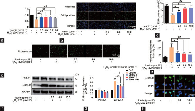
Icariin is a pure compound derived from Epimedium brevicornu Maxim, and it helps the regulation of male reproduction. Nevertheless, the role and underlying mechanisms of Icariin in mediating male germ cell development remain to be clarified. Here, we have demonstrated that Icariin promoted proliferation and DNA synthesis of mouse spermatogonial stem cells (SSCs). Furthermore, surface plasmon resonance iron (SPRi) and molecular docking (MOE) assays revealed that phosphodiesterase 5A (PDE5A) was an important target of Icariin in mouse SSCs. Mechanically, Icariin decreased the expression level of PDE5A. Interestingly, hydrogen peroxides (H 2 O 2 ) enhanced the expression level of phosphorylation H2A.X (p-H2A.X), whereas Icariin diminished the expression level of p-H2A.X and DNA damage caused by H 2 O 2 in mouse SSCs. Finally, our in vivo animal study indicated that Icariin protected male reproduction. Collectively, these results implicate that Icariin targets PDE5A to regulate mouse SSC viability and DNA damage and improves male reproductive capacity. This study thus sheds new insights into molecular mechanisms underlying the fate decisions of mammalian SSCs and offers a scientific basis for the clinical application of Icariin in male reproduction.

Keywords: DNA damage; Icariin; PDE5A; proliferation; spermatogonial stem cells.


Conflict of interest statement

All authors declared no competing interests.

Figures

Figure 1
Figure 1
Icariin enhances the viability and DNA synthesis of mouse SSCs. (a) MTT assay was utilized to measure viability of C18-4 cells after 3 days of culture under different concentrations of Icariin. (b) Western blot was used to check the expression level of PCNA in C18-4 cells with treatment of Icariin at different concentrations for 24 h. (c) Statistical analysis of PCNA expression in b. (d) EdU incorporation assay showed the EdU-positive cells affected by Icariin at 2.5–15 µmol l−1 in C18-4 cells. (e) Statistical analysis of EdU-positive cells in d. *P < 0.05 and **P < 0.01, the value of the indicated group compared with that in the control group (n = 3 for each group). MTT: methyl thiazolyl tetrazolium; SSC: spermatogonial stem cell; PCNA: proliferating cell nuclear antigen; EdU: 5-ethynyl-2’-deoxyuridine.
Figure 2
Figure 2
The effect of PDE5A silencing on the viability and DNA synthesis of C18-4 cells. (a) The qPCR assay was used to assess the impact of two PDE5A siRNAs on PDE5A mRNA of C18-4 cells. (b) Western blot demonstrated the influence of two PDE5A siRNAs on PDE5A protein of C18-4 cells. GAPDH served as a control of loading protein. (c) Statistical analysis of PDE5A protein expression in b. (d) The influence of PDE5A silencing on the viability of C18-4 cells. (e) EdU incorporation assay illustrated the impact of PDE5A siRNA-2 on DNA synthesis of C18-4 cells. (f) Statistical analysis of EdU assay of EdU-positive cells in e. *P<0.05 and **P < 0.01, the value of the indicated group compared with that in the control group. Ctrl: control; PDE5A: phosphodiesterase 5A; siRNA: small interfering RNA; GAPDH: glyceraldehyde-3-phosphate dehydrogenase; EdU: 5-ethynyl-2’-deoxyuridine; mRNA: messenger RNA.
Figure 3
Figure 3
The influence of Icariin and PDE5A silencing on PDE5A and p-PDE5A expression levels of C18-4 cells. (a) The qPCR was utilized to detect the effect of Icariin and PDE5A siRNA-2 on the level of PDE5A mRNA in C18-4 cells. (b) Western blot was employed to determine the influence of Icariin and PDE5A siRNA-2 on the level of PDE5A protein in C18-4 cells. (c) Statistical analysis of the expression of PDE5A protein in b. (d) Western blots showed the changes in the p-PDE5A and PDE5A expression levels of C18-4 cells treated with different concentrations of Icariin for 24 h. GAPDH was used as a loading control of proteins. (e) Statistical analysis of p-PDE5A and PDE5A protein expression in d. *P < 0.05 and **P < 0.01, the value of the indicated group compared with that in the control group (n = 3 for each group). Ctrl: control; PDE5A: phosphodiesterase 5A; p-PDE5A: phosphorylation of PDE5A; siRNA: small interfering RNA; GAPDH: glyceraldehyde-3-phosphate dehydrogenase; mRNA: messenger RNA; EdU: 5-ethynyl-2’-deoxyuridine; NS: no statistical difference.
Figure 4
Figure 4
H2O2 inhibits viability and causes DNA damage in C18-4 cells. (a) MTT assay showed the effect of H2O2 at different concentrations on cell viability of C18-4 cells for 24 h. (b) EdU incorporation assay was utilized to detect EdU-positive cells in C18-4 cells affected by H2O2 at different concentrations. (c) Statistical analysis of EdU-positive cells in b. (d) The ROS kit was used to determine the changes in ROS levels of C18-4 cells after 2 h of treatment with H2O2 at different concentrations. (e) The ROS relative fluorescence intensity was calculated from the results of the five independent experiments in d. (f) Western blot showed p-H2A.X expression changes in C18-4 cells after 2 h of H2O2 treatment at different concentrations. (g) Statistical analysis of p-H2A.X protein expression in f.. (h) Western blot displayed the changes in expression levels of p-PDE5A and PDE5A in C18-4 cells after 2 h treatment of H2O2 at different concentrations. (i) Statistical analysis of p-PDE5A and PDE5A protein expression in h. *P < 0.05, **P < 0.01, and ***P < 0.001, the value of the indicated group compared with that in the control group (n = 3 for each group). NS: no statistical difference; MTT: methyl thiazolyl tetrazolium; PDE5A: phosphodiesterase 5A; p-PDE5A: phosphorylation of PDE5A; p-H2A.X: phosphorylation of H2A.X; H2O2: hydrogen peroxides; siRNA: small interfering RNA; GAPDH: glyceraldehyde-3-phosphate dehydrogenase; mRNA: messenger RNA; ROS: reactive oxygen species.
Figure 5
Figure 5
Icariin protects DNA damage of C18-4 cells caused by H2O2. (a) MTT assay showed the effect of Icariin combination with H2O2 at different concentrations for 24 h on the viability of C18-4 cells. (b) EdU incorporation assay detected the EdU-positive cells in C18-4 cells with Icariin at different concentrations for 24 h and followed by H2O2 at 200 µmol l−1 for 24 h. (c) Statistical analysis of EdU-positive cells in b. (d) Changes of ROS levels in C18-4 cells treated with Icariin for 24 h and followed by H2O2 at 200 µmol l−1 for 2 h. (e) The ROS relative fluorescence intensity was calculated from the results of the five independent assays in d. (f) Western blot showed the changes of PDE5A and p-H2A.X protein expression in C18-4 cells treated with Icariin for 24 h and followed by H2O2 at 200 µmol l−1 for 2 h. (g) Statistical analysis of PDE5A and p-H2A.X protein expression in f. (h) Immunocytochemical staining illustrated that the fluorescence intensity of p-H2A.X in C18-4 cells treated with Icariin for 24 h and followed by H2O2. *P < 0.05 and **P < 0.01, the value of the indicated group compared with that in the control group (n = 3 for each group). #P < 0.05 and ##P < 0.01, vs H2O2 treatment (n = 3 for each group). NS: no statistical difference; MTT: methyl thiazolyl tetrazolium; PDE5A: phosphodiesterase 5A; p-PDE5A: phosphorylation of PDE5A; p-H2A.X: phosphorylation of H2A.X; H2O2: hydrogen peroxides; GAPDH: glyceraldehyde-3-phosphate dehydrogenase; ROS: reactive oxygen species; DAPI: 4’,6-diamidino-2-phenylindole.
Figure 6
Figure 6
The influence of Icariin on reproductive reproduction of male mice in vivo. (a) Body and (b) testicular weight changes of male mice after being treated with H2O2, Icariin, and H2O2 plus Icariin for 21 consecutive days. (c) The representative testes of male mice after being treated with H2O2, Icariin, and H2O2 plus Icariin for 21 consecutive days (n = 8). (d) The abnormal rate of sperm of mice after being treated with H2O2, Icariin, and H2O2 plus Icariin for 21 consecutive days. (e) The appearance of sperm abnormalities in representative male mice after being treated with H2O2 for 21 consecutive days. (f) H&E staining of testicular seminiferous tubules of mice after being treated with H2O2, Icariin, and H2O2 plus Icariin for 21 consecutive days. (g) Western blot detected the expression changes of PDE5A and p-H2A.X proteins in the testis tissues of male mice after being treated with H2O2, Icariin, and H2O2 plus Icariin for 21 consecutive days. (h) Statistical analysis of PDE5A and p-H2A.X expression in g. *P < 0.05 and **P < 0.01, the value of the indicated group compared with that in the control group (n = 3 for each group). #P < 0.05 H&E, vs H2O2 treatment (n = 3 for each group). NS: no statistical difference; MTT: methyl thiazolyl tetrazolium; PDE5A: phosphodiesterase 5A; p-PDE5A: phosphorylation of PDE5A; p-H2A.X: phosphorylation of H2A.X; H2O2: hydrogen peroxides; GAPDH: glyceraldehyde-3-phosphate dehydrogenase; H&E: hematoxylin and eosin.

Similar articles

References

    1. Practice Committee of the American Society for Reproductive Medicine. Diagnostic evaluation of the infertile male:a committee opinion. Fertil Steril. 2015;103:e18–25. - PubMed
    1. Levine H, Jørgensen N, Martino-Andrade A, Mendiola J, Weksler-Derri D, et al. Temporal trends in sperm count:a systematic review and meta-regression analysis. Hum Reprod Update. 2017;23:646–59. - PMC - PubMed
    1. Bisht S, Faiq M, Tolahunase M, Dada R. Oxidative stress and male infertility. Nat Rev Urol. 2017;14:470–85. - PubMed
    1. Kumar N, Singh AK. Trends of male factor infertility, an important cause of infertility:a review of literature. J Hum Reprod Sci. 2015;8:191–6. - PMC - PubMed
    1. Steiner AZ, Hansen KR, Barnhart KT, Cedars MI, Legro RS, et al. The effect of antioxidants on male factor infertility:the males, antioxidants, and infertility (MOXI) randomized clinical trial. Fertil Steril. 2020;113:552–60.e3. - PMC - PubMed

MeSH terms

LinkOut - more resources